Engineered biological pacemakers

a technology of biological pacemakers and implantable pacemakers, applied in the field of biological pacemakers, can solve the problems of limited lifetime, implantable pacemakers may not directly respond to physiological signals, and limitations, and achieve the effect of treating or preventing arrhythmias

Active Publication Date: 2012-05-31
UNIVERSITY OF PITTSBURGH +1
View PDF2 Cites 1 Cited by
  • Summary
  • Abstract
  • Description
  • Claims
  • Application Information

AI Technical Summary

Problems solved by technology

While effectively improving the lives of many patients, implantable pacemakers have certain limitations.
For example, implantable pacemakers rely on a self-contained power source such as a battery and consequently have a limited lifetime before the power source is in need of replacement.
Also, implantable pacemakers may not directly respond to physiological signals similar to the way the SA node responds to such signals.
Furthermore, there is the risk of infections associated with implantable pacemakers, which, while infrequent, can be catastrophic.
Also, there is the potential for interference from other devices.
While many cell types and gene targeting strategies have been suggested, the progress has been limited.
But targeting HCN channels cannot generate “funny” current (If) in adult ventricular myocytes because these cells do not express HCN channels and generate only a negligible If current.
There are two more major problems with this approach: absence of sustainability and absence of rhythmicity of spontaneous excitations.
Previous studies showed that fast pacing in the presence of β-AR stimulation can indeed activate Ca2+ cycling in ventricular myocytes, but this spontaneous activity is not sustained; it is only temporary, just a few beats, and cannot be rhythmic because it is associated with arrhythmogenic full-cell-length Ca2+ waves.
The Ca2+ waves in ventricular or atrial myocytes are known to produce cardiac arrhythmia and therefore cannot provide a pacemaker function.
While these results show that these cells can potentially function as biological pacemakers, there are still some fundamental problems remaining with this approach.
One major problem is that ESCs are highly heterogeneous cells; they express major cardiac ion currents and Ca2+ cycling proteins, but many ESCs generate irregular APs that could be proarrhythmic and, therefore, cannot be used in humans.
Currently, there is no effective means to identify and select so called “sinus-node-like” cells generating rhythmic APs other than to directly measure those APs and / or chronotropic responses to neuromediators.

Method used


Image

  • Engineered biological pacemakers
  • Engineered biological pacemakers
  • Engineered biological pacemakers

Examples


example 1

Culture and Differentiation of ES Cells

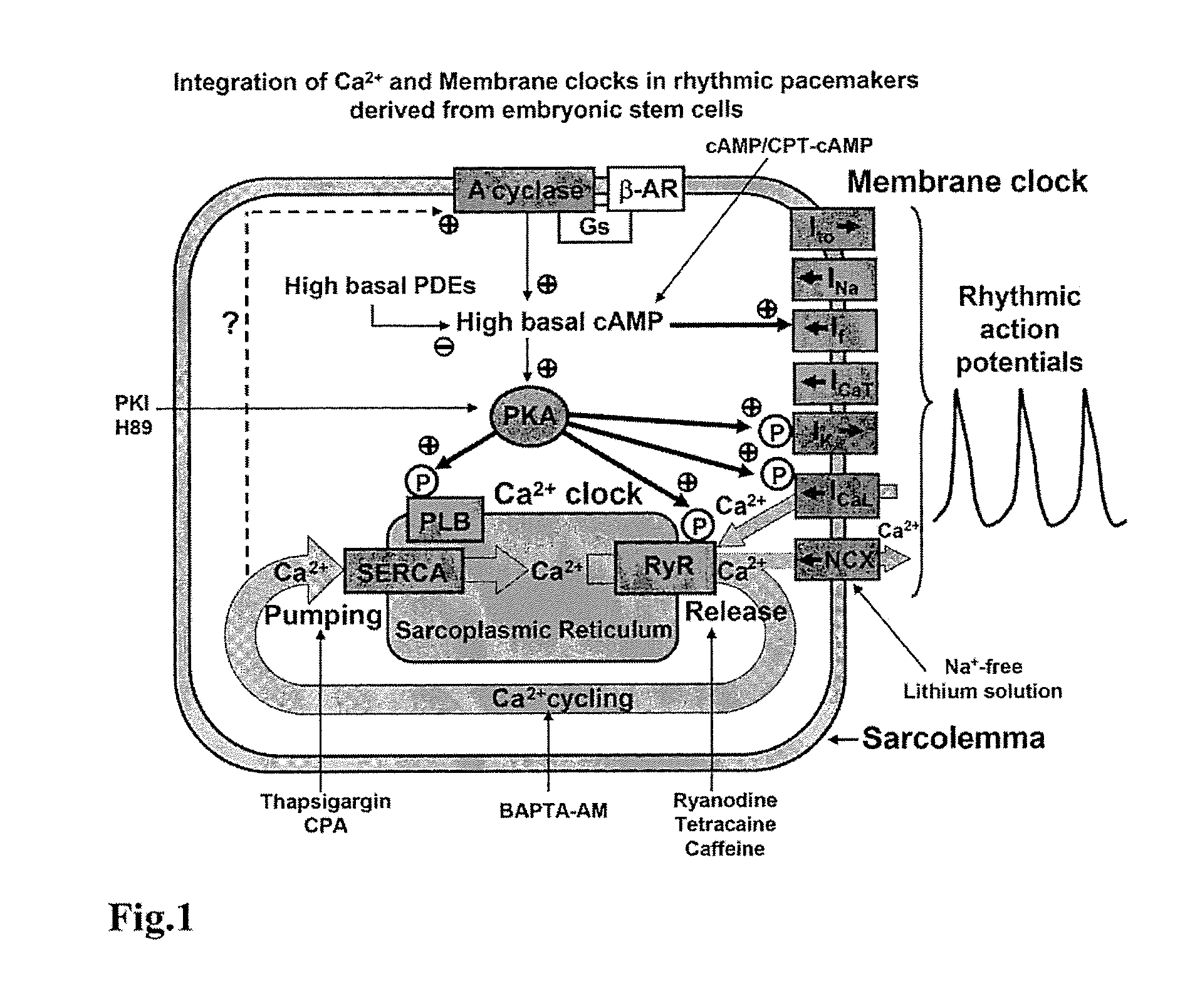
[0073]Only late-stage differentiated ESCs (7+9 to 14 days) were used. These cells possess both major ion channels (ICaL, If, INa, Ito, IK)(4), exchangers (NCX), and Ca2+ cycling proteins (RyR2, SERCA2A, calcequestrin, and phospholamban). Late-stage ESCs have been successfully used as temporary biological pacemakers in whole animal model experiments.

[0074]Mouse ES cells of line R1 were differentiated into cardiac cells using the “hanging drop” technique. ESCs were isolated from in vitro-grown embryoid bodies by microdissection followed by collagenase treatment. ES cells of line R1 were cultivated on inactivated feeder layer of primary mouse embryonic fibroblasts on gelatin (0.1%)-coated petri dishes (Fischerbrand) in DMEM supplemented with 15% heat-inactivated fetal bovine serum (FBS), 2 mmol / L L-glutamine, 0.1 mmol / L γ-mercaptoethanol, non-essential amino acids (NEAA, stock solution diluted 1:100), penicillin-streptomycin (stock solution dilute...

example 2

Electrophysiology

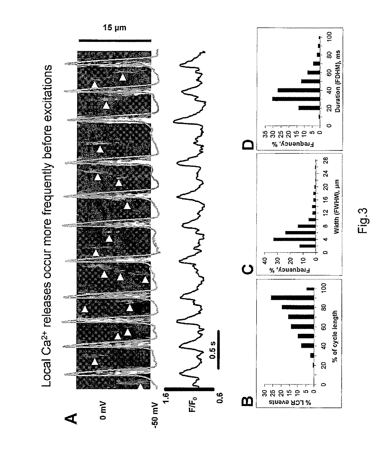
[0075]ESCs differentiated in vitro for 9 to 20 days after plating of 7 days old embryoid bodies were studied during continuous superfusion with solution containing in mmol / L: 140 NaCl, 5.4 KCl, 5 HEPES, 2 MgCl2, 1.8 CaCl2, 10 glucose, (pH adjusted to 7.4 with NaOH). Selection of the cells for patch-clamp technique was based on spontaneous beating. Action potentials were measured at physiological temperature (35±2° C.) with standard current-clamp technique (Axopatch 200, Axon Instruments, Foster City, Calif., USA), pCLAMP7 software was used for data acquisition and analysis (Axon Instruments). Perforated patch-clamp technique was performed with β-escin added to the electrode solution. Patch pipette solution contained in mmol / L: 120 K-gluconate, 5 NaCl, 5 Mg-ATP, 5 HEPES, 20 KCl, 3 Na2ATP (pH adjusted to 7.2 with NaOH). Recording of L-type Ca2+ current (ICaL) simultaneously with Ca2+ signal imaging (see Example 3) were performed under whole cell voltage clamp. Patch p...

example 3

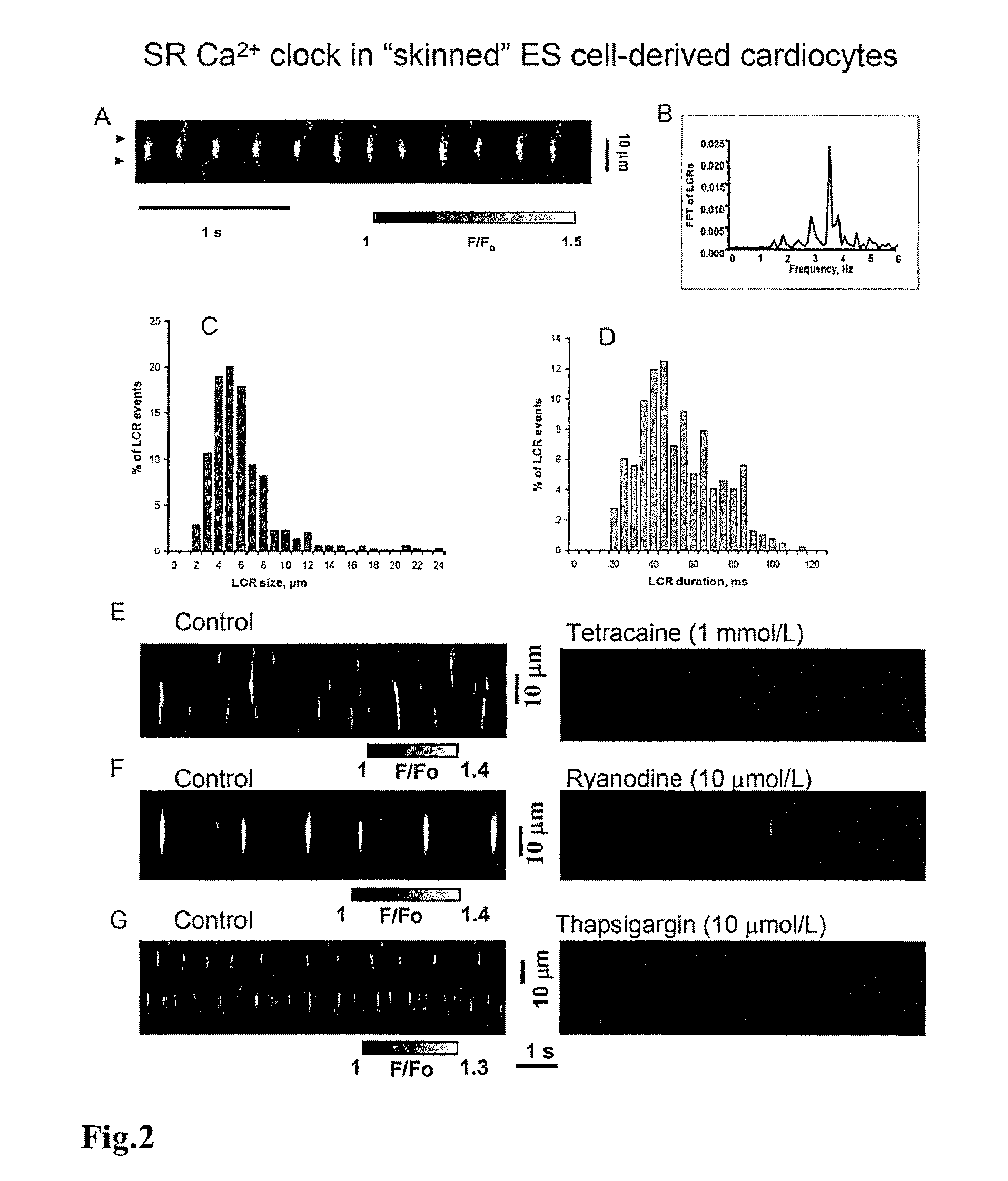
Ca2+ Imaging in Intact Cells

[0076]Confocal imaging of Ca2+ release in ESCs was performed by placing cells on the stage of a inverted confocal microscope and loaded for 15 min with 10 μmol / L fluo-4 AM in dimethyl sulfoxide (Molecular Probes, Eugene, Oreg.) in perforated patch experiments. In whole cell configuration, fluo-4 was added directly to the pipette solution.


PUM

PropertyMeasurementUnit
membrane potentialaaaaaaaaaa
volumesaaaaaaaaaa
volumesaaaaaaaaaa

Abstract

Biological pacemakers engineered to intrinsically generate rhythmic excitations are disclosed. In addition, methods of producing the biological pacemakers are disclosed. Methods of treating or preventing arrhythmia and heart disease associated with a defective pacemakers are also disclosed.

Description

CROSS REFERENCE TO RELATED APPLICATION[0001]This application claims the benefit of U.S. Provisional Application No. 61 / 180,491 filed May 22, 2009, herein incorporated by reference in its entirety.STATEMENT OF FEDERALLY SPONSORED RESEARCH[0002]The U.S. Government may have certain rights in the invention described herein, which was made in part with funds from NIH Contract No. HHSN263200700019I.FIELD OF THE INVENTION[0003]The present invention relates to biological pacemakers. More specifically, the invention relates to biological pacemakers engineered to intrinsically generate rhythmic electric excitations, methods of producing the biological pacemakers, and methods of treating or preventing arrhythmia and heart disease associated with a defective pacemaker.BACKGROUND OF THE INVENTION[0004]Cardiac contraction in a healthy human heart is initiated by spontaneous excitation of the sinoatrial (“SA”) node located in the right atrium. The electric impulse generated by the SA node travels ...

Claims


Application Information

Patent Timeline
Patent Type & Authority Applications(United States)
IPC IPC(8): A61K35/34A61P9/06C12N5/10
CPCA61K48/005C12N5/0657C12N15/86C12N2500/14C12N2830/002C12N2506/02C12N2510/00C12N2740/15043C12N2501/01A61P9/06
Inventor MALTSEV, VICTORLAKATTA, EDWARD G.ZAHANICH, IHORSIRENKO, SYEVDAMILKHEEV, MAXIMVODOVOTZ, YORAM
Owner UNIVERSITY OF PITTSBURGH
Why Eureka
Try Eureka