Compositions and Methods for Implantation of Processed Adipose Tissue and Processed Adipose Tissue Products

a technology of adipose tissue and adipose tissue products, applied in the field of regenerative medicine, can solve the problems of requiring significant time for new tissue formation, method cost, and procedure limitations, and achieve the effect of reducing the lifetime of implanted processed adipose tissu

Inactive Publication Date: 2012-07-26
THE JOHN HOPKINS UNIV SCHOOL OF MEDICINE
View PDF5 Cites 60 Cited by
  • Summary
  • Abstract
  • Description
  • Claims
  • Application Information

AI Technical Summary

Benefits of technology

[0104]As used herein, a “substantial immune response” is understood as an immunological response of a subject after implantation of a processed adipose tissue of the invention that requires intervention by a medical professional (e.g., need for removal of the processed adipose tissue, administration of immunosuppressive drugs); or results in substantially reduced lifetime of the implanted processed adipose tissue, for example the duration of the implanted processed adipose tissue is decreased by 50% or more, at least 60% or more, at least 70% or more, at least 80% or more, or at least 90% or more, and wherein the implanted processed adipose tissue is infiltrated with at least one type of inflammatory cell including, but not limited to, macrophages, neutrophils, and eosinophils. A substantial immune response does not include temporary and transient (e.g., one week or less, 6 days or less, 5 days or less, 4 days or less, 3 days or less, 2 days or less, or one day or less) of general redness, irritation, and swelling at the site of implantation that is reduced over time, and may not be a response to the implanted tissue per se, but instead to the disruption of the skin or otherwise adjacent tissue due to the disruption or stretching of the skin or tissue associated with implantation.

Problems solved by technology

While biomaterials and cells are often employed to regenerate new tissues, these methods tend to be costly and require significant time for new tissue formation.
However, such procedures also have their limitations.
Such flaps are necessarily limited in size by the amount of tissue present in the woman for use, and transfer of muscle from the abdomen or back can extend recovery time and result in donor site morbidity.
Such loss of persistence requires multiple procedures to maintain the desired correction.
In addition, implanted adipose tissue often leads to post-operative calcifications.
This phenomena is of particular importance for women with a history of breast cancer undergoing breast reconstruction following mastectomy, as the calcifications may interfere with mammography readings and result in multiple, unnecessary breast biopsies and anxiety.

Method used


Image

  • Compositions and Methods for Implantation of Processed Adipose Tissue and Processed Adipose Tissue Products
  • Compositions and Methods for Implantation of Processed Adipose Tissue and Processed Adipose Tissue Products
  • Compositions and Methods for Implantation of Processed Adipose Tissue and Processed Adipose Tissue Products

Examples


example 1

Preparation of a Cellular Biocompatible Biomaterial

[0196]A lipoaspirate was obtained using standard minimally invasive surgical techniques. Tumescent fluid was removed and the lipoaspirate was placed on ice until next step. The lipoaspirate was combined with various surfactants and scaffolds for the preparation of the biocompatible biomaterial.

[0197]1. Hyaluronic acid was emulsified with cellularized lipoaspirate in a 1:1 ratio with the addition of 5% Pluronic surfactant.

[0198]2. A 10% weight per volume PEG-DA was dissolved in a 50:50 ratio of PBS:Lipoaspirate.

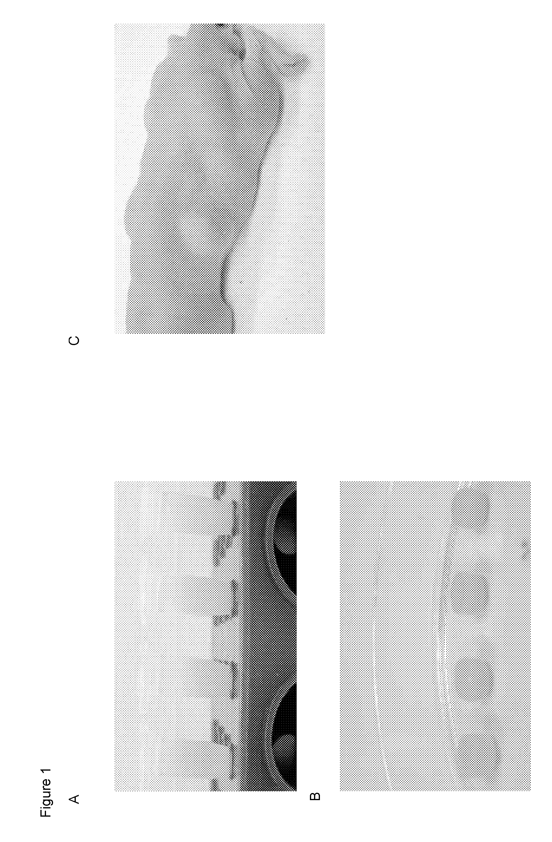
[0199]FIG. 1 shows emulsification of the lipoaspirate and aqueous PEG mixture in the absence or presence of surfactant. In FIG. 1A, the far left tube contains no surfactant. Phase separation between the aqueous and lipid layers of the mixture is evident. Moving to the right, increased concentrations of surfactant were added, and improved emulsification was observed.

[0200]The HA used was a commercially available crosslinked HA ...

example 2

Preparation of Acellular Biomaterial / Processed human Adipose Tissue (PhAT)

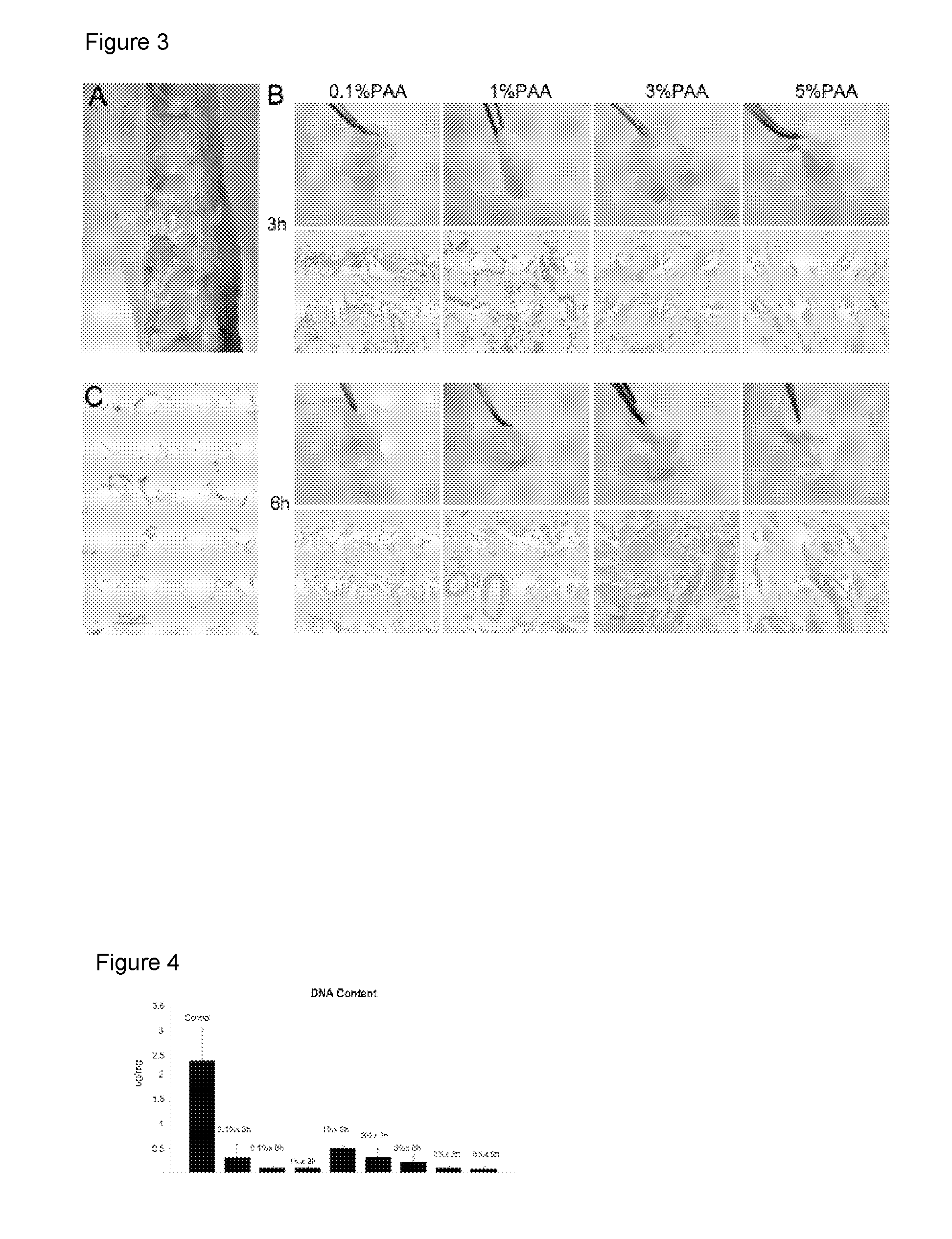
[0206]Tissue acquisition and processing. Tissue was acquired from fresh surgical and cadaveric sources with appropriate consent of the donors. A representative sample of subcutaneous fat is shown in FIGS. 3A and 9A. A representative histological section showing nuclei, lipid vacuoles, and extracellular matrix is provided in FIGS. 3C and 9C.

[0207]Subcutaneous fat was isolated from the sample by scraping. The scraped adipose tissue was homogenized in a blender. The homogenate was then placed on a strainer and washed for 5 minutes under deionized water to wash lipid and cellular debris. This was repeated three times. An equal weight of the homogenized and washed tissue was placed in 0.1%, 1%, 3%, or 5% peracetic acid for 3 or 6 hours on a shaker at 37° C. As adipocytes die, oil is released from the cells. To fully infiltrate the adipose tissue and remove the oil, the material was manually manipulated with a morta...

example 3

Analysis of Acellular Biocompatible Biomaterial

[0215]The acellular biocompatible biomaterial (processed human adipose tissue or PhAT) was tested using a number of assays to demonstrate that the material is acellular, lipid-free, includes intact extracellular matrix (ECM), and the appropriate dynamic stiffness. Exemplary assays used are provided. Other methods to determine if the material has the desired characteristics are known in the art.

[0216]Cell Free. Hematoxylin and eosin (H&E) staining was performed on paraffin-embedded sections of PAT prepared by at least one of the methods of the previous example to determine the presence of nuclei (cells) (compare FIGS. 7C and 9C to FIGS. 7B and 9D). No cellular material or nuclei were observed after processing with PAA and DNAse (FIG. 7B) or with PAA, TX-100, and DNAse (FIG. 9D) per the methods of the invention.

[0217]In addition, MHC class I immunostaining can be performed to evaluate the presence of antigens.

[0218]DNA content: To demonst...


PUM

PropertyMeasurementUnit
elastic modulusaaaaaaaaaa
diameteraaaaaaaaaa
thicknessaaaaaaaaaa

Abstract

The invention provides compositions and methods for the preparation of processed adipose tissue. The invention further provides methods of use of the processed adipose tissue.

Description

CROSS REFERENCE TO RELATED APPLICATIONS[0001]This application claims priority to Provisional Patent Application Ser. No. 61 / 232,915 filed on Aug. 11, 2009. This application is related to U.S. Provisional Patent Application Ser. No. 61 / 065,322 filed on Feb. 11, 2008 and PCT application PCT / US2009 / 00887 filed on Feb. 11, 2009 and published on Aug. 20, 2009 as WO 2009 / 102452. The applications are all incorporated herein by reference in their entirety.BACKGROUND[0002]The field of regenerative medicine aims to provide tissue substitutes for reconstruction secondary to trauma, disease or congenital abnormalities. While biomaterials and cells are often employed to regenerate new tissues, these methods tend to be costly and require significant time for new tissue formation. Restoration of soft tissue form is critical for a number of applications including trauma reconstruction, breast reconstruction, and cosmetics (nasolabial folds, wrinkles, etc). In general there are two approaches today;...

Claims


Application Information

Patent Timeline
Patent Type & Authority Applications(United States)
IPC IPC(8): A61K35/12A61P31/00A61P29/00A61K38/18C12P1/00A61K35/35
CPCA61K31/00A61L27/3633A61L2430/40C12N5/0653C12N2533/90A61K35/35A61K45/06A61K2300/00A61L2430/34A61P29/00A61P31/00
Inventor NAHAS, ZAYNAELISSEEFF, JENNIFER H.WU, IWEN
Owner THE JOHN HOPKINS UNIV SCHOOL OF MEDICINE
Why Eureka
Try Eureka