
Methods for diagnosis and treatment of cellular proliferative disorders

a technology for cellular proliferative disorders and diagnosis, applied in the field of cancer, can solve the problems of presenting a barrier to the success of these efforts, and achieve the effects of preventing nuclear export, promoting apoptosis, and high levels of pk

Inactive Publication Date: 2012-11-01
SANFORD BURNHAM MEDICAL RES INST
View PDF0 Cites 2 Cited by
  • Summary
  • Abstract
  • Description
  • Claims
  • Application Information

AI Technical Summary

Benefits of technology

[0012]The present invention is based on the seminal discovery of the role of ATF2 at the outer membrane of mitochondria, where it is recruited following genotoxic stress in order to promote apoptosis. The ability of ATF2 to mediate this newly discovered function depends on PKC-activity. Constitutively high levels of PKCε in melanoma, prevent nuclear export of ATF2 and subsequent mitochondrial-dependent tumor suppressor function.
[0017]Also provided herein is a method for diagnosis of or prediction of risk of a cellular proliferative disorder in a subject. The method includes obtaining a biological sample from the subject; contacting the sample with an antibody that recognizes phosphorylated ATF2; and detecting the level of phosphorylated ATF2, wherein an increased level of phosphorylated ATF2 in the sample compared to a healthy control indicates a presence or increased risk of a cellular proliferative disorder, thereby providing a diagnosis of or prediction of risk of the disorder in the subject. A finding of increased phosphorylation at T52 is indicative of increased resistance to genotoxic stress and increased survival of the cell. In one embodiment, the phosphorylated ATF2 is located primarily in the nucleus. An increased level of phosphorylated ATF2 in the nucleus may, for example, be indicative of a likelihood of resistance of the cell to genotoxic stress and cell survival.

Problems solved by technology

Although clinical trials of inhibitors for B-Raf and MEK have shown promising results against melanoma, the development of resistance to these monotherapies presents a barrier to the success of these efforts.

Method used


Image

  • Methods for diagnosis and treatment of cellular proliferative disorders
  • Methods for diagnosis and treatment of cellular proliferative disorders
  • Methods for diagnosis and treatment of cellular proliferative disorders

Examples


example 1

Genotoxic Stress Induces ATF2 Nuclear Export and Mitochondrial Localization

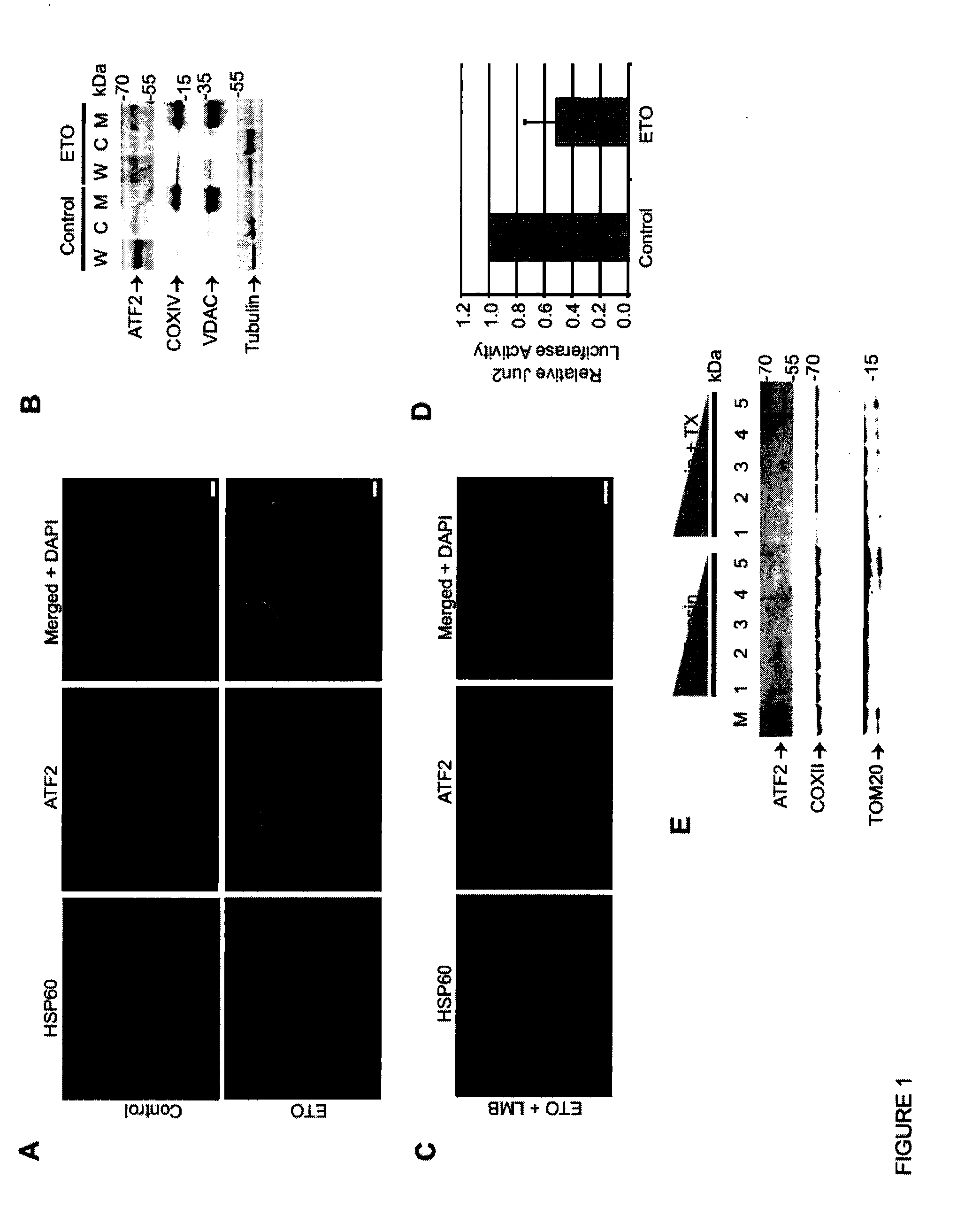
[0061]This example demonstrates that ATF2 localizes at the mitochondrial outer membrane in response to genotoxic stress.

[0062]While ATF2 cytoplasmic localization has been previously reported in various tissues, the function of cytoplasmic ATF2 has not yet been described. Based on the observed cytosolic localization of ATF2 in squamous cell carcinoma (SCC) tumors, an SCC line (SCC9 ) was employed to analyze potential ATF2 cytoplasmic function. Mass spectrometric analysis of cytosol-localized ATF2-bound proteins in SCC9 cells identified a cluster of mitochondria-outer membrane related proteins, suggestive of an ATF2 mitochondrial interaction (FIG. 15). Indeed, while SCC9 cells grown under non-stressed conditions exhibit predominantly nuclear localization of ATF2 , genotoxic stress induced by etoposide resulted in its accumulation at the mitochondria in ˜64% of the cells subjected to this treatment (FIG. 1A). Si...

example 2

ATF2 Interacts with HK1:VDAC1 Complexes Following Genotoxic Stress

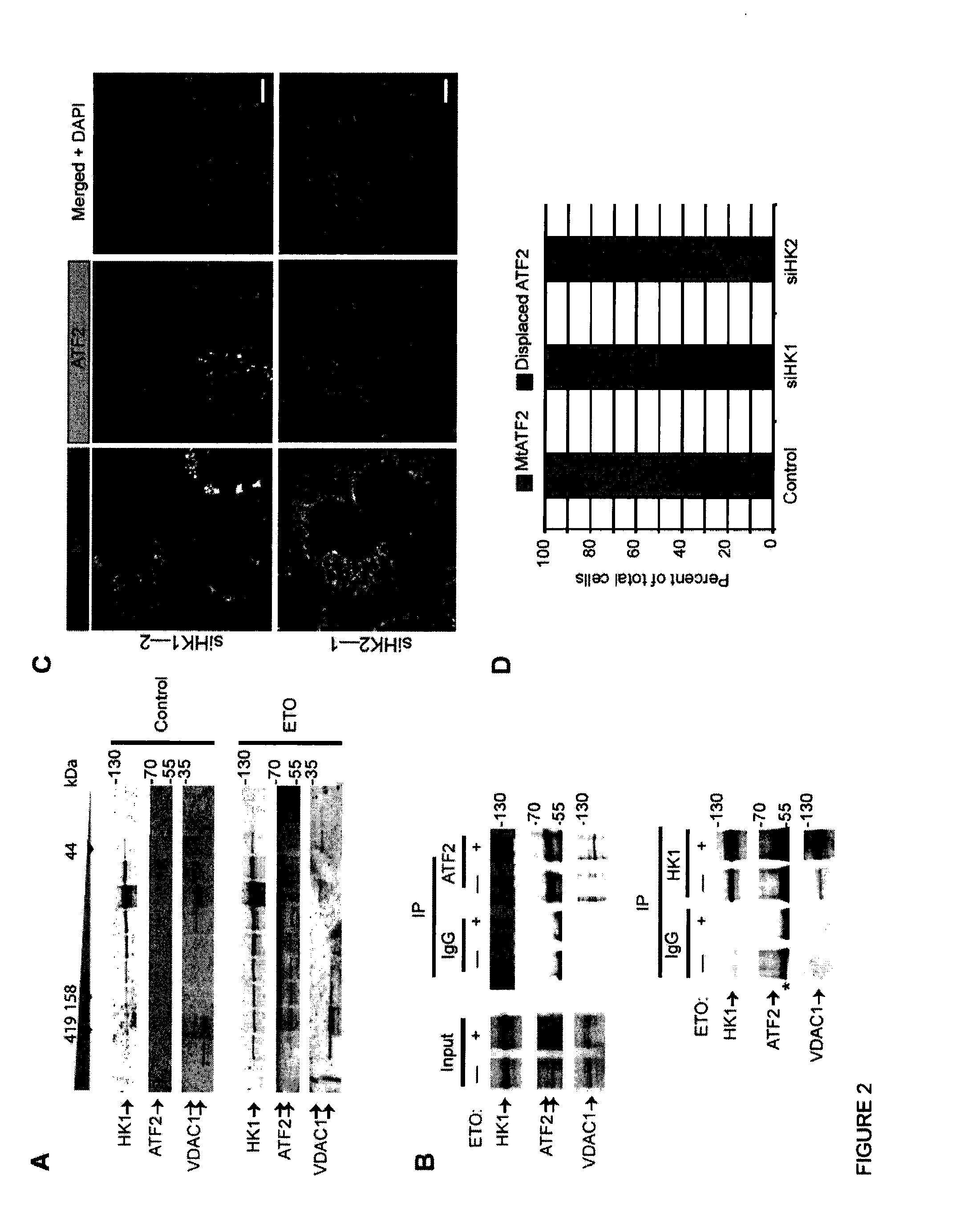
[0065]This example illustrates that ATF2 interacts with VDAC1 and HK1 complex at the mitochondria.

[0066]To determine ATF2 function at mitochondria, interaction of various MOM protein(s) with ATF2 was examined, with a focus on proteins identified in a mass spectrometry analysis (FIG. 15). Among the top-ranking candidates was a group of proteins known to form MOM complexes following exposure to genotoxic stress, including Hexokinase-1 (HK1), and Voltage-Dependent Anion Channel 1 (VDAC1) (FIG. 8).

[0067]In response to various forms of stress, HK1 and HK2 bind to VDAC1, which oligomerizes to form large molecular weight complexes. To determine whether ATF2 is part of these complexes, an assessment was made of the distribution of ATF2, HK1 and VDAC1 in fractions obtained following fast protein liquid chromatographic (FPLC) analysis using a gel filtration column. Whereas prior to stress, ATF2 did not co-distribute to fraction...

example 3

PKCε-Phosphorylation Negatively Regulates ATF2 Mitochondrial Localization

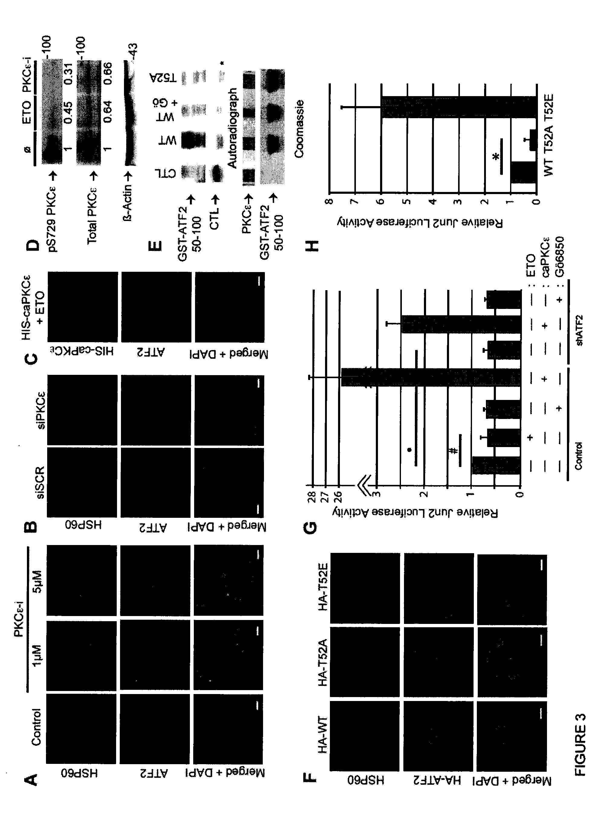
[0069]This example illustrates that PKCε phosphorylation of ATF2 on Thr52 negatively regulates its mitochondrial localization.

[0070]Phosphorylation of ATF2 by p38 or JNK on Thr69 / 71 is required for its dimerization with members of the AP-1 transcription factor family and for transcriptional activity. However, phosphorylation of Thr69 / 71 was not required for ATF2 mitochondrial localization, as inhibition of either p38 or JNK activity, either by specific pharmacological inhibitors did not alter its nuclear localization (FIG. 9B).

[0071]To identify kinases that might affect ATF2 mitochondrial localization, the ATF2 protein sequence was subjected to phosphorylation site scanning (http: / / scansite.mit.edu). This analysis predicted an uncharacterized, but highly probable PKCε / ∂ / ζ phosphorylation site at ATF2 residue Thr52. To evaluate the possible role of PKCε in the mitochondrial function of ATF2, immunofluorescent st...


PUM

PropertyMeasurementUnit
molecular weightaaaaaaaaaa
disorderaaaaaaaaaa
genotoxic stressaaaaaaaaaa

Abstract

Methods for the diagnosis or prognosis of cellular proliferative disorders by detecting expression of PKC in cancer cells or tumor cells are provided herein. Also provided are methods for treating a melanocyte proliferative disorder with agents that modulate the translocational activity of ATF2 and / or PKCε activity.

Description

CROSS REFERENCE TO RELATED APPLICATION(S)[0001]This application claims the benefit of priority under 35 U.S.C. §119(e) of U.S. Ser. No. 61 / 408,913, filed Nov. 1, 2010, the entire content of which is incorporated herein by reference.GRANT INFORMATION[0002]This invention was made in part with government support under NCI Grant Nos. CA099961 and CA051995. The United States government has certain rights in this invention.BACKGROUND OF THE INVENTION[0003]1. Field of the Invention[0004]The invention relates generally to cancer and more specifically to diagnosis and treatment of tumors and melanoma through activating transcription factor 2 (ATF2) and PKCε.[0005]2. Background Information[0006]Malignant melanoma, the leading cause of death from skin cancer, exhibits a remarkable resistance to chemotherapy and, therefore, remains an area of significant unmet need in oncology. Current understanding of mechanisms underlying melanoma development and progression has enabled development of specifi...

Claims


Application Information

Patent Timeline
Patent Type & Authority Applications(United States)
IPC IPC(8): A61K39/395G01N21/64C07K16/18C12Q1/68G01N33/53A61P35/00G01N33/573C40B30/06
CPCC12Q1/6886C12Q2600/106G01N33/5011G01N33/5743G01N33/57484C07K16/40G01N2333/91205G01N2440/14G01N2510/00G01N2800/50C07K16/18G01N2333/4706A61P35/00
Inventor RONAI, ZE'EV
Owner SANFORD BURNHAM MEDICAL RES INST
Learn More