Removal of virulence factors through extracorporeal therapy

a technology of virulence factor and extracorporeal therapy, which is applied in the direction of antibacterial agents, extracellular fluid disorders, separation processes, etc., can solve the problems of increased paralysis, limited success, and increased paralysis

Inactive Publication Date: 2012-12-06
EXTHERA MEDICAL
View PDF1 Cites 33 Cited by
  • Summary
  • Abstract
  • Description
  • Claims
  • Application Information

AI Technical Summary

Problems solved by technology

For example, Guillian-Barre syndrome is currently understood to be an autoimmune disorder triggered by viral infection that stimulates the body's immune system to over produce antibodies or other proteins which can attack the patient's nervous system, causing increasing levels of paralysis, Most patients recover over time, though such patients appear to be susceptible to recurrence of the condition from subsequent viral infections.
Although this approach has been investigated by many scientists, only limited success has been reported to date.
The most common problem has been bleeding complications associated with the large amounts of free heparin introduced into the blood stream to achieve a clinically-useful reduction of pathogenic microbes.
The disease can be fatal to animals.
When inhaled, the body's immune system can quickly become overwhelmed and go into shock.
The use of the attenuated live vaccines may have local adverse responses and are not very effective.
Unrecognized infections usually are fatal.

Method used


Image

  • Removal of virulence factors through extracorporeal therapy
  • Removal of virulence factors through extracorporeal therapy
  • Removal of virulence factors through extracorporeal therapy

Examples


example 1

6.1. Example 1

Preparation of Heparin Column

[0092]Polyethylene (PE) beads, with an average diameter of 0.3 mm (lot no. 180153), are supplied by the Polymer Technology Group (Berkeley, USA) and the columns (Mobicol, 1 mL) are obtained from MoBiTec (Germany). Heparin and polyethyleneimine (PEI) are purchased from Scientific Protein Laboratories (Waunakee, Wis., USA) and BASF (Ludwigshafen, Germany) respectively. All chemicals used are of analytical grade or better.

[0093]Immobilization of heparin onto the beads are performed as described by Larm et al. (Larm O, Larsson R, Olsson P. A new non-thrombogenic surface prepared by selective covalent binding of heparin via a modified reducing terminal residue. Biomater Med Devices Artif Organs 1983; 11: 161-173).

[0094]The polymeric surface is heparinized using the general procedure described below.

[0095]The polymeric surface is etched with a oxidizing agent (potassium permanganate, ammoniumperoxidisulfate) in order to introduce hydrophilic char...

example 2

6.2. Example 2

Removal of Toxins

[0103]Arterial blood is drawn from the hemodialyzers of patients. The blood is collected in EDTA vacuum tubes and immediately 1 mL is applied to the previously prepared columns and passed through using a roller-pump at for example, one of 1, 5 and 10 mL / min. Blood that has passed through the columns is immediately collected at the other end and cold-centrifuged (4500 G). The supernatants are subsequently collected and frozen at −80° C. for later analysis.

[0104]Briefly, passage through the heparinised beads results in a significantly bigger decrease in blood toxins as compared to non-heparinized beads.

example 3

6.3. Example 3

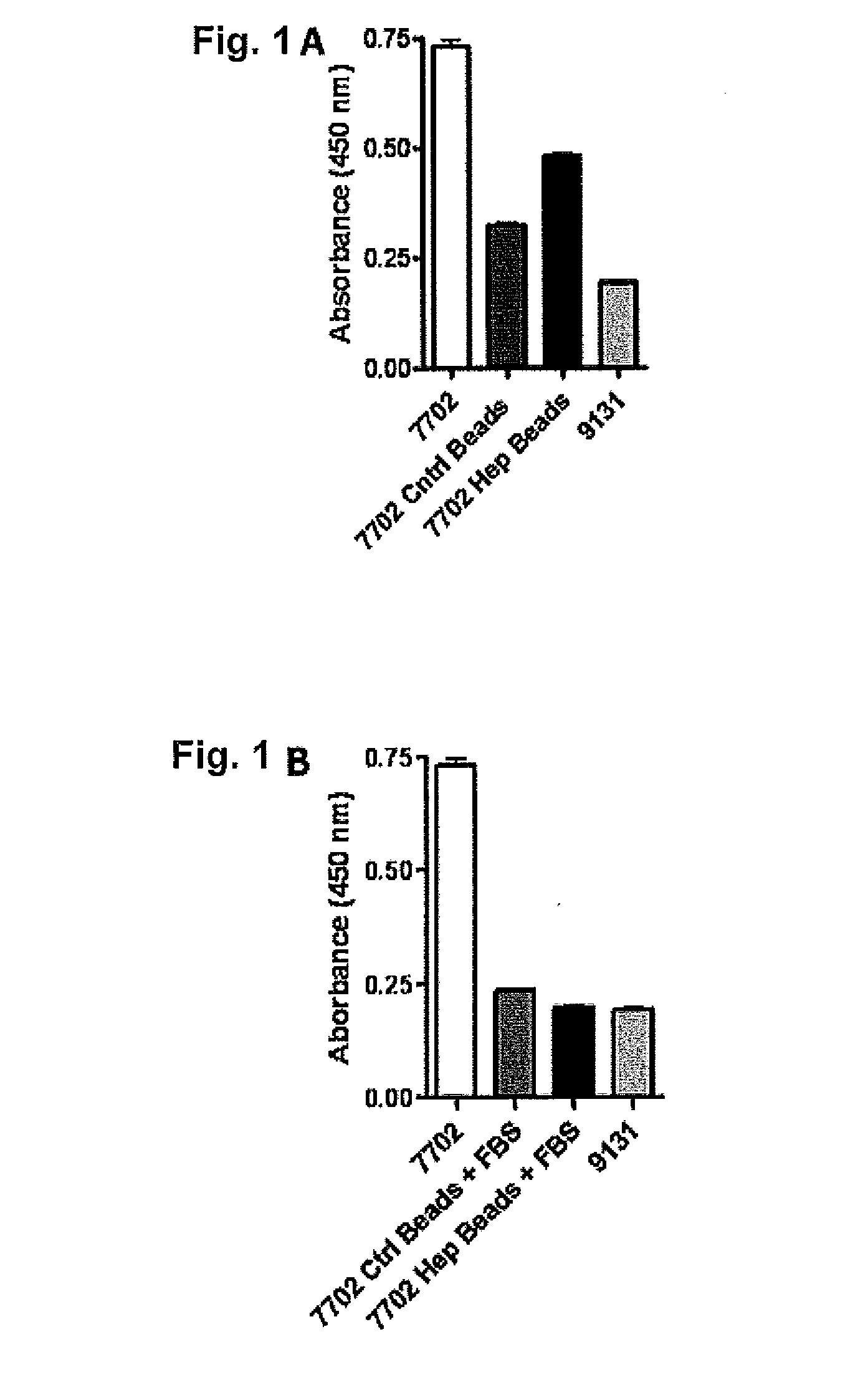
In Vitro Removal of B. anthracis PA

[0105]Bacterial Supernatants: Overnight cultures of B. anthracis 7702 or 9131 were cultured in Luria Broth (LB) at 37° C. while shaking at 250 RPM. 20 mLs of LB with 0.8% sodium bicarbonate (NaHCO3) at pH 8 (pH with 1 M HEPES) was inoculated with 1 mL overnight 7702 culture and grown until late exponential phase (approximately 7 hours) while shaking at 250 RPM at 37° C. Cultures were centrifuged for 5 minutes at 3500 RPM to remove bacterial cells and debris. Supernatants were collected and passed through a 0.2 μm filter and used immediately or stored at −20° C.

[0106]Preparation of Beads: 1 g heparin or control beads were added to syringes with a filter placed in the bottom and on top of the beads. Prior to experiments, beads were prepped by the addition of 2 mLs Tris-buffered Saline (TBS). Where Fetal Bovine Serum (FBS) was used, the beads were prepped by the addition of 2 mLs TBS followed by 2 mLs FBS, which was passaged over the bea...


PUM

PropertyMeasurementUnit
flow rateaaaaaaaaaa
diameteraaaaaaaaaa
diameteraaaaaaaaaa

Abstract

A method to remove virulence factors from infected blood by passing the blood through a surface cartridge with immobilized carbohydrates, such as heparin, wherein the virulence factors are toxins released from pathogens such as B. anthracis, S. aureus, and P. aeruginosa.

Description

FIELD OF THE INVENTION[0001]The present invention is directed to a method for removing pathogens and / or toxins released from pathogens from blood or serum (blood) by contacting the blood with a solid, essentially nonporous substrate which has been surface treated with heparin, heparin sulfate and / or other molecules or chemical groups (the adsorbent media or media) having a binding affinity for the pathogens and / or toxins to be removed (the adsorbents). The invention can be used to remove virulence factors, e.g. toxins, that are released from pathogens such as Bacillus anthracis, Pseudomonas aureginosa, and Staphylococcus aureus. In one aspect, the size of the interstitial channels within said media is balanced with the amount of media surface area and the surface concentration of binding sites on the media to provide adequate adsorptive capacity while also allowing relatively high flow rates of blood past the media.[0002]The present invention also provides a method of treating a dis...

Claims


Application Information

Patent Timeline
Patent Type & Authority Applications(United States)
IPC IPC(8): A61M1/36B01D15/08
CPCA61K31/727A61M1/362A61M1/3679A61P31/00A61P31/04A61P7/00A61P7/08A61M1/38
Inventor MCCREA, KEITHWARD, ROBERT S.
Owner EXTHERA MEDICAL
Why Eureka
Try Eureka