Remedy for cartilage-related diseases

a cartilage-related disease and treatment technology, applied in the direction of biocide, drug composition, peptide/protein ingredients, etc., can solve the problems of not being established as an effective treatment method, and activities other than the aimed activity become side effects, etc., to inhibit cartilage degradation, inhibit cartilage calcification, and stimulate chondrogenesis

Inactive Publication Date: 2012-12-13
ONO PHARMA CO LTD
View PDF3 Cites 2 Cited by
  • Summary
  • Abstract
  • Description
  • Claims
  • Application Information

AI Technical Summary

Problems solved by technology

Since the articular cartilage tissue is poor in repairing ability, it is known that even a microscopic lesion is difficult to be treated, gradually progresses and finally results in osteoarthritis.
In recent years, transplantation of self-chondrocytes into damaged part of cartilage has been carried out as a new therapeutic method, but has not been established yet as an actually effective therapeutic method because of the certain reasons such as limitation of the object to partial lesions, future problems of the cartilage-collected parts, necessity of a strictly managed culturing facility for the operation and the like.
However, since PGE2 itself has a variety of physical activity, there is a fault that the activities other than the aimed activity become side effects.

Method used


Image

  • Remedy for cartilage-related diseases
  • Remedy for cartilage-related diseases
  • Remedy for cartilage-related diseases

Examples


example 1

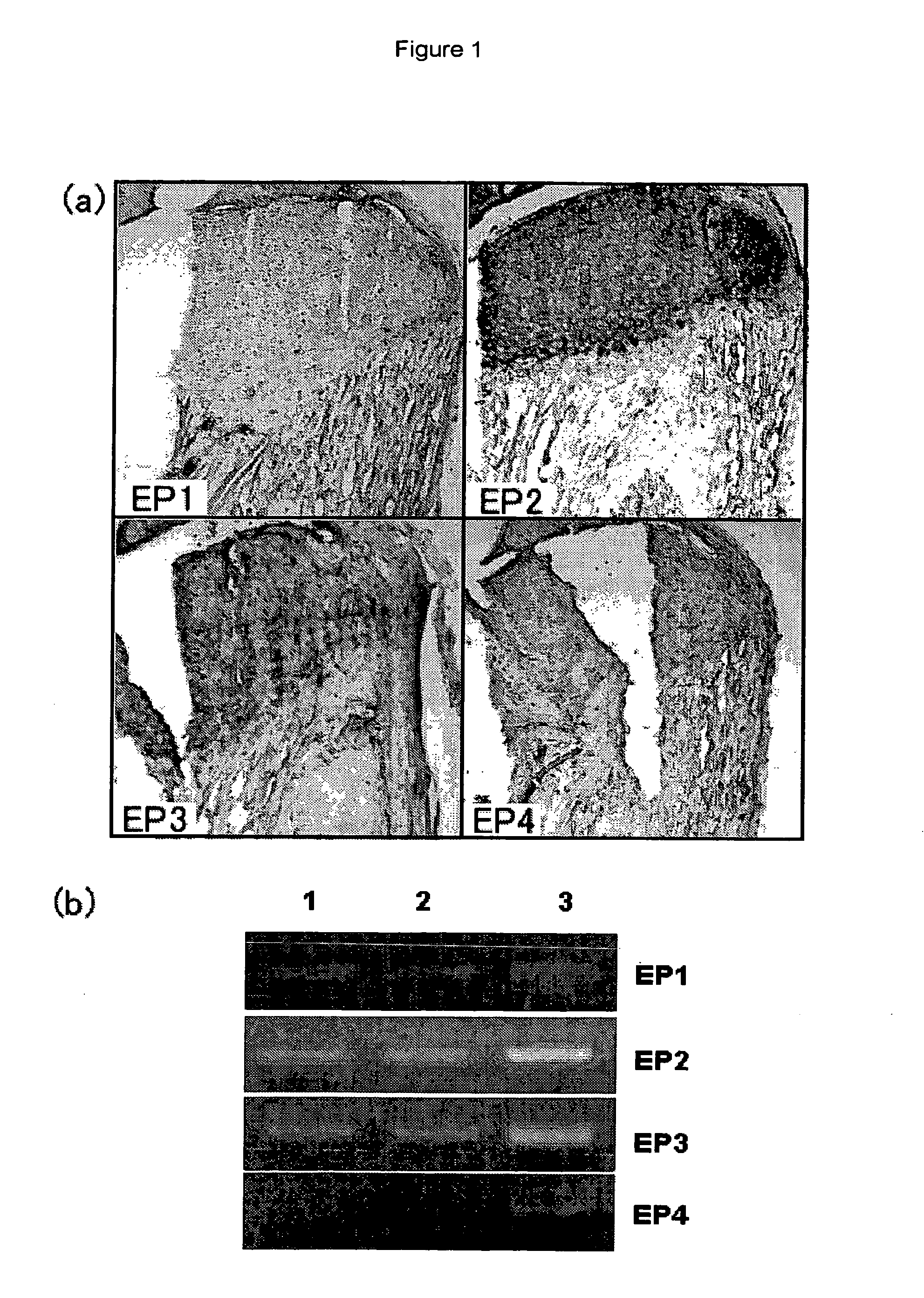
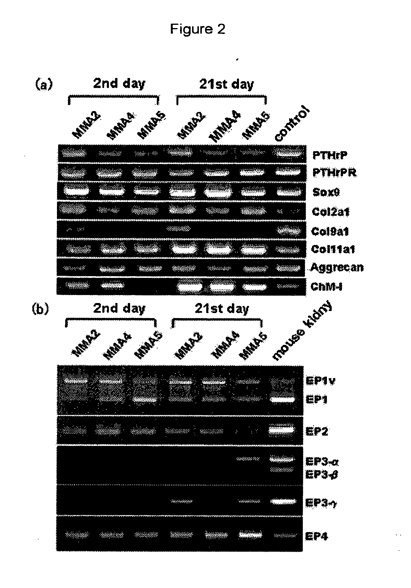
[0391]Cartilage tissues of a femur and the shinbone collected from a p53 defective mouse of 4 weeks of age (Tukada, T., Oncogene, 1992, vol. 8, pp. 3313-3322) were cut into pieces, treated with 0.1% collagenase and then cultured (culture conditions; 5% CO2, 37° C., under humidification, hereinafter, this was carried out under the same conditions) using DMEM / Ham's F12 (1:1) medium (contains 10% fetal bovine serum (FBS) and antibiotics (to be referred to as DMEM / Ham's F12 medium hereinafter)). By carrying out dilution sub-culturing from the cell group under a 80 to 90% confluent state, a chondrocyte strain MMA2 recognized by an international deposition number FERM BP-10029 was isolated. Expression analysis of the chondrocyte strain by RT-PCR method revealed that it expressed articular cartilage-related genes of type II collagen, aggrecan and the like (FIG. 2 (a)). Also, this was a cell having characters as an articular chondrocyte, such as formation of no calcified node even after a l...

example 2

[0393]Each of the chondrocytes was cultured at a cell density of 3×10⁵ cells / 100 mm culture dish (contains 5 μM indometacin). By respectively adding (17S)-2,5-ethano-6-oxo-17,20-dimethyl-PGE1 as a selective EP1 agonist, (5Z,9β,11α,13E)-17,17-propano-11,16-dihydroxy-9-chloroprostar-5,13,19-trienoic acid as a selective EP2 agonist, 11α,15α-dimethoxy-9-oxoprosta-5Z,13E-dienoic acid as a selective EP3 agonist, and 11α,15α-dihydroxy-9-oxo-16-(3-methoxymethylphenyl)-17,18,19,20-tetranol-3,7-dithiaprost-13E-enoic acid (obtained from ONO PHARMACEUTICAL CO., LTD.) as a selective EP4 agonist, respective actions after 72 hours were evaluated.

example 3

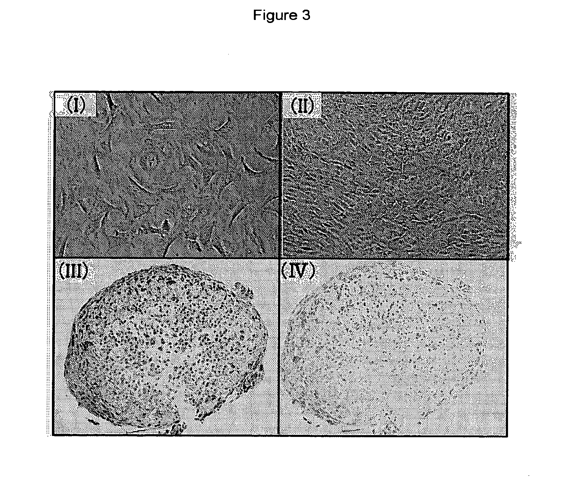
[0394]Monolayer culturing of each of the chondrocytes was started using DMEM / Ham's E12 medium containing 50 mg / ml of ascorbic acid. Medium exchange was not carried out for the first 6 days after commencement of the culturing, but carried out thereafter on every other day, and cultured cell pellet recovered on the 21st day was fixed with 20% formaldehyde and embedded in paraffin. Its section of 6 μm in thickness was stained with hematoxylin-eosin (0.1 N) hydrochloric acid solution or 0.1% alcian blue (0.1 N) hydrochloric acid solution and photographed using an optical microscope.


PUM


Abstract

The present invention relates an agent for treating cartilage-related disease comprising as an active ingredient a substance having an EP2 and / or EP3 agonist activity. A substance having an agonist activity to EP2 and / or EP3 has effects of stimulating chondrogenesis, stimulating chondrocyte growth, stimulating chondrocyte differentiation, inhibiting cartilage calcification and inhibiting cartilage degradation, or effects of stimulating integrin mRNA expression, stimulating fibronectin mRNA expression, stimulating D1 mRNA expression and inhibiting osteopontin mRNA expression, and, therefore, is useful as an agent for treating cartilage-related disease.

Description

[0001]This is a Divisional application of U.S. application Ser. No. 10 / 565,831 filed Jan. 25, 2006, which is the National Stage Entry under 35 U.S.C. §371, of PCT / JP04 / 010890 filed on Jul. 23, 2004, which claims priority to Japanese Patent Application 2003-280191 filed on Jul. 25, 2003, all of which are incorporated herein by reference.TECHNICAL FIELD[0002]The present invention relates to an agent for treating cartilage-related disease and an agent for producing cartilage graft comprising as an active ingredient a substance having a selective EP2 and / or EP3 agonist activity.BACKGROUND ART[0003]As the articular cartilage lesions which cause pain, movable region limitation and the like, there are various lesions such as osteoarthritis, rheumatoid arthritis, traumatic or osteonecrosis-accompanied osteochondritis dissecans and the like, and particularly, the number of patients of osteoarthritis has been considerably increased with the advance of aging society. Since the articular cartil...

Claims


Application Information

Patent Timeline
Patent Type & Authority Applications(United States)
IPC IPC(8): A61K31/201A61K31/202A61P19/04A61K31/4406A61K31/5575A61K38/18A61K38/27A61K45/06A61P19/02A61P43/00
CPCA61K31/4406A61K31/5575A61K38/18A61K38/27A61K45/06A61K38/30A61K2300/00A61P19/00A61P19/02A61P19/04A61P19/08A61P43/00
Inventor TOGUCHIDA, JUNYA
Owner ONO PHARMA CO LTD
Why Eureka
Try Eureka