Low coherence enhanced backscattering tomography and techniques

a backscattering tomography and low coherence technology, applied in the field of biophotonics or biomedical optics, can solve the problems of low reproducibility, difficult for oct to image structures, and current approaches such as diffuse optical tomography (dot) suffer from poor spatial resolution (5-10 millimeters), and achieve low coherence light, high resolution advantage, and enhanced backscattering tomography

Active Publication Date: 2013-03-14
FAIRFIELD UNIVERSITY
View PDF6 Cites 7 Cited by
  • Summary
  • Abstract
  • Description
  • Claims
  • Application Information

AI Technical Summary

Benefits of technology

[0007]The present disclosure describes a method for depth-selective sensing of the superficial layer of tissue. In accordance with the disclosure, a 3D tomographic tool images the microarchitecture and molecular conformation of the superficial layer of tissue. The method, referred to herein as low coherence enhanced backscattering tomography (LEBT), combines the high resolution advantage of low coherence light and the high sensitivity advantage of light scattering to tissue structure and composition, and furthermore offers the following advantages:

Problems solved by technology

Traditional histopathological examination has several limitations, including: (1) it can only acquire the two dimensional information from the cells or nuclei of interest; (2) it is qualitative or semi-quantitative; (3) it requires tissue excision and processing for examination; and (4) it is frequently subject to the inter- and intra-personal variability in interpretation, thus with a relatively low reproducibility.
Current approaches such as diffuse optical tomography (DOT) suffer from poor spatial resolution (5-10 millimeters) due to light diffusion.
However, it is difficult for OCT to image structures such as nuclei for various technical reasons.

Method used


Image

  • Low coherence enhanced backscattering tomography and techniques
  • Low coherence enhanced backscattering tomography and techniques
  • Low coherence enhanced backscattering tomography and techniques

Examples


Embodiment Construction

[0025]LEBT and Biomedical Imaging

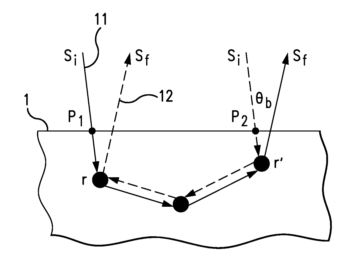
[0026]A LEBT technique according to the disclosure can image intact biological tissues at the microscopic scale ex vivo and in vivo based on optical contrast, extending LEBS to a three dimensional (3D) tomographic imaging modality. By detecting only low-order backscattering light via spatial coherence gating, LEBT solves the low spatial resolution problem due to light diffusion and achieves excellent depth selection. At the same time, low-order scattering light is sensitive to the microarchitecture and the molecular conformation of biological tissues, relating to physiological states such as the morphological alteration due to carcinogenesis and the oxygenation of hemoglobin.

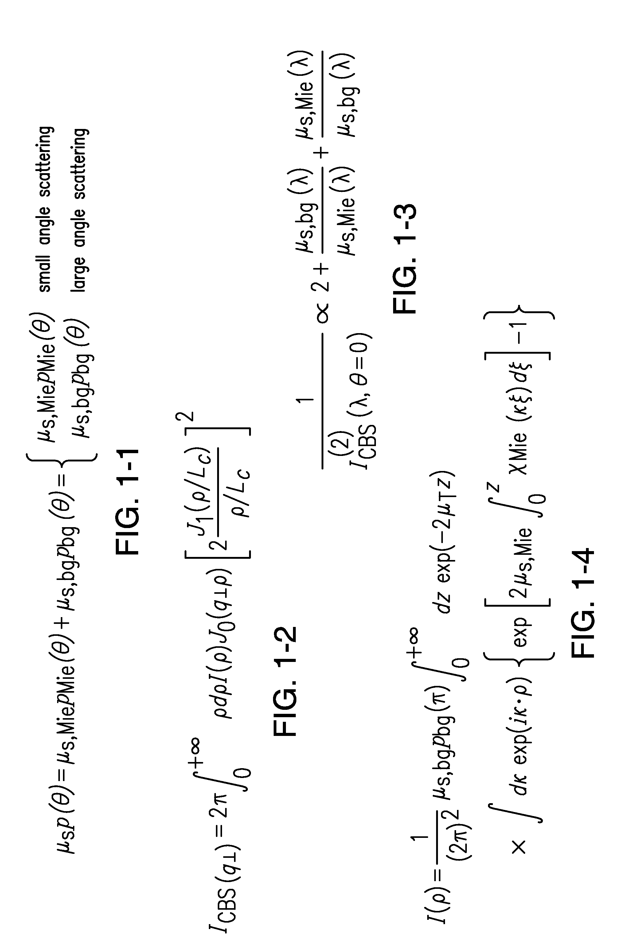
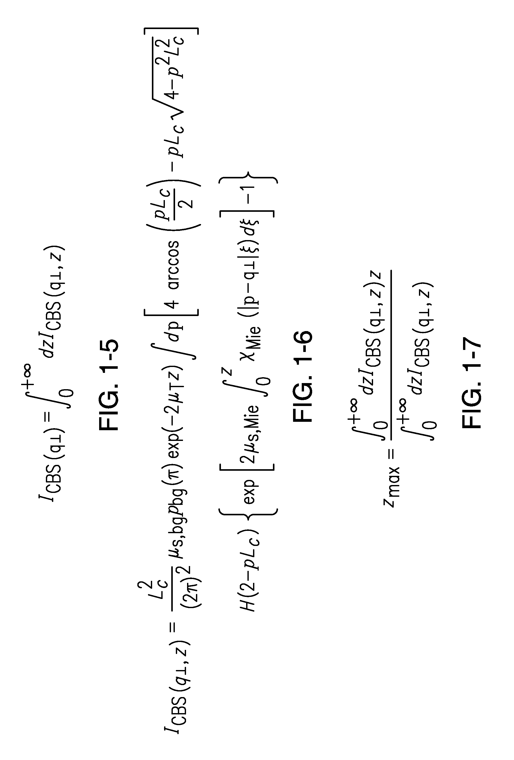
[0027]Epithelial tissues have a multi-layered structure composed of a superficial cellular layer (epithelium) with a characteristic thickness of ˜100 μm. The main characteristics of light propagation in a turbid medium can be summarized by a set of length scales: the scattering m...


PUM


Abstract

A low coherence enhanced backscattering tomography (LEBT) method is disclosed for depth-selective sensing of the superficial layer of tissue. 3D images of the microarchitecture and molecular conformation of the superficial layer of tissue are obtained. The method combines the high resolution advantage of low coherence light and the high sensitivity advantage of light scattering to tissue structure and composition. Intact tissue can be examined without the need of excision or processing. The method can be applied in in situ measurements. According to the method, 3D images of the nuclear morphology and cellular structure for the superficial layer of the tissue are generated; this is particularly useful in detecting cancer and precancer at the earliest stage of carcinogenesis.

Description

FIELD OF THE DISCLOSURE[0001]This disclosure relates to biophotonics or biomedical optics, and more particularly to characterization of the superficial layer of tissue using low coherence enhanced backscattering of light.BACKGROUND OF THE DISCLOSURE[0002]Histopathological examination of tissue has been an essential component of pathology. Current knowledge about carcinogenesis, cancer diagnostics and prognostication is predominantly based on histological study on tissue, cells and nuclei. Morphometric information, especially nuclear morphometry, has been shown to have great potential in assisting cancer screening, diagnosis, grading and classification, prognosis, analysis of angiogenesis, and evaluating the efficacy of therapy.[0003]Traditional histopathological examination has several limitations, including: (1) it can only acquire the two dimensional information from the cells or nuclei of interest; (2) it is qualitative or semi-quantitative; (3) it requires tissue excision and pr...

Claims


Application Information

Patent Timeline
Patent Type & Authority Applications(United States)
IPC IPC(8): G01B9/02
CPCA61B5/0066G01N21/4795G01B11/14G01B11/24G01B9/02G01N2021/4709
Inventor XU, MIN
Owner FAIRFIELD UNIVERSITY
Why Eureka
Try Eureka