Image processing method in microscopy

a microscopy and image processing technology, applied in image enhancement, image analysis, instruments, etc., can solve the problems of difficult to achieve good performance with reasonable optimization, low rendering performance in terms of display speed, and large size of digital images, so as to improve image registration efficiency and reduce image dimensions , the effect of faster computation of spatial transformation

Active Publication Date: 2013-04-11
KONINKLIJKE PHILIPS ELECTRONICS NV
View PDF7 Cites 24 Cited by
  • Summary
  • Abstract
  • Description
  • Claims
  • Application Information

AI Technical Summary

Benefits of technology

[0049]In a further embodiment, each digital image is provided from a multi-resolution image data structure. In the step of computing a transform function, any digital image used therein is obtained from a low resolution level in the image data structure. And in a step of retrieving a sub-region from an image, this image is preferably obtained from a higher resolution level in the image data structure. This provides for further improvement of the image registration efficiency, notably because it is substantially faster to compute the spatial transformation on a lower resolution image (e.g. the low resolution image has smaller image dimensions than the high resolution image). Moreover, as can be understood from before, computing the spatial transformation on the low resolution image, but applying it on the high resolution image, produces high quality output images with smaller computational overhead.

Problems solved by technology

A problem however with such image processing algorithms is that, usually in digital pathology, the size of digital images are far bigger than that of digital images taken from other usual medical imaging instruments such as ultrasound, MRI, CT, PET, or X-ray.
As a consequence, in digital pathology, the large size of the digital images often require much more intensive computation and often lead to a low rendering performance notably in terms of display speed.
However, it is still difficult to achieve good performance with reasonable optimizations.
Among others, such solutions again require a large amount of storage space and large bandwidth consumption notably between the storage in PACS and the computation device.
Even though this solution may provide real time performance, the down sampled image displayed to the pathologist may not have sufficient quality.
As mentioned before, image processing also refers to image registration and here again the above-described problems may occur.
However, due to the nature of the slide preparation and due to the digitization process, the tissue and its features may not have the exact same shape, appearance or spatial alignment, making it difficult to find the same region on different slides of adjacent tissue.
Similar to MR, the pixel intensity distribution of digital microscopy images depends on a large variety of factors, which are often difficult to control and possibly not uniform for the entirety of the image or between scans.
The three differences explained above (i.e. extreme image size, non-absolute pixel intensity distribution, and multi-modal acquisition of adjacent tissue cross-sections) means that registration of digital microscopy images, in particular of multi-modal images, for whole slide microscopy is not trivial and again requires intensive computation which limits the performance of the digital pathology systems in terms of speed.
Again, even though the two solutions described above (hardware or image processing algorithm optimization, and down sampling the whole slide image) may be used to improve the performance of the digital pathology system, this may be not be sufficient.

Method used


Image

  • Image processing method in microscopy
  • Image processing method in microscopy
  • Image processing method in microscopy

Examples


Embodiment Construction

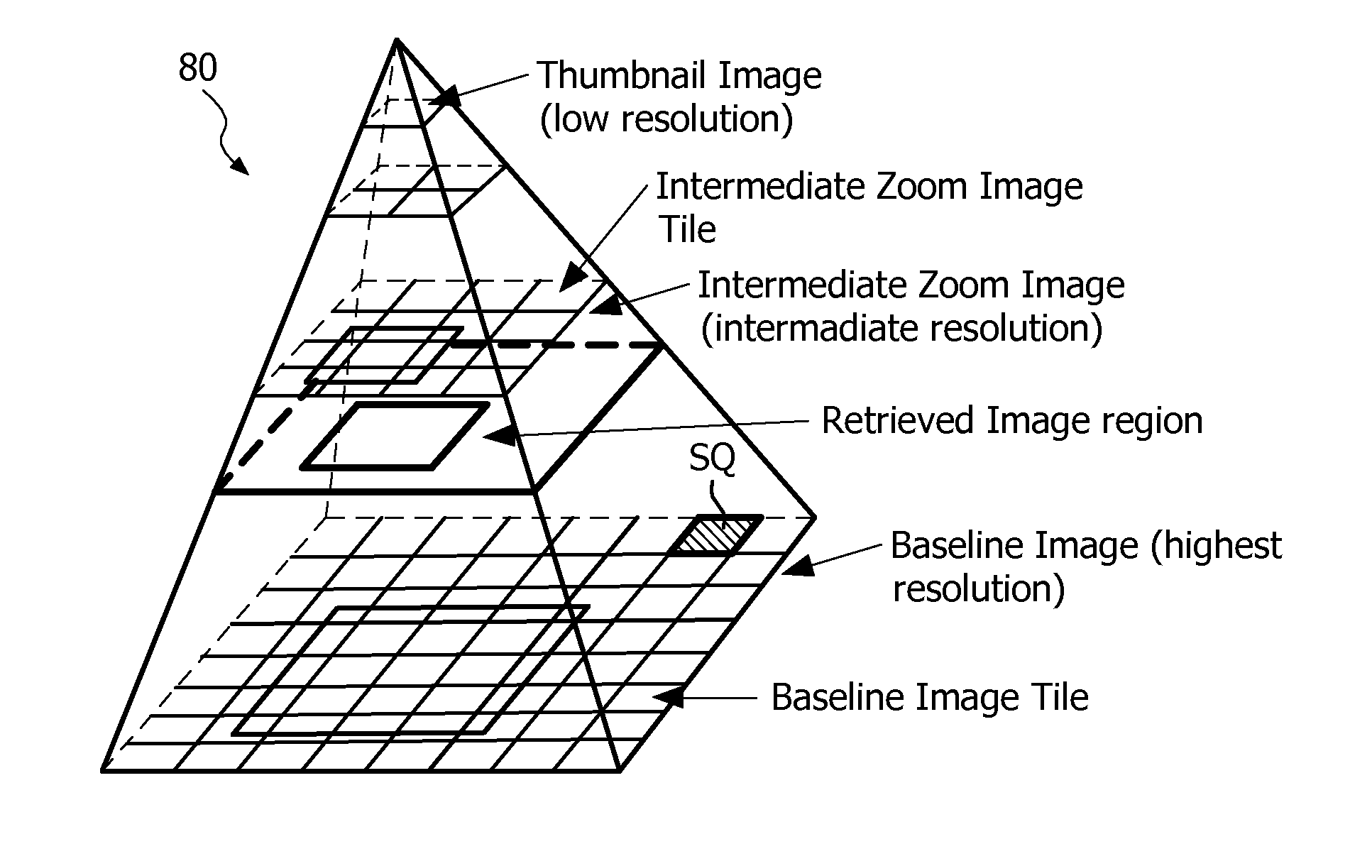
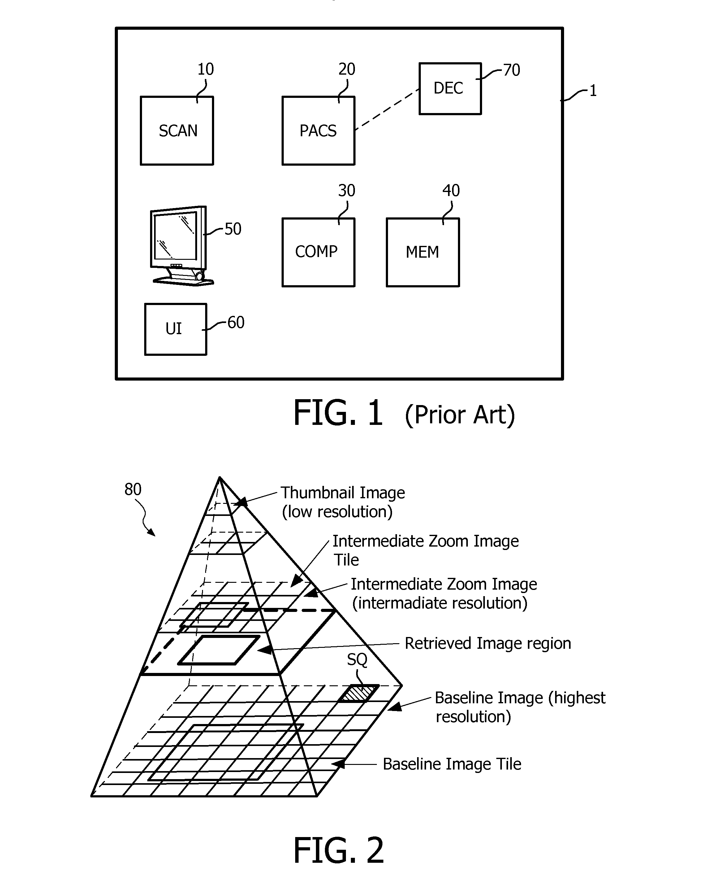
[0060]Preliminarily it should be understood that the term “content”, when referring to an image, indicates any kind of information that can be derived in this image. Such information may typically correspond to certain biological features present in the sample and may typically correspond to information derived from pixel data.

[0061]The terms “transform”, “transform function, “transformation” shall have the same definition.

[0062]The term “area” may preferably refer to a physical portion of the sample under investigation, while the term “region” or “sub-region” may preferably refer to a portion of the image in the digital world.

[0063]The term “sub-region” shall define a portion of a “region”.

[0064]Further, “providing an image” shall encompass various possibilities known in the art such as receiving the image from a scanner, from a storage memory, from a telecommunication link like an intranet network or like internet, from a data structure as described herein after, etc.
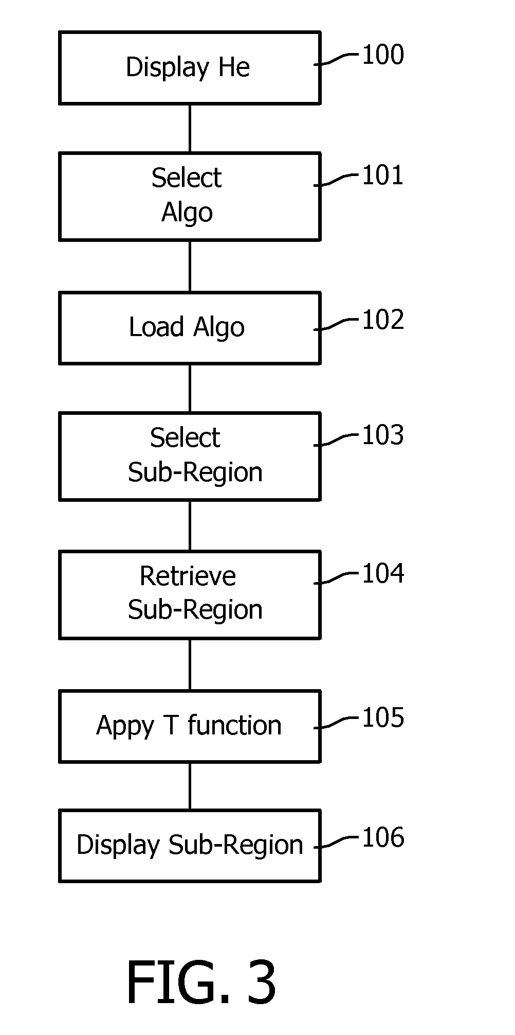
[0065]Accordi...


PUM


Abstract

The invention pertains to the field of image processing in digital pathology. It notably proposes a method for processing a first digital image, representing a sample in a region, and which image has been acquired from the sample by means of a microscopic imaging system (1) and is stored in a multi-resolution image data structure (80), comprising the steps of:—retrieving (104) a sub-region of the first digital image at a first resolution, —executing (105) a transform function on the retrieved sub-region, the transform function modifying a content of the sub-region according to at least one metric derived from a second resolution representation of the first digital image.

Description

FIELD[0001]The invention relates to the field of digital pathology, in particular to technology for whole slide scanning.[0002]In particular the invention relates to an image processing method that enhances / modifies a digital microscopy image.BACKGROUND[0003]Digital pathology provides new ways to visualize tissue slides and enables new workflows for analyzing these slides.[0004]As illustrated in FIG. 1, a typical system 1 for digital pathology may notably comprise a scanner 10 for scanning a sample, for example a tissue sample and create a digital image thereof. Many types of scanner have already been proposed, such as 1D line scanner, 2D array scanner, so called Time Delay Integration (TDI) scanner, etc.[0005]The system may further comprise a PACS 20 (Picture Archiving communication System) for archiving and communicating information such as a digital image of the tissue sample. The system may further comprise a computing device 30 for executing computer implemented instructions, a...

Claims


Application Information

Patent Timeline
Patent Type & Authority Applications(United States)
IPC IPC(8): G06K9/00
CPCG06T3/0068G06T2200/24G06K9/0014G06T2207/20016G06T2207/20104G06T2207/10056G06V20/695
Inventor MUELLER, DANIEL CARLVOSSEN, DIRK LEO JOEPHULSKEN, BAS
Owner KONINKLIJKE PHILIPS ELECTRONICS NV
Why Eureka
Try Eureka