Therapeutic uses of microvesicles and related micrornas

a technology of microvesicles and micrornas, which is applied in the field of therapeutic uses of microvesicles and related micrornas, can solve the problems of difficult to predict the biological activity of microvesicles, time-consuming and inefficient, and many largely unexplained problems, so as to achieve predictable and effective therapeutic results.

Inactive Publication Date: 2013-06-06
THE UNIV COURT OF THE UNIV OF GLASGOW
View PDF5 Cites 26 Cited by
  • Summary
  • Abstract
  • Description
  • Claims
  • Application Information

AI Technical Summary

Benefits of technology

[0006]The present invention provides improved methods and compositions based on microvesicles for the treatment of various diseases, disorders and conditions. In particular, the present invention encompasses the recognition that microvesicles contain specific microRNAs which may function as intercell

Problems solved by technology

While such proposed uses provide at least some promising perspectives, several largely unexplained problems remain.
For example, biological activity of microvesicles is often difficult to predict.
Moreover, currently

Method used


Image

  • Therapeutic uses of microvesicles and related micrornas
  • Therapeutic uses of microvesicles and related micrornas
  • Therapeutic uses of microvesicles and related micrornas

Examples


example 1

Morphological Examination of Pancreas-Derived Pathfinder Cells (PDPC) and Identification of Microvesicles (MVs)

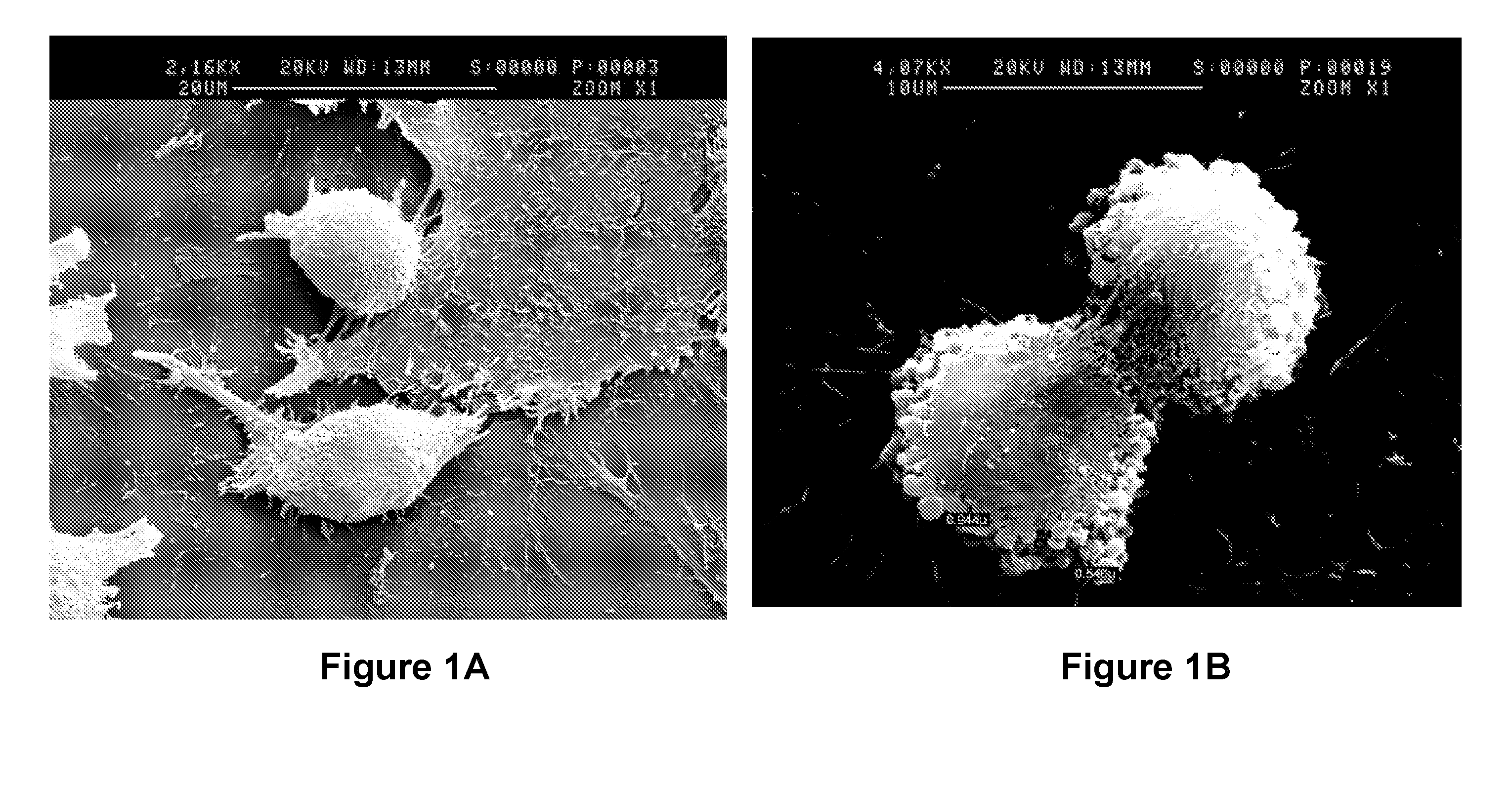
[0171]In the present Example, morphological studies of rat pancreas-derived pathfinder cells (PDPC) were conducted by scanning electron microscopy (EM). Scanning EM images revealed protrusions from surfaces of PDPCs that are provisionally identified as nascent microvesicles (MVs).

[0172]Pathfinder cells were isolated from rat pancreas cultured as previously described. (See, e.g., International Patent Publication No. WO2006 / 120476 A1, the entire contents of which are herein incorporated by reference.) These rat PDPCs were grown in medium containing fetal bovine serum (FBS) that was depleted of bovine microvesicles.

[0173]Pictures of a subconfluent culture of rat PDPCs were taken by a scanning electron microscope. FIG. 1A shows a representative picture, showing PDPCs of both the fibroblastoid and small round cell types. As can be seen in FIG. 1A, both cell types have very great...

example 2

Analysis of miRNA Expression in Rat PDPCs and in MVs Isolated from Rat PDPCs

[0176]Results from Example 1 may shed light into the mechanism of PC action on other cells and tissues. To further investigate the mechanism of PC action, microvesicles obtained from PDPCs were studied in further detail.

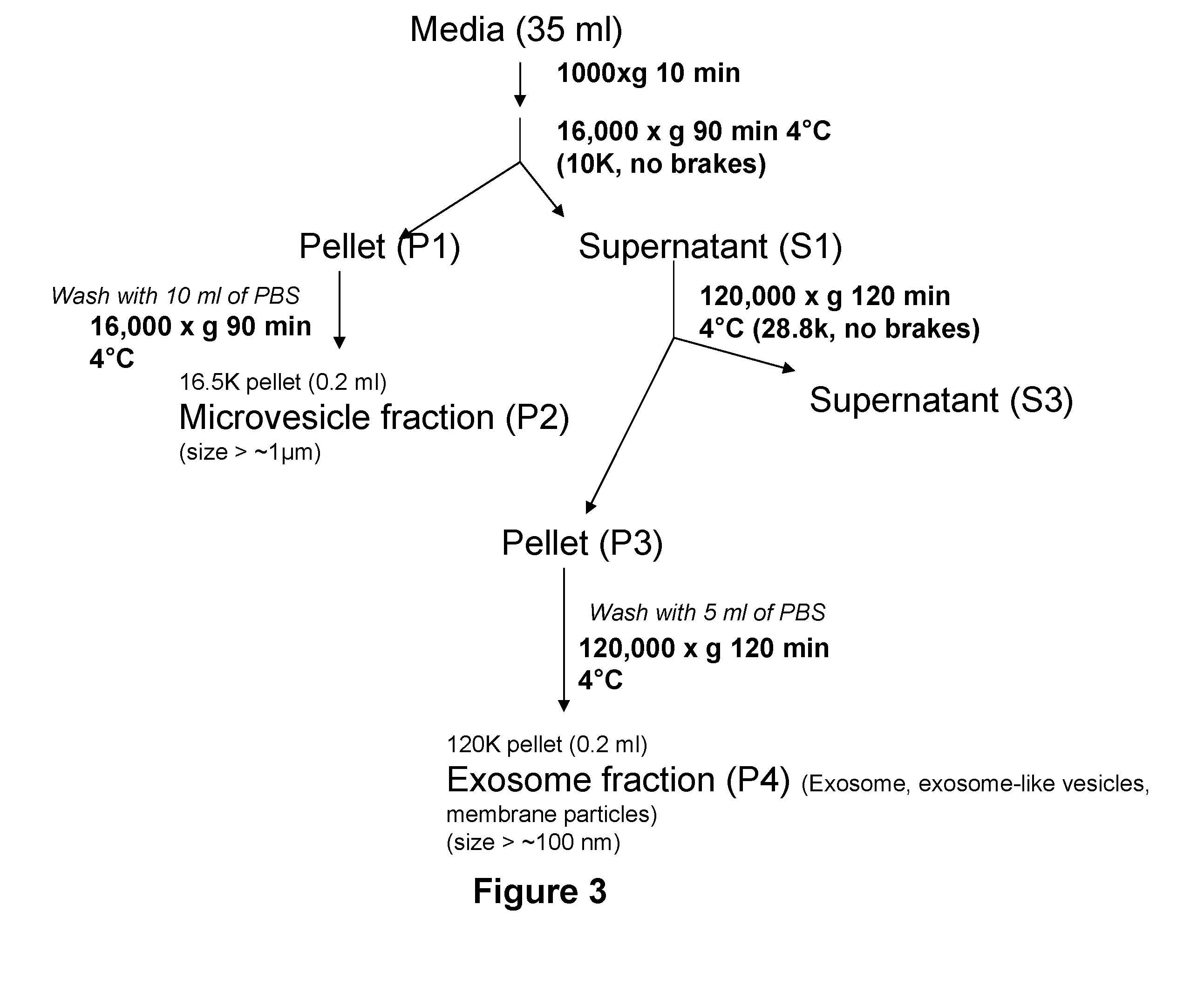
[0177]In the present Example, MVs were purified from supernatants of rat PDPC cultures in medium with serum depleted of bovine microvesicles using a differential centrifugation protocol. RNA was prepared from both MVs and PDPCs using standard procedures. RNA samples were reverse-transcribed (RT) and amplified in a quantitative PCR assay in order to analyze expression of miRNAs.

Materials and Methods

[0178]RNA extraction. RNA from cells and microvesicles (MVs) was extracted using TRI Reagent (Sigma), with the following modifications to the manufacturer's protocol. After addition of ⅕th volume chloroform to the TRI Reagent, samples were spun at 6° C. for 15 minutes at 16,000×g. Aqueous phases wer...

example 3

Assays for Characterizing Effects of MVs or miRNAs on Cell Growth

[0185]The present Example demonstrates the effects of MVs on growth of rat PDPCs.

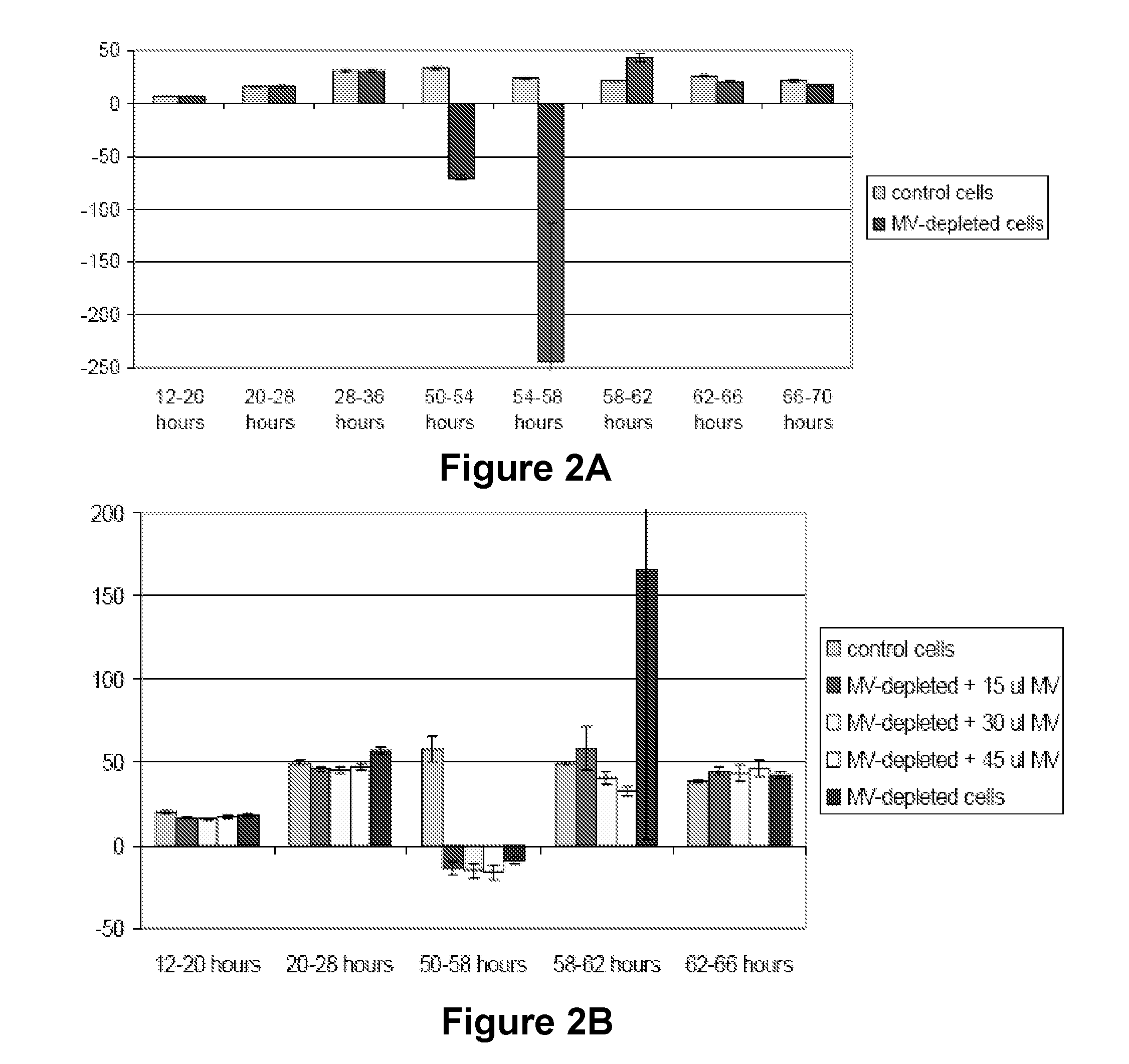
[0186]An XCELLINGENCE™ machine was used to measure cell growth in rat PDPC cultures that were depleted of bovine MVs, or depleted of MVs and then had rat PDPC MVs added back.

[0187]Rat PDPCs were cultured in medium containing bovine serum, and then at 43 hours were switched to bovine MV-depleted medium. Depleting MVs resulted in a decrease in cell proliferation, with a doubling time slowing to 31 hours (FIG. 2A). A negative effect on doubling time was seen, with a later recovery.

[0188]In a separate set of experiments, cultures were MV-depleted at 48 hours, and then exogenous MVs are added 10 hours later. A dose-dependent recovery of rat PDPC doubling time (i.e., increase in cell proliferation) was observed after addition of rat PDPC-derived MVs (FIG. 2B). The increase in cell proliferation persisted for 48 hours and then faded. The rapid re...


PUM

PropertyMeasurementUnit
Massaaaaaaaaaa
Massaaaaaaaaaa
Diameteraaaaaaaaaa

Abstract

The present invention provides improved methods and compositions based on microvesicles for the treatment of various diseases, disorders and conditions. In particular, the present invention encompasses the recognition that microvesicles contain specific microRNAs which may function as intercellular regulators involved in cell or tissue regeneration, remodeling, reconstruction, reprogramming or transdifferentiation. Thus, among other things, the present invention provides methods and compositions based on microvesicles and/or associated microRNAs that provide more predictable and effective therapeutic results.

Description

CROSS REFERENCE TO RELATED APPLICATIONS[0001]This patent application is a continuation of international application No. PCT / IB2011 / 002028 filed on Aug. 12, 2011, which claims the benefit of U.S. Provisional Patent Application Ser. Nos. 61 / 373,715, filed Aug. 13, 2010 and 61 / 380,766, filed Sep. 8, 2010, the entirety of each of which is incorporated herein by reference.[0002]This application relates to international application PCT / IB2011 / 002048, and entitled “Cellular and Molecular Therapies” filed on Aug. 12, 2011, the entirety of which is incorporated herein by reference.SEQUENCE LISTING[0003]The present specification makes reference to a Sequence Listing (submitted electronically as a .txt file named “Sequence Listing.txt on Feb. 13, 2013). The .txt file was generated on Feb. 13, 2013 and is 93.1 kb in size. The entire contents of the Sequence Listing are herein incorporated by reference.BACKGROUND[0004]Microvesicles were historically regarded as cellular debris with no apparent f...

Claims


Application Information

Patent Timeline
IPC IPC(8): C12N15/85C07H21/04A61K35/22A61K35/39
CPCA61K35/22A61K35/39C12N15/88G01N33/5061G01N33/507G01N33/5073A61K2035/124C12N2320/11C12N15/113A61K35/28A61K9/5068C12N2310/141C12N2330/10A61P13/12A61P17/02A61P25/00A61P25/02A61P43/00A61P9/10A61P3/10A61K35/14A61K35/407C12N2320/30
Inventor SHIELS, PAULDAVIES, WAYNE
Owner THE UNIV COURT OF THE UNIV OF GLASGOW
Why Eureka
Try Eureka