Image-overlay medical evaluation devices and techniques

a medical evaluation device and image overlay technology, applied in the field of medical imaging systems, can solve the problems of inability to accurately and predict the accuracy of bench-top surgical guides, prohibitively expensive and time-consuming to use non-ionizing radiation, and repeated doses of medical patients and medical personnel, and achieves the effects of low cost, short time, and low cos

Inactive Publication Date: 2013-07-04
GOLE PHILIP D
View PDF7 Cites 116 Cited by
  • Summary
  • Abstract
  • Description
  • Claims
  • Application Information

AI Technical Summary

Benefits of technology

[0008]The image-overlay techniques and related systems of the present invention provide for simple, expedient, and reliable surgical guide fabrication and evaluation, which is sufficiently low in cost and short in time for its use to be justified in most cases where a surgical implant is desired. Desirable characteristics for surgical guide include precision, low cost, easy fabrication by substantially any clinician, use as a diagnostic adjunct, and facilitating the reduction of radiation (particularly ionizing radiation) exposure to the patient. The systems and techniques of the present invention facilitate the pre-operative, mid-operative, and post-operative evaluation of proposed, ongoing, and completed medical procedures, as well as the fabrication of precise surgical guides for routine use, and in substantially all cases in which surgical implants are desired, to help ensure favorable treatment outcomes for medical patients. Moreover, the techniques of the present invention generally do not require any changes to the actual surgical or other medical procedures that are used on the patient, and can be used at substantially any stage of a surgical procedure and while using standard surgical equipment.
[0009]The image-overlay techniques and systems of the present invention have the ability to achieve these benefits, including the ability quickly produce an accurate surgical guide, and to “CT-confirm” the accuracy of such guides. With access to a pre-surgical CT image and the means to create a digital image of a working model, substantially any appropriately equipped dentist or laboratory technician can create a “CT-confirmed” surgical guide, sometimes within a matter of hours. The combination of a “CT-confirmed” surgical guide and the related image-overlay techniques of the present invention reduce exposure of patients to mid-surgery and post-surgery radiation. The techniques may also be used to evaluate the actual location of an osteotomy or surgical implant within patient tissue, without the use of ionizing radiation other than an initial pre-surgical scan. The use of these techniques can have the immediate impact of reducing the radiation exposure to a patient by at least 50% during a given surgical procedure.
[0035]Thus, the present invention provides techniques and systems that provide the benefits of mid-surgical 3-dimensional radiographic scans with a CT, CBCT, X-ray, MRI, or similar device, but without the mid-surgical use of ionizing radiation, and typically more quickly and at lower cost than would be the case when using equipment that produced ionizing radiation. The techniques may also be used to create accurate surgical guides, to confirm the accuracy of surgical guides prior to and / or during a surgical procedure, and to evaluate the result of the surgical procedure, all without the use of additional ionizing radiation. In addition, the techniques may be used without alteration to the preferred surgical methods and surgical equipment of a surgeon or other medical professional.

Problems solved by technology

However, repeated scans expose medical patients and medical personnel to repeated doses of radiation.
It also can be prohibitively costly and time-consuming to use non-ionizing radiation, such as magnetic resonance imaging (“MRI”), for such evaluations.
Although such methods typically provide improved accuracy over medical procedures (e.g., drilling osteotomies) free-hand or without the use of a guide, such bench-top surgical guides can still be inaccurate and unpredictable.
However, typical commercial methods of surgical guide fabrication can be tedious and expensive for the practicing clinician, and can take days or weeks to complete, especially when some of the steps are completed at an off-site location and require shipping of casts, surgical guides, and the like.
Moreover, even after a clinician uses a surgical guide to place one or more dental implants, confirming the accuracy of placement has typically required exposing the patient to a post-surgical X-ray or CBCT or CT scan, resulting in additional radiation exposure to the patient.
While conventional radiographic methods can determine depth and mesial-distal dimensions, a true 3-dimensional assessment is difficult to achieve using known methods.
However, the access to a CBCT or CT scan during surgery, additional radiation exposure, and cost to perform these procedures can be prohibitive.

Method used


Image

  • Image-overlay medical evaluation devices and techniques
  • Image-overlay medical evaluation devices and techniques
  • Image-overlay medical evaluation devices and techniques

Examples


Embodiment Construction

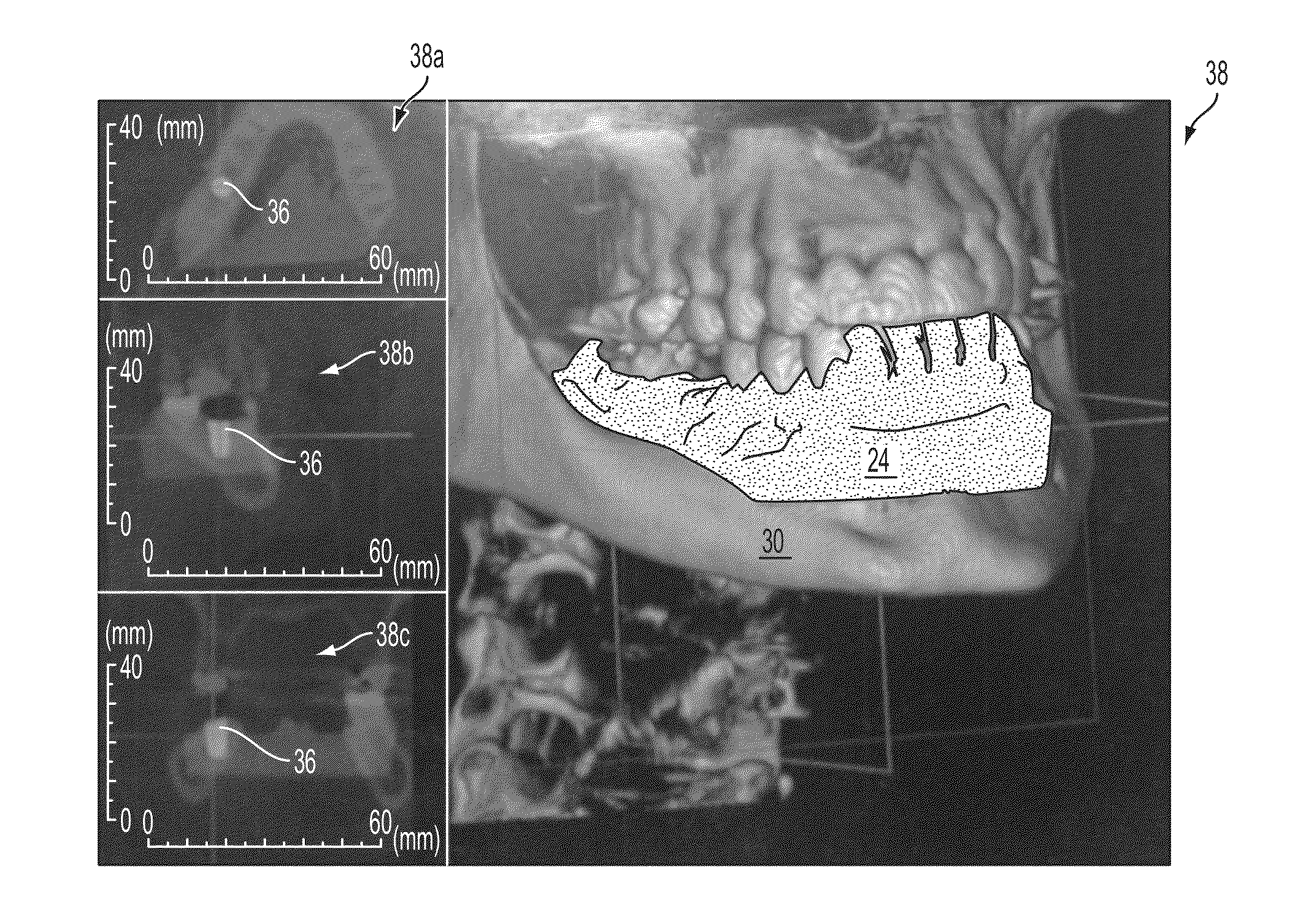
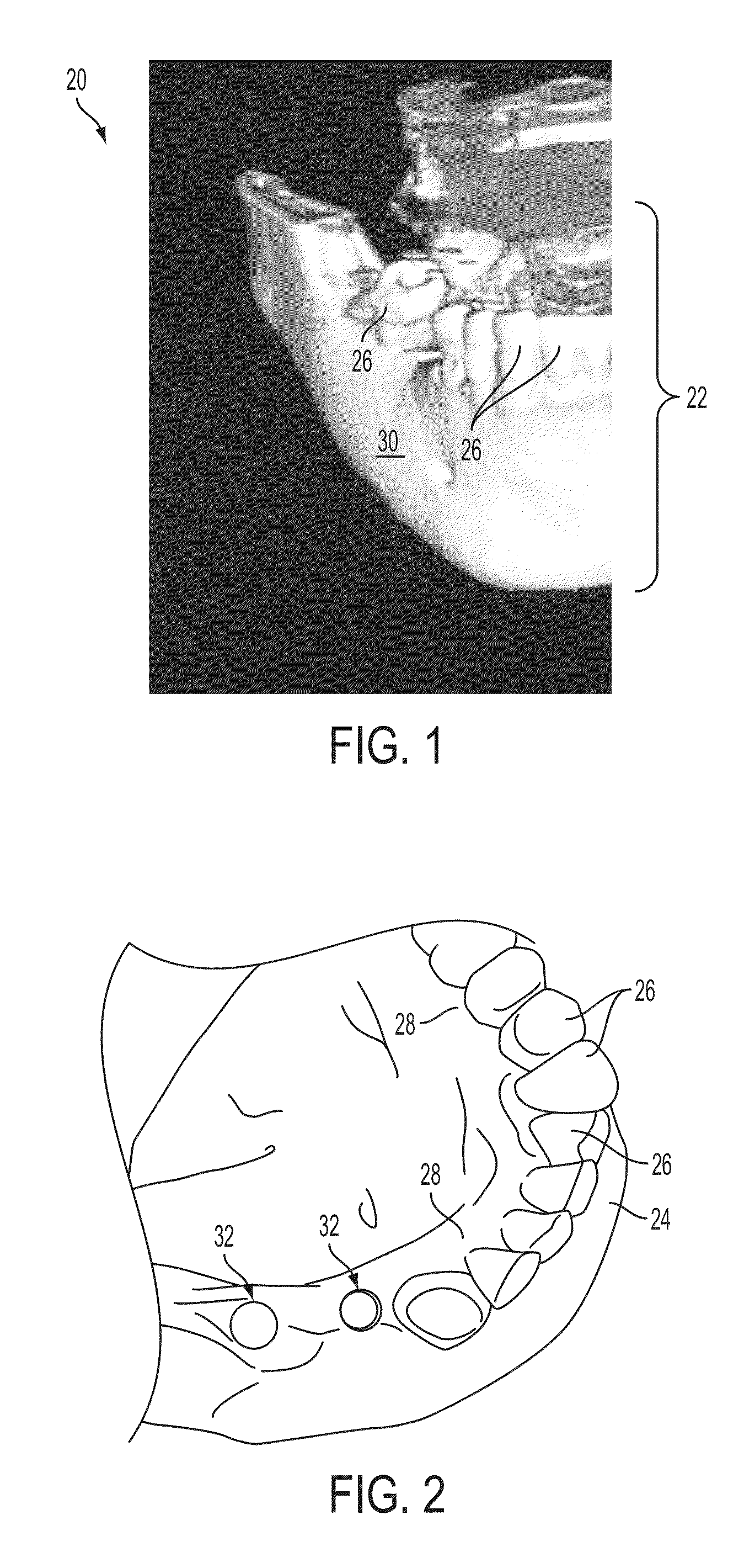
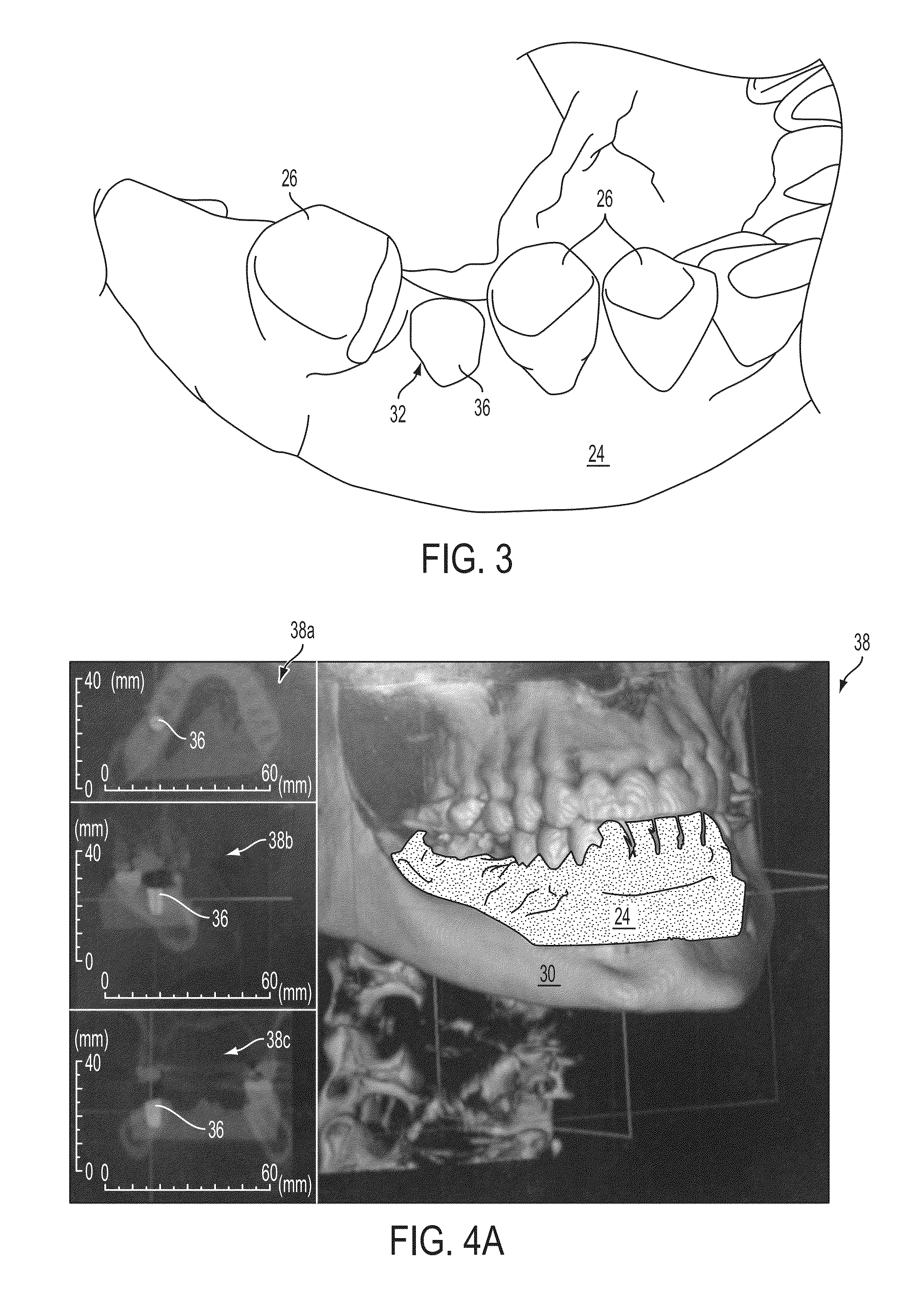
[0064]The systems and image-overlay techniques of the present invention include devices and methods used to determine the precise anatomical position of surgical implants or prostheses, as well as osteotomies or other modifications to body tissues. 3-dimensional volumetric rendering software of pre-surgical patient DICOM (Digital Imaging and Communications in Medicine) image files are superimposed with either DICOM images of physical models or optical (such as laser) scans of patient anatomy or models, or negative DICOM images of an impression of the patient's anatomy, to accurately reveal precise anatomical or potential anatomical implant positions. The image-overlay techniques allow a clinician to fabricate, evaluate, and confirm accuracy of a surgical guide using computed tomography (“CT”) scanning, cone beam computed tomography (“CBCT”), laser scanning, or the like, prior to or during placement of a dental implant (or other surgical implant).

[0065]In accordance with current sugg...


PUM


Abstract

A system and methods are provided for evaluating the position of surgical implants or openings formed in anatomical tissue during medical procedures, in which an initial 3-dimensional image of the anatomical tissue is combinable with one or more subsequent 3-dimensional images of the same or correlating tissue, and without the use of ionizing radiation on a patient for at least the subsequent images. The system includes a software program, a computer with display, a radiographic image scanning device, and a non-radiographic image scanning device. The computer accesses a medical patient information database and a medical implant database for images used by the software program. A pre-operative image is combined with subsequent images to facilitate evaluation of the proposed placement of a surgical implant or opening relative to tissues of the patient anatomy. Methods of evaluating the accuracy of surgical guides and of fabricating surgical guides are also disclosed.

Description

CROSS REFERENCE TO RELATED APPLICATIONS[0001]The present application claims the priority benefit of U.S. provisional application Ser. No. 61 / 582,007, filed Dec. 30, 2011, and of U.S. provisional application Ser. No. 61 / 597,494, filed Feb. 10, 2012, which are hereby incorporated herein by reference in their entireties.FIELD OF THE INVENTION[0002]The present invention relates to medical imaging systems and methods for use in visually confirming the location or proposed location of prostheses or implants to body tissues, or for modifying body tissues such as osteotomies, osteoplasty, or modifications to softer tissues, and for the manufacturing and evaluation of surgical guides used in modifying body tissues including for attaching prostheses or implants.BACKGROUND OF THE INVENTION[0003]Typical methods for attaching prostheses or implants (such as dental implants, for example) to body tissues (such as bone) involve multiple radiographic scans of at least a portion of the body with diff...

Claims


Application Information

Patent Timeline
Patent Type & Authority Applications(United States)
IPC IPC(8): A61B5/00
CPCA61B5/0035A61B5/055A61B5/1077A61C1/084A61B6/506A61B6/508A61C9/0046A61B5/1079A61B5/7425B33Y80/00
Inventor GOLE, PHILIP D.
Owner GOLE PHILIP D
Why Eureka
Try Eureka