Disintegrating stent and method of making same

a technology of disintegrating stents and medical prostheses, which is applied in the field of disintegrating implantable medical prostheses, can solve the problems of bioabsorbable stents having their own disadvantages, affecting the quality of life of patients,

Inactive Publication Date: 2013-07-18
BOSTON SCI SCIMED INC
View PDF5 Cites 5 Cited by
  • Summary
  • Abstract
  • Description
  • Claims
  • Application Information

AI Technical Summary

Benefits of technology

[0019]While all bioabsorbable stents “disintegrate” during the absorption process, one object of the present invention is to purposely design the implant to cause fracture and / or disintegration to happen, preferably at specific sites and the pieces eliminated from the body, rather than rely on complete bioabsorbtion of the stent within the body. In particular, the invention relates to engineered biodegradation to promote predictable and designed fracture and / or disintegration of the stent so that small stent pieces may be transported out of the body, rather than relying on the full bioabsorbability of the material to allow complete dissolution of the stent.
[0024]The conventional objective of materials and implant design engineers has been to control or manipulate the manufacturing process to avoid early disintegration from fast-degrading amorphous regions by trying to increase the crystallinity of the polymer. The result is that bioabsorbable stents of the prior art are comprised of polymer monofilaments that are generally 40%-60% crystalline. By contrast, the present invention relates to thermomechanically processing a bioabsorbable polymer to produce more fast-degrading amorphous regions in the material to create predictable and / or more numerous sites of polymer fragmentation. Amorphous regions may be created by fast cooling of melt-spun polymer extrudate to prevent nucleation and growth of significant crystalline regions.
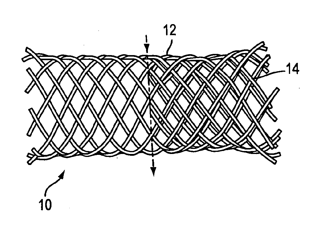
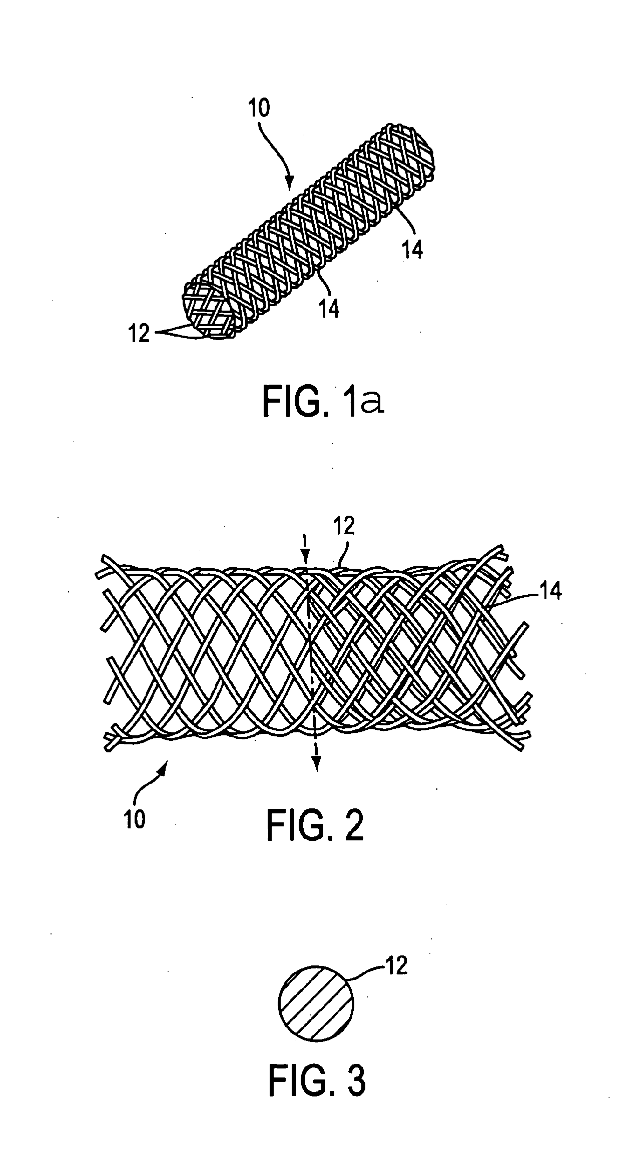
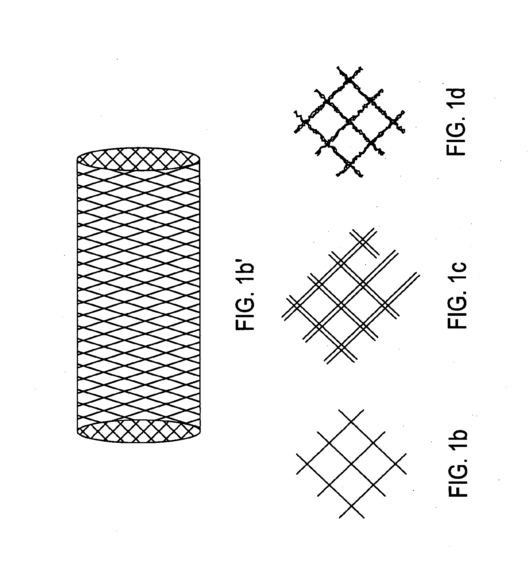
[0029]Alternatively, unmodified bioabsorbable polymers may be used in the form of thin monofilaments or cable strands to construct the stent Thinner monofilaments disintegrate into finer particles than the thicker (0.25 mm diameter) monofilaments. Thus, one embodiment of the invention relates to replacing the standard 24 single strands of 0.25 mm diameter bioabsorbable polymer monofilament with 24 paired strands of 0.12 mm diameter monofilament or, alternatively, 24 strands of pre-braided cable each containing two or more monofilaments of very fine diameter, about 0.05 mm or less in diameter. This embodiment of the present invention may also be extrapolated for use in large stents such as 22 mm diameter esophageal or colonic stents where large diameter monofilaments would traditionally be used in the braids. It is beneficial to control the disintegration particle size in these large stents by using multiple, small diameter filaments in their design so that the fragments are be less harmful and more easily passed through the vessel system out of the body.
[0030]Additionally, the disintegrating stents of the present invention may be designed so that they are strained by the body to promote disintegration at purposely created weak spots within the material. An example would be to significantly oversize all or portions of a self-expanding stent within a vessel. Prior to implantation the device would be unstrained. After delivery to and release within the target vessel, the stent may be strained, as a result of its larger size relative to the vessel diameter. This strain will facilitate fracture of the stent at the pre-manufactured weak spots after a pre-determined amount of time and the stent will break into small soft particles to be carried away by the vessel contents.

Problems solved by technology

The disadvantages of perm anent metal expandable or solid plastic tubular removable stents are addressed in part by bioabsorbable stents, but bioabsorbable stents can have their own disadvantages.
The “burst” of degradation products from fast-absorbing stents can potentially cause significant inflammation and hyperplasia leading to obstruction or clinical complications.
In both the cases of fast- and slow-absorbing stents, the implants can break apart into large pieces at locations of high stress or strain when degradation occurs.
Such large pieces can cause tissue damage, if the fracture surfaces are sharp, and may lead to luminal obstruction.

Method used


Image

  • Disintegrating stent and method of making same
  • Disintegrating stent and method of making same
  • Disintegrating stent and method of making same

Examples


example 1

[0067]One method of creating multiple fracture initiation sites in a biodegradable polymer is to create a structure having more amorphous and less crystalline regions in the material.

[0068]Methods for making polycrystalline monofilaments are generally known. For example, methods for making PLA monofilaments are described in detail in U.S. patent application Ser. No. 08 / 08 / 904,467, filed Aug. 1, 1997. Generally, PLA monofilaments may be produced by a process involving seven general steps as summarized herein. Methods of making monofilaments from other polycrystalline polymers, including but not limited to the polymers enumerated hereinabove, are equally well known to those of ordinary skill in the art, and this example is not intended to limit the present invention in any way.

[0069]First, a polymer formed of poly-L-lactic acid is brought to an elevated temperature above the melting point, preferably 210.degree.-230.degree. C. Second, the material is then extended at the elevated temp...

example 2

[0073]Mechanical properties generally increase with increasing molecular weight. For instance, the strength and modulus of polycrystalline polymers generally increase with increasing molecular weight. Conversely, degradation time generally decreases with decreasing initial molecular weight (i.e., a stent made of a low molecular weight polymer is bioabsorbed more quickly than a stent made of a high molecular weight polymer). Moreover, the molecular weight and mechanical properties of the material generally decreases as degradation progresses. Accordingly, in addition to, or as an alternative to, the creation of a heterogeous molecular structure to promote controlled disintegration and fracture, the stent material may be subjected to post-extrusion or molding operations to create pre-selected “weak spots,” localized pre-degradation of the molecular weight of the crystalline structure of the polymer.

[0074]One method of creating pre-degraded regions in a monofilament is to mask some por...

example 3

[0075]In addition to manipulating the molecular structure during or after extrusion, mechanical features such as stress concentrations, fissures, notches grooves, indentations or surface contours may be designed into the implant to cause predictable, controlled fracture and / or disintegration. FIGS. 12a-c and 13 illustrate types of mechanical features may be introduced into the surfaces of stent materials to facilitate planned and controlled fracture.

[0076]Such periodic fracture initiation sites are designed such that they are not deleterious enough to initiate fracture in full-strength material. However, when degradation occurs and the material loses strength, the stress concentrations or fissures become more significant relative to the strength of the material and serve as points of weakness in the device in order to facilitate disintegration. The methods of making the features include lathe turning, milling, drilling, die-forming, laser curing, and chemical etching. Die chatter ma...


PUM

PropertyMeasurementUnit
sizeaaaaaaaaaa
sizeaaaaaaaaaa
sizeaaaaaaaaaa

Abstract

A temporary stent endoprosthesis that does not require an interventional procedure for removal. The disintegrating stent is preferably made from a bioabsorbable polymer, such as by braiding polymer monofilaments into a tubular mesh shape, and the polymer has fracture initiation sites within it that promotes the disintegration of the stent into small pieces that are harmlessly transported out of the body by the vessel contents. Fracture initiation sites may be created by controlling the heterogenous structure of amorphous and crystalline regions, by introducing internal or surface fracture initiation sites, or use of multiple strands with small section size.

Description

CROSS-REFERENCE TO RELATED APPLICATIONS[0001]This application is a continuation of application Ser. No. 10 / 742,943, filed Dec. 23, 2003, which is a divisional of application Ser. No. 09 / 592,413, filed Jun. 13, 2000, the entire contents of which are herein incorporated by reference.BACKGROUND OF THE INVENTION[0002]1. Field of the Invention[0003]The present invention relates generally to implantable temporary medical prostheses that do not require interventional procedures for removal. In particular, the present invention is a disintegrating implantable medical prosthesis that disintegrates into small pieces that are harmlessly transported out of the body by normal body function.[0004]2. Related Technology[0005]Medical prostheses frequently referred to as stents are well known and commercially available. They are, for example, disclosed generally in the Wallsten U.S. Pat. No. 4,655,771, the Wallsten et al. U.S. Pat. No. 5,061,275 and in Hachtmann et al., U.S. Pat. No. 5,645,559. Devic...

Claims


Application Information

Patent Timeline
Patent Type & Authority Applications(United States)
IPC IPC(8): A61F2/82A61F2/00A61F2/02A61F2/88A61F2/90A61F2/92A61L31/14
CPCA61F2/88A61F2/90A61F2/92A61F2/95A61F2/966A61F2230/0078A61F2250/0071A61L31/148A61F2/82A61F2230/005A61F2210/0004
Inventor STINSON, JONATHAN S.
Owner BOSTON SCI SCIMED INC
Why Eureka
Try Eureka