
Method of determination of autoantibody level by means of enzyme immunoassay

a technology of enzyme immunoassay and autoantibody level, applied in the field of medicine, can solve the problems of reducing the sensitivity of eia method, and achieve the effect of increasing the sensitivity of the claimed method, reducing the sensitivity of eia method, and extending the functional capabilities of the claimed method

Inactive Publication Date: 2013-07-25
SERGEEVA SVETLANA ALEXANDROVNA +3
View PDF4 Cites 18 Cited by
  • Summary
  • Abstract
  • Description
  • Claims
  • Application Information

AI Technical Summary

Benefits of technology

The patent describes a method for improving the sensitivity and accuracy of detecting natural autoantibodies in biological fluids. The method involves coating a solid phase with a protein and using biotinylated agents to capture the autoantibodies. An enzyme-labeled antibody is then used to determine the level of certain isotypes of autoantibodies. This method minimizes the likelihood of non-specific binding and ensures that all epitopes of the antigen remain free for binding with natural autoantibodies. The addition of iron-containing oxidizer and heat treatment further enhances the method's sensitivity and accuracy in determining the total level of natural autoantibodies and reducing the possibility of obtaining false negative results. The use of individual control solid phase for each investigated specimen of biological fluid also improves the accuracy of the method. Overall, this patent offers a reliable and sensitive method for quantitative determination of natural autoantibodies.

Problems solved by technology

Due to combined application of the physical sorption solid phase, coated with streptavidin, and biotinylated agents at the physical sorption solid phase, all epitopes of the immobilized antigen remain free for binding with natural autoantibodies, thus increasing sensitivity of the claimed method, while direct immobilization of an antigen results in conformational changes and destruction of antigen epitopes, which reduces sensitivity of EIA method.

Method used


Image

  • Method of determination of autoantibody level by means of enzyme immunoassay
  • Method of determination of autoantibody level by means of enzyme immunoassay
  • Method of determination of autoantibody level by means of enzyme immunoassay

Examples


example 1

[0020]In order to confirm the possibility of obtaining the technical effect while implementing the claimed method of quantitative determination of the level of natural autoantibodies to human gamma interferon, serum specimens were taken from twenty healthy donors who were not subjected to interferon therapy. Hence, their serum does not contain autoantibodies to human gamma interferon, yet natural autoantibodies to human gamma interferon are present.

[0021]A portion of wells of 96-well plate, coated with streptavidin from Greiner bio-one GmbH (Catalog 655990), were inoculated with 100 μl of the recombinant human gamma interferon from eBiosciences (Catalog 34-8319-85), and the other portion (control wells for investigated specimens) were inoculated with bovine serum albumin from Sigma Aldrich (Catalog A-3803), both biotinylated according to the standard procedure of U-CyTech Bioscience Company (Standard Operating Procedure UCT-127) at the rate of 100 μl / well at 100% humidity (the p...

example 2

[0039]Serum specimens were taken from twenty patients with infectious mononucleosis, at which the level of natural autoantibodies to human gamma interferon increases.

[0040]All stages of determining the level of natural autoantibodies to human gamma interferon were similar to those described in Example 1, except for the fact that the investigated specimens were diluted by 1000 times, human serum albumin was used as a blocking agent, and the incubation temperature was 56° C.

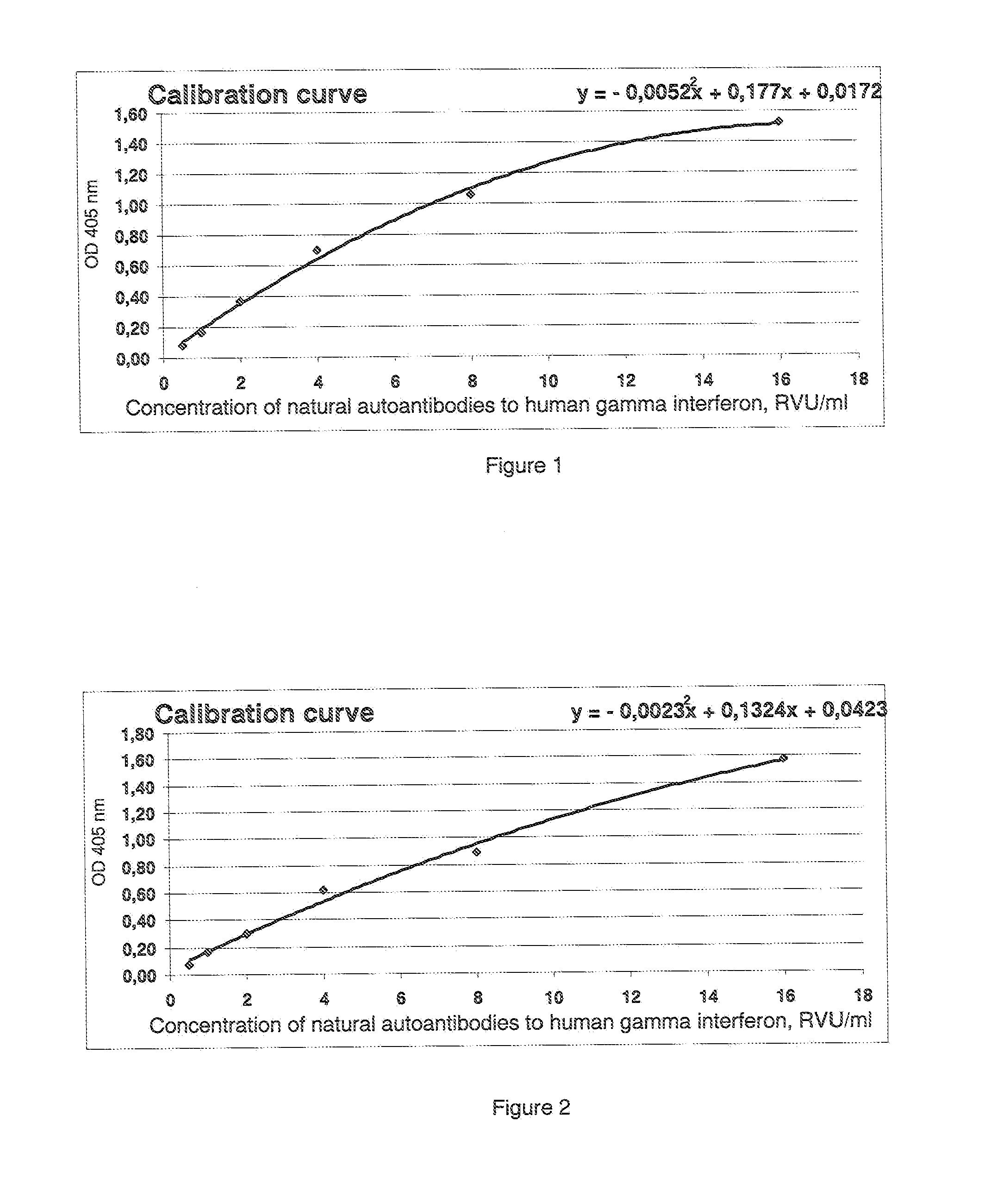
[0041]The calibration curve is shown in FIG. 2, and the obtained results are presented in Table 4.

TABLE 4InvestigatedConcentration of natural autoantibodiesspecimento human gamma interferon, RVU / mlS11675.9S21591.7S31897.5S41480.0S51924.4S61974.6S71537.4S81221.1S91624.5S101569.9S111874.6S121530.2S131870.8S141798.9S152151.8S161882.2S171505.0S181810.2S191526.6S201451.4

example 3

[0042]To verify the possibility of obtaining the technical effect when implementing the claimed method for quantitative determination of the level of natural autoantibodies to human gamma interferon, where prior to heat treatment, the tested biological fluid is additionally treated with iron-containing oxidizer, specimens were taken from twenty healthy donors who were not subjected to interferon therapy and, therefore, there is no autoantibodies to human gamma interferon in serum, yet there are natural autoantibodies to human gamma interferon.

[0043]A portion of wells of 96-well plate, coated with streptavidin from Greiner bio-one GmbH (Catalog 655990), were inoculated with 100 μl of the recombinant human gamma interferon from eBiosciences (Catalog 34-8319-85), and the other portion (control wells for investigated specimens) were inoculated with bovine serum albumin from Sigma Aldrich (Catalog A-3803), both biotinylated according to the standard procedure of U-CyTech Bioscience Co...


PUM

PropertyMeasurementUnit
temperatureaaaaaaaaaa
humidityaaaaaaaaaa
wavelengthaaaaaaaaaa

Abstract

The method for quantitative determination of the level of natural autoantibodies in human biological fluids, when as a solid phase of physical sorption is used the solid phase of physical sorption, coated with streptavidin, and the solid phase of physical sorption is treated with preliminary biotinylated antigen and blocking agent for closing the sites of nonspecific binding at the solid phase of physical sorption, for which purpose are used proteins, biotinylated according to standard procedure. As the conjugate-containing solution are used enzyme-labeled monoclonal and polyclonal antibodies, which react with one or all isotypes of human immunoglobulins. In addition, the tested biological fluid is preliminary diluted in a buffer, containing proteins which are used for closing the sites of nonspecific binding at solid phase of physical sorption, and also substances protecting natural autoantibodies from destruction during heat treatment, and subjected to heat treatment. For each tested specimen of biological fluid, a control solid phase of physical sorption is used, and the number of natural autoantibodies is determined with the use of a calibration curve which is plotted using monoclonal or polyclonal antibodies to antigen.

Description

TECHNICAL FIELD[0001]The invention is related to the field of medicine, in particular to laboratory diagnostics, and it can be used to improve efficiency and reliability of quantitative determination of natural autoantibody concentration in human biological fluids.PREVIOUS TECHNICAL LEVEL[0002]There is a known technical method for quantitative determination of autoantibody level to endogenous proteins in human biological fluids by means of solid phase enzyme immunoassay (EIA) (RU 2137134 C1, G01N33 / 53, 1999). However, this method is of little use for the quantitative determination of natural autoantibody level in human biological fluids.[0003]A method of enzyme immunoassay is known, which includes adsorption of antigens at a solid phase of physical sorption, incubation of tested biological specimens, incubation of a conjugate-containing solution, and spectrophotometric analysis of the reaction of extinction of a chromatic agent (RU 2014610 C1, G01N33 / 535, 1994).[0004]In addition, th...

Claims


Application Information

Patent Timeline
Patent Type & Authority Applications(United States)
IPC IPC(8): G01N33/543
CPCG01N33/564G01N33/54393
Inventor SERGEEVA, SVETLANA ALEXANDROVNATARASOV, SERGEI ALEXANDROVICHTARASOV, ALEXANDER VLADIMIROVICHVAN DER MEIDE, PETER H.
Owner SERGEEVA SVETLANA ALEXANDROVNA
Learn More