Vibrational spectroscopy for quantitative measurement of analytes

a vibrational spectroscopy and quantitative measurement technology, applied in the field of structural biology, can solve the problems of limiting the usefulness of linear infrared spectroscopy as a structural tool, difficult characterization of specific classes of proteins, and difficult to solve the relative few crystal structures that have been solved, etc., to achieve simple and reliable analysis

Active Publication Date: 2013-08-29
MASSACHUSETTS INST OF TECH
View PDF3 Cites 21 Cited by
  • Summary
  • Abstract
  • Description
  • Claims
  • Application Information

AI Technical Summary

Benefits of technology

[0009]The present invention provides a simple and reliable analysis to quantify the percentage of amino-acids in α-helix or β-sheet conformations of proteins in solution. Multidimensional spectroscopy has been developed as a technique to characterize complex systems in solution. In particular, coherent two-dimensional infrared (2DIR) spectroscopy has the ability to directly measure the structure, static disorder, conformational flexibility, and solvent exposure of residues within a protein. 2DIR spec

Problems solved by technology

Despite the abundance, diversity, and biological importance of membrane proteins, relatively few crystal structures have been solved due to the difficulty in crystallizing the proteins.
Specific classes of proteins such as fibrous proteins, intrinsically disordered proteins, gels, amyloids, and aggregates,

Method used


Image

  • Vibrational spectroscopy for quantitative measurement of analytes
  • Vibrational spectroscopy for quantitative measurement of analytes
  • Vibrational spectroscopy for quantitative measurement of analytes

Examples


Embodiment Construction

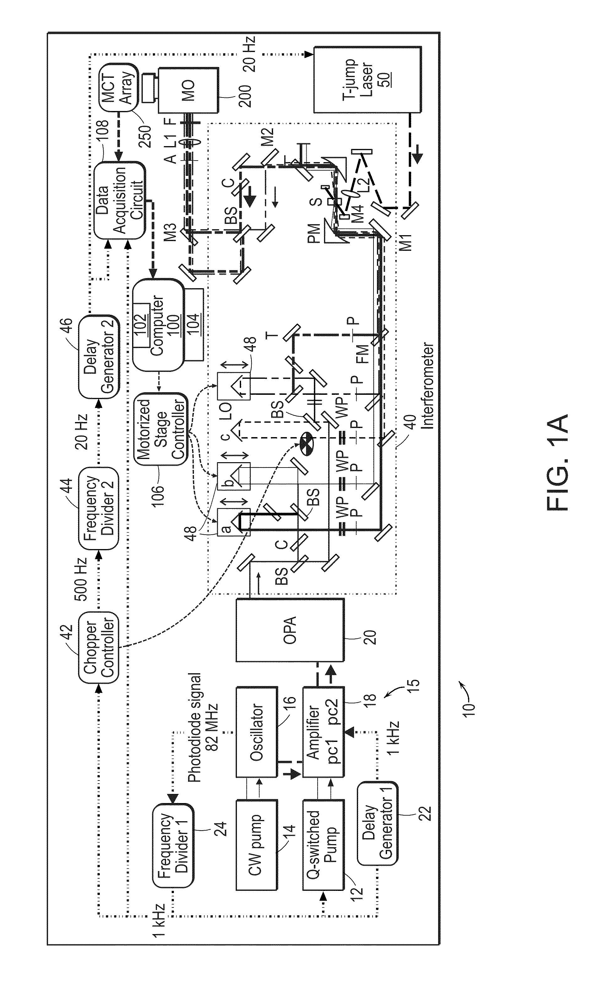
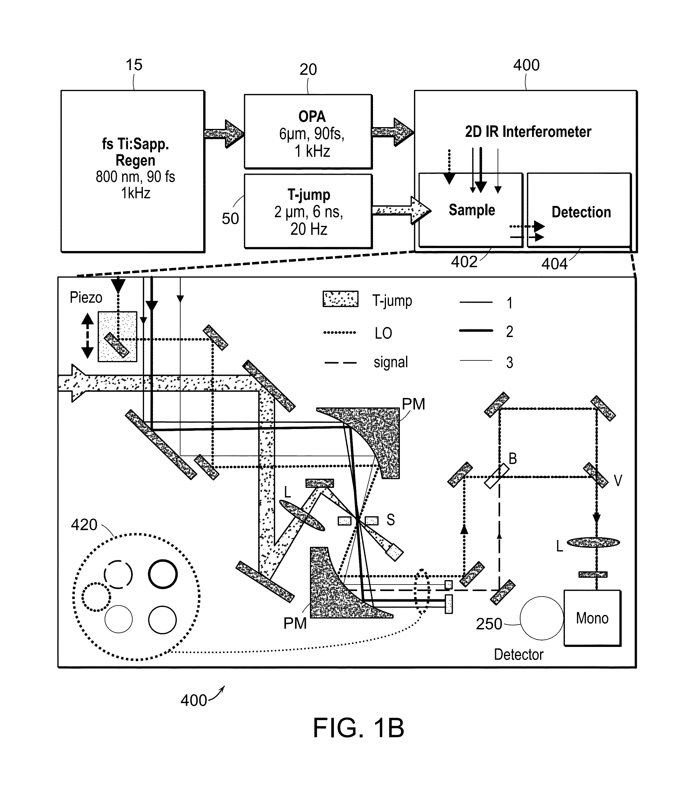
[0030]The two-dimensional spectrometer system 10 is illustrated in FIG. 1A. The system provides for the acquisition of 2D IR spectra and related dispersed vibrational echo (DVE) spectra, the synchronization of femtosecond 15 and T-jump laser 50 systems, and the acquisition of transient 2D IR and DVE spectra. The optical components used to generate and detect the nonlinear signal is shown in FIG. 1A. A Ti:sapphire oscillator system 14, 16 (Tsunami, Spectra-Physics) is used to obtain an initial femtosecond pulse, which is amplified by a regenerative amplifier 18 (Spitfire, Spectra-Physics). The amplified pulse pumps an optical parametric amplifier 20 (OPA). The signal and idler fields are focused onto the AgGaS2 crystal (C) to generate a 90 fs [full width at half maximum] (FWHM) mid-IR pulse centered at 6 um (FWHM 160 cm'1) by difference frequency mixing. A single IR pulse is divided into four identical pulses by 4-mm-thick 50:50 ZnSe beam splitters (BS) (BS1516Z50050S, Rocky Mountain...


PUM


Abstract

The present invention relates to systems and methods for the determination of the secondary structure composition of proteins using coherent two-dimensional infrared (2DIR) spectroscopy of backbone amide I vibrations (1580-1720 cm−1). Fractions of α-helix, β-sheet, and unassigned regions in globular proteins were determined by singular value decomposition using basis spectra from sixteen commercially-available proteins with known crystal structures. Preferred methods included removing each protein from the set and comparing the predicted composition against the crystal structure. The root-mean-squared (RMS) errors of the predicted secondary structure compositions were found to be 7.9% for α-helix, 5.5% for β-sheet, and 7.6% for unassigned regions. The structure analysis can also be performed using one-dimensional absorption spectra and the RMS errors are compared with those obtained from 2DIR.

Description

STATEMENT REGARDING FEDERALLY SPONSORED RESEARCH OR DEVELOPMENT[0001]This invention was made with government support under Grant No. CHE-0911107 awarded by the National Science Foundation. The government has certain rights in this invention.BACKGROUND OF THE INVENTION[0002]Modern structural biology seeks to establish a molecular understanding of biological function by exploring structure-function relationships in biomolecules. The mechanisms for protein folding, protein-protein interactions, ligand binding and catalysis are fundamentally linked to structure. Membrane proteins, in particular, drive key biological processes such as cell-signaling, and ion transport through the cell membrane. Despite the abundance, diversity, and biological importance of membrane proteins, relatively few crystal structures have been solved due to the difficulty in crystallizing the proteins. Modern tools such as cryo-electron-microscopy or x-ray crystallography enable detailed measurements of protein s...

Claims


Application Information

Patent Timeline
IPC IPC(8): G01J5/02G16B15/20
CPCG01N21/35G01N21/3577G01N2201/1293G01N21/636G16B99/00G16B15/20
Inventor BAIZ, CARLOS R.JONES, KEVIN C.TOKMAKOFF, ANDREI
Owner MASSACHUSETTS INST OF TECH
Why Eureka
Try Eureka