Method of examining polycystic kidney disease and method of screening for therapeutic agent of the disease

a polycystic kidney and disease marker technology, applied in the field of examining polycystic kidney disease, can solve the problem of not having established an effective therapeutic method

Inactive Publication Date: 2013-08-29
KYOTO UNIV
View PDF3 Cites 59 Cited by
  • Summary
  • Abstract
  • Description
  • Claims
  • Application Information

AI Technical Summary

Problems solved by technology

Although a causative gene of the autosomal dominant polycystic kidney disease has been specified, no effective therapeutic method has been established.

Method used


Image

  • Method of examining polycystic kidney disease and method of screening for therapeutic agent of the disease
  • Method of examining polycystic kidney disease and method of screening for therapeutic agent of the disease
  • Method of examining polycystic kidney disease and method of screening for therapeutic agent of the disease

Examples


example 1

Establishment of iPS Cell Lines

[0280]Fibroblasts were established by culturing skin samples obtained via biopsies from four autosomal dominant polycystic kidney disease patients with onset of cerebral aneurysm as a complication and from three autosomal dominant polycystic kidney disease patients not developing cerebral aneurysm under agreement. The resultants were each designated PK-ane fibroblasts and PK-free fibroblasts, respectively, and then used in this Example. Meanwhile, a fibroblast cell line of three healthy persons not developing autosomal dominant polycystic kidney disease and cerebral aneurysm is designated nonPK fibroblast and then used in this Example.

[0281]Human cDNAs corresponding to Oct3 / 4, Sox2, Klf4, and c-Myc were introduced into the above fibroblasts using retrovirus according to the method described by Takahashi, K. et al. (Cell, 131(5), 861, 2007). Similarly, human cDNAs corresponding to Oct3 / 4, Sox2, and Klf4 were introduced into the above fibroblasts using a...

example 2

Induction of Differentiation into Vascular Endothelial Cells

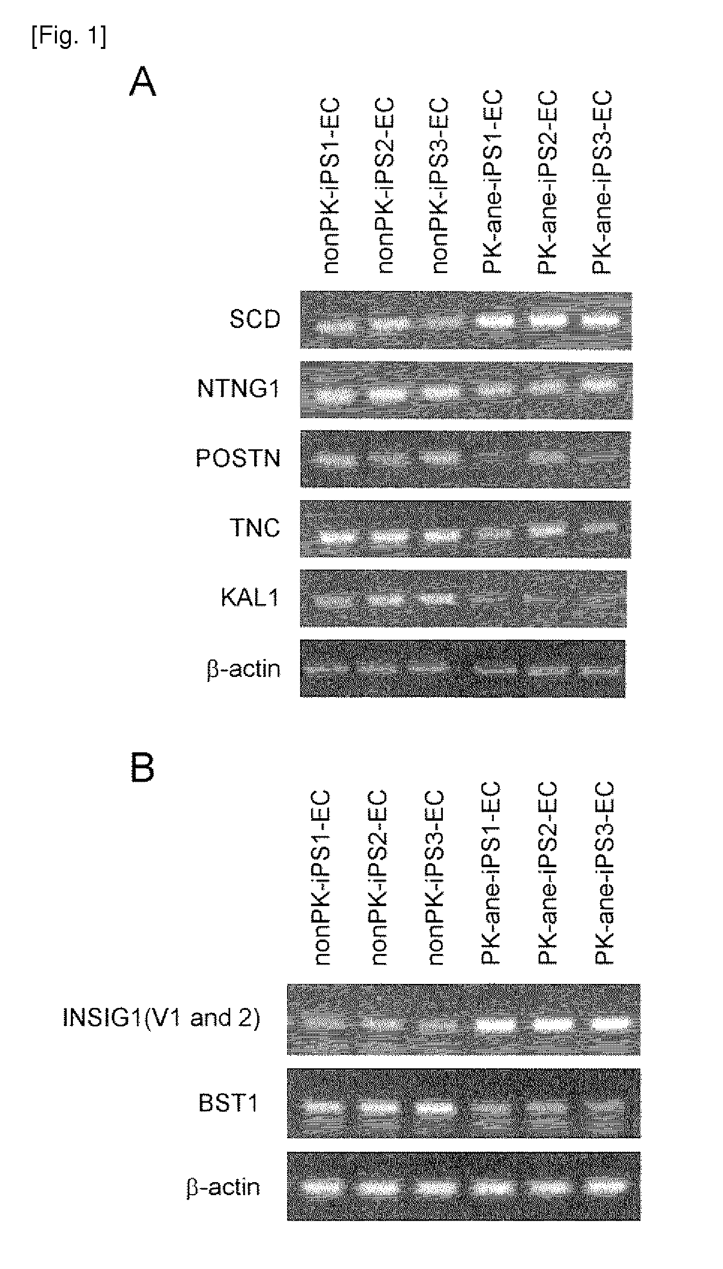
[0282]Each iPS cell line colony prepared as described in Example 1 was separated into pieces with an appropriate size, sprayed over a type-I collagen coating dish (Becton Dickinson), followed by 1 day of culture in a medium for primate ES / iPS cells (ReproCELL) to adhere the cells to the dish surface. On day 2, a GSK-3 alpha / beta inhibitor (Sigma), N2 supplement, and B27 supplement (both, Invitrogen) were added and then cells were cultured for further 3 days. Then the medium was exchanged with a serum free medium for human hematopoietic stem cells (Invitrogen), and then 50 ng / ml VEGF (Peprotec Inc) was added. After further 5 days of culture, cells were dissociated, and then VEGFR2-positive, TRA1-negative, and VE-cadherin-positive cells were separated by FACS. Subsequently, the separated cells were sprayed over type-IV collagen coating dishes (Becton Dickinson), and then cultured in a growth medium for vascular endothelial ce...

example 3

Induction of Differentiation into Vascular Mural Cells

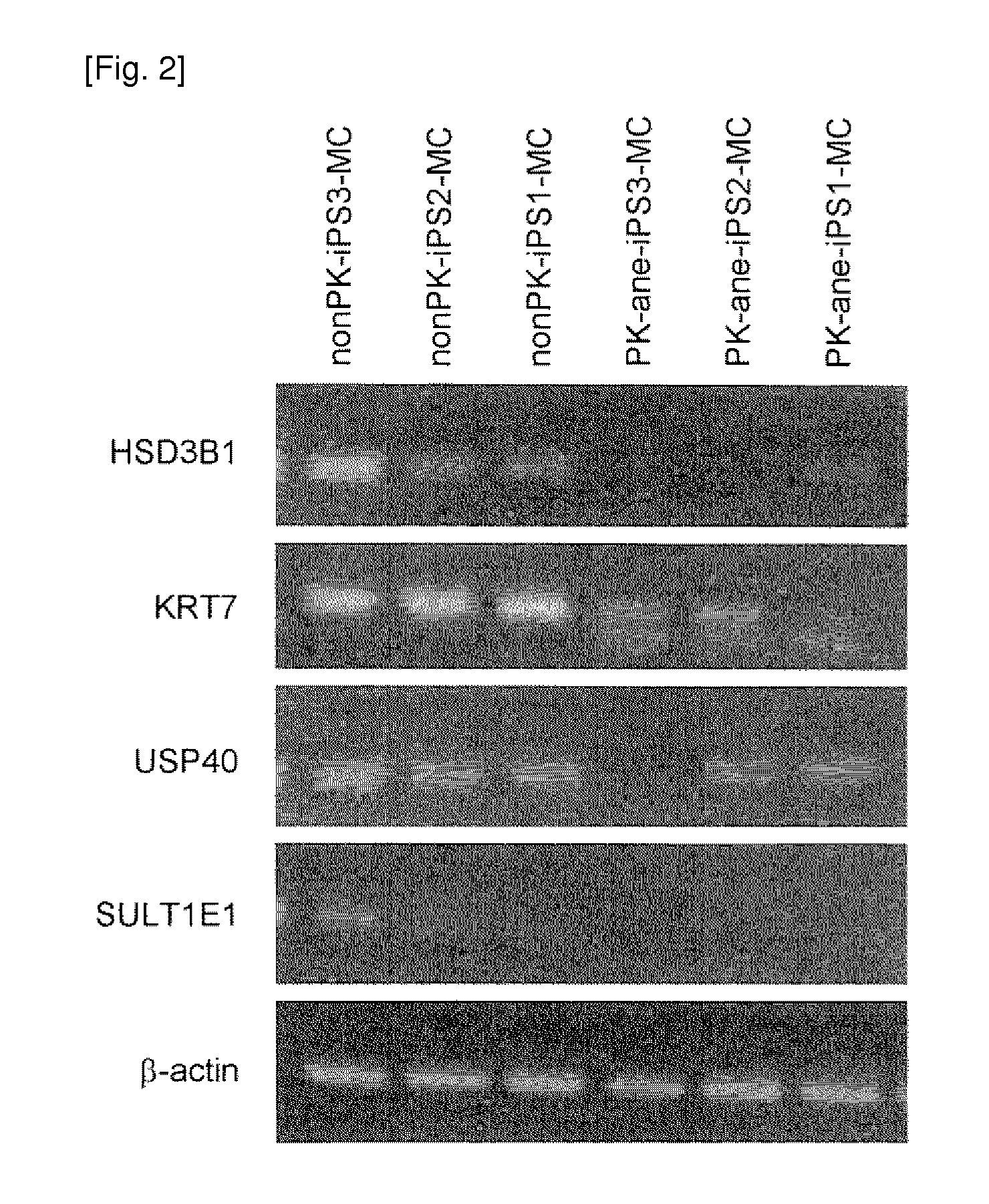
[0283]Each of the above prepared iPS cell line colonies was separated into pieces with an appropriate size, sprayed over a type-I collagen coating dish (Becton Dickinson), followed by 1 day of culture with a medium for primate ES / iPS cells (ReproCELL) to adhere the cells to the dish surface. On day 2, a GSK-3 alpha / beta inhibitor (Sigma), N2 supplement, and B27 supplement (both, Invitrogen) were added, and then cells were cultured for further 3 days. Then the medium was exchanged with a serum free medium for human hematopoietic stem cells (Invitrogen). After 5 days of culture, cells were dissociated, and then VEGFR2-positive, TRA1-negative, and VE-cadherin-negative cells were separated by FACS. Subsequently, the thus separated cells were sprayed over a type-IV collagen coating dish (Becton Dickinson) and further cultured in MEM containing 2-% FCS and 20 ng / ml PDGF-BB (Peprotech Inc). Thus, cells were induced to differentiate into...


PUM

PropertyMeasurementUnit
timeaaaaaaaaaa
timeaaaaaaaaaa
timeaaaaaaaaaa

Abstract

The present invention provides a method of examining polycystic kidney disease or a complication of polycystic kidney disease using a gene(s) selected from the group consisting of NTNG1, POSTN, TNC, KAL1, BST1, ACAT2, INSIG1, SCD, HSD3B1, KRT7, USP40, SULT1E1, BMP6, CD274, CTGF, E2F7, EDN1, FAM43A, FRMD3, MMP10, MYEOV, NR2F1, NRCAM, PDCK1, PLXNA2, SLC30A3, SNAI1, SPOCD1, MMP1, TFPI2, HMGA2, KRTAP4-7, KRTAP4-8, KRTAP4-9, MYPN, RPPH1, and SIAE, and a method of screening for a therapeutic agent or a preventive agent therefore, and further vascular endothelial cells or vascular mural cells obtained via differentiation induction from iPS cells formed from a somatic cell of a subject suffered from polycystic kidney disease and having cerebral aneurysm as a complication.

Description

TECHNICAL FIELD[0001]The present invention relates to a method of examining polycystic kidney disease, a disease marker, and a method of screening for a therapeutic agent for the disease.BACKGROUND ART[0002]Polycystic kidney disease is classified into autosomal dominant polycystic kidney disease (ADPKD) and autosomal recessive polycystic kidney disease (ARPKD). ADPKD is developed by about one in 4000 people in Japan. The assumed number of ADPKD patients is said to range from 20,000 to 50,000. ADPKD is causative disease of end-stage chronic renal failure that results in dialysis introduction, and it is the fourth most common disease following diabetic nephropathy, primary glomerular nephritis, and hypertensive nephrosclerosis.[0003]This disease is an autosomal dominant disease due to a genetic mutation of PKD1 or PKD2 (JP Patent Publication (Kohyo) No. 2001-520502 A, JP Patent Publication (Kohyo) No. 2004-504038 A, and JP Patent Publication (Kokai) No. 2009-065988 A). Also, such an a...

Claims


Application Information

Patent Timeline
Patent Type & Authority Applications(United States)
IPC IPC(8): C07K16/18C12N15/113C12Q1/68C12N15/11
CPCC12N5/069C12N2501/135C12N2501/165C12N2506/45C12Q1/6883C12Q2600/158C12N15/113G01N2800/2871G01N2800/347G01N2800/56C12Q2600/136C07K16/18C12N15/11G01N33/6893A61P13/12
Inventor OSAFUNE, KENJITOYODA, TAROSHIOTA, FUMIHIKONAKAO, KAZUWASONE, MASAKATSUTAURA, DAISUKE
Owner KYOTO UNIV
Why Eureka
Try Eureka