Reconstituted polypeptides

a polypeptide and polypeptide technology, applied in the field of reconstituted polypeptides, can solve the problems of limiting the level of uptake, the size of the antibody and the complexity of the six loops is a major design hurdle,

Inactive Publication Date: 2013-09-12
RES CORP TECH INC
View PDF1 Cites 35 Cited by
  • Summary
  • Abstract
  • Description
  • Claims
  • Application Information

AI Technical Summary

Benefits of technology

The patent text describes a new method for creating a protein binding pair using two polypeptides that can associate with each other. These polypeptides have specific binding domains that can bind to each other with a high degree of specificity. The binding pair can be used to create a complex with another protein or peptide. The method involves creating two polypeptides that have complementary binding domains and then combining them to form a complex. The complex can be used for various applications such as protein-protein interactions and protein-peptide interactions. The patent also describes a way to enhance the stability of the complex by using disulfide bonds between certain amino acids. Overall, this method allows for the creation of a stable and specific protein binding pair.

Problems solved by technology

However, the size of the antibodies and the complexity of six loops represents a major design hurdle if the end result is to be a relatively small peptide ligand.
These antibody fragments are not suitable for structural analysis using NMR spectroscopy due to their size, low solubility or low conformational stability.
However, the short persistence of scFvs in the circulation limits the exposure of tumor cells to the scFvs, placing limits on the level of uptake.
Since X-ray crystallography is not suited for characterizing flexible parts of molecules, structural studies in the solution state have not been possible to provide dynamic pictures of the conformation of antigen-binding sites.
However, as expected, they are too small and too flexible to maintain full affinity and specificity.
Mouse CDRs have been grafted onto the human Ig framework without the loss of affinity (Jones et al., 1986; Riechmann et al., 1988), though this “humanization” does not solve the above-mentioned problems specific to solution studies.
Thus, while Tendamistat may be antigenic in humans, its small size may reduce or inhibit its antigenicity.
Also, Tendamistat's stability is uncertain.
Disulfide bonds, however, are a significant disadvantage to such molecules in that they can be broken under reducing conditions and must be properly formed in order to have a useful protein structure.
Further, the size of the loops in Tendamistat are relatively small, thus limiting the size of the inserts that can be accommodated in the scaffold.
Moreover, it is well known that forming correct disulfide bonds in newly synthesized peptides is not straightforward.
When a protein is expressed in the cytoplasmic space of E. coli, the most common host bacterium for protein overexpression, disulfide bonds are usually not formed, potentially making it difficult to prepare large quantities of engineered molecules.

Method used


Image

  • Reconstituted polypeptides
  • Reconstituted polypeptides
  • Reconstituted polypeptides

Examples


example i

Construction of the Fn3 Gene

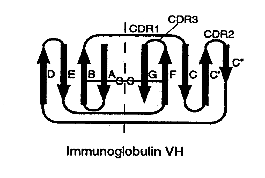
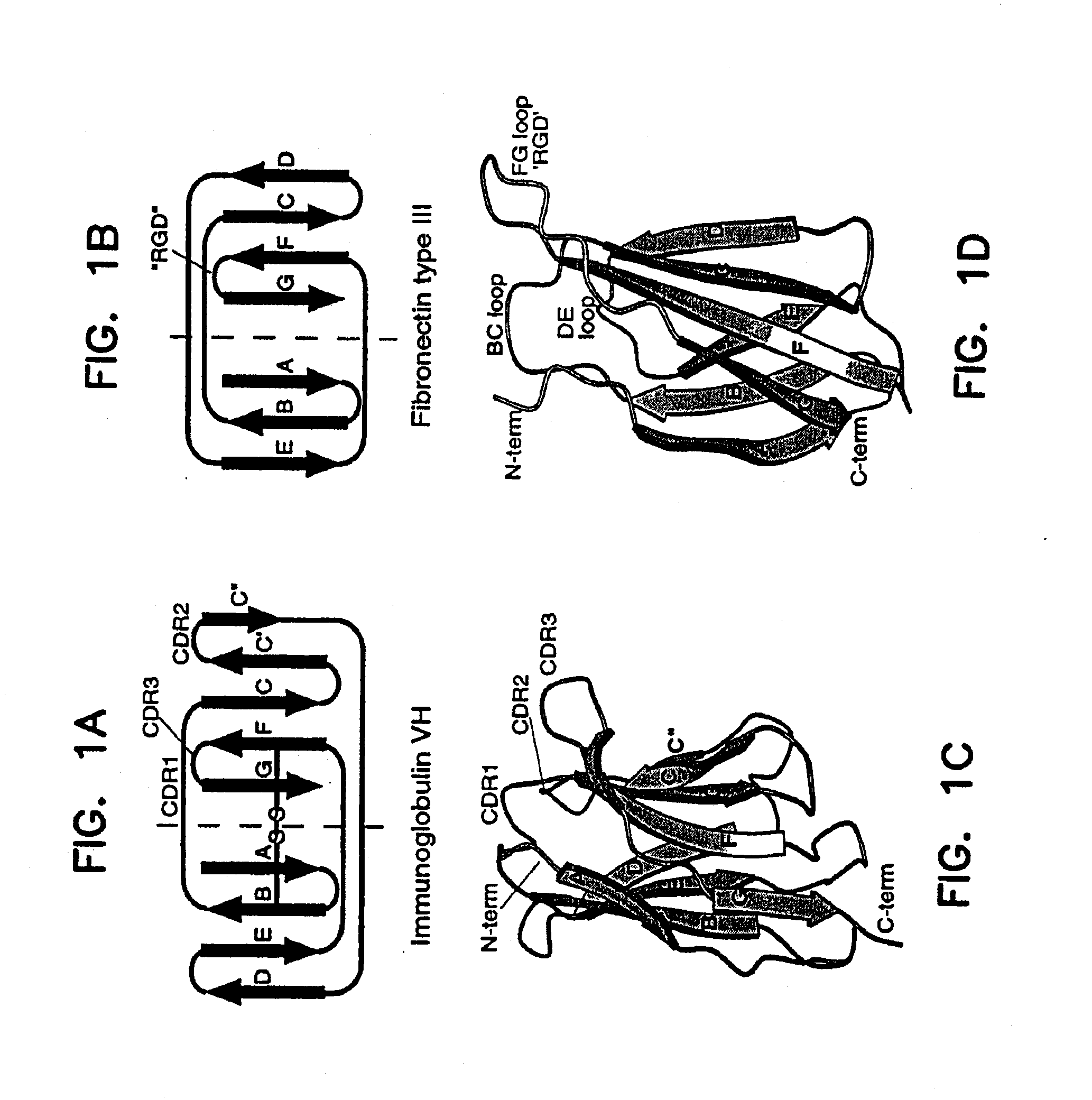
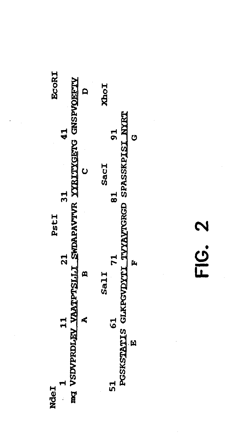
[0173]A synthetic gene for tenth Fn3 of fibronectin (FIG. 1) was designed on the basis of amino acid residue 1416-1509 of human fibronectin (Komblihtt, et al., 1985) and its three dimensional structure (Main, et al., 1992). The gene was engineered to include convenient restriction sites for mutagenesis and the so-called “preferred codons” for high level protein expression (Gribskov, et al., 1984) were used. In addition, a glutamine residue was inserted after the N-terminal methionine in order to avoid partial processing of the N-terminal methionine which often degrades NMR spectra (Smith, et al., 1994). Chemical reagents were of the analytical grade or better and purchased from Sigma Chemical Company and J. T. Baker, unless otherwise noted. Recombinant DNA procedures were performed as described in “Molecular Cloning” (Sambrook, et al., 1989), unless otherwise stated. Custom oligonucleotides were purchased from Operon Technologies. Restriction and modifica...

example ii

Modifications to Include Restriction Sites in the Fn3 Gene

[0179]The restriction sites were incorporated in the synthetic Fn3 gene without changing the amino acid sequence Fn3. The positions of the restriction sites were chosen so that the gene construction could be completed without synthesizing long (>60 bases) oligonucleotides and so that two loop regions could be mutated (including by randomization) by the cassette mutagenesis method (i.e., swapping a fragment with another synthetic fragment containing mutations). In addition, the restriction sites were chosen so that most sites were unique in the vector for phage display. Unique restriction sites allow one to recombine monobody clones which have been already selected in order to supply a larger sequence space.

example iii

Construction of M13 Phage Display Libraries

[0180]A vector for phage display, pAS38 (for its map, see FIG. 8) was constructed as follows. The XbaI-BamHI fragment of pET12a encoding the signal peptide of OmpT was cloned at the 5′ end of the Fn3 gene. The C-terminal region (from the FN5F and FN5R oligonucleotides, see Table 2) of the Fn3 gene was replaced with a new fragment consisting of the FN5F and FN5R′ oligonucleotides (Table 2) which introduced a MluI site and a linker sequence for making a fusion protein with the pIII protein of bacteriophage M13. A gene fragment coding the C-terminal domain of M13 pIII was prepared from the wild-type gene III of M13 mp18 using PCR (Corey, et al., 1993) and the fragment was inserted at the 3′ end of the OmpT-Fn3 fusion gene using the MluI and HindIII sites.

[0181]Phages were produced and purified using a helper phage, M13K07, according to a standard method (Sambrook, et al., 1989) except that phage particles were purified by a second polyethylene...


PUM


Abstract

The present invention provides modified fibronectin type III (Fn3) molecules, and nucleic acid molecules encoding the modified Fn3 molecules. Also provided are methods of preparing these molecules, and kits to perform the methods.

Description

CLAIM OF PRIORITY[0001]This application claims priority under 35 U.S.C. §119(e) from U.S. Provisional Application Ser. No. 60 / 386,991, filed Jun. 6, 2002 which application is incorporated herein by reference.GOVERNMENTAL RIGHTS[0002]Portions of the present invention were made with support of the United States Government via a grant from the National Institutes of Health under grant numbers R29-GM 55042 and R01-DK63090, and via a grant from the Department of Defense under grant number DAMD17-01-1-0385. The U.S. Government therefore may have certain rights in the invention.BACKGROUND OF THE INVENTION[0003]U.S. patent application Ser. Nos. 09 / 096,749, which corresponds to Publication No. US 2002 0019517, and 09 / 903,412 are hereby incorporated by reference in their entirety.Antibody Structure[0004]A standard antibody (Ab) is a tetrameric structure consisting of two identical immunoglobulin (Ig) heavy chains and two identical light chains. The heavy and light chains of an Ab consist of d...

Claims


Application Information

Patent Timeline
Patent Type & Authority Applications(United States)
IPC IPC(8): C07K16/00C07K14/78C07K16/18
CPCC07K16/00C07K16/18C07K14/78C07K2318/20C07K2317/565
Inventor KOIDE, SHOHEI
Owner RES CORP TECH INC
Why Eureka
Try Eureka