

Method for delineation of tissue lesions
- Summary
- Abstract
- Description
- Claims
- Application Information
AI Technical Summary
Benefits of technology
Problems solved by technology
Method used
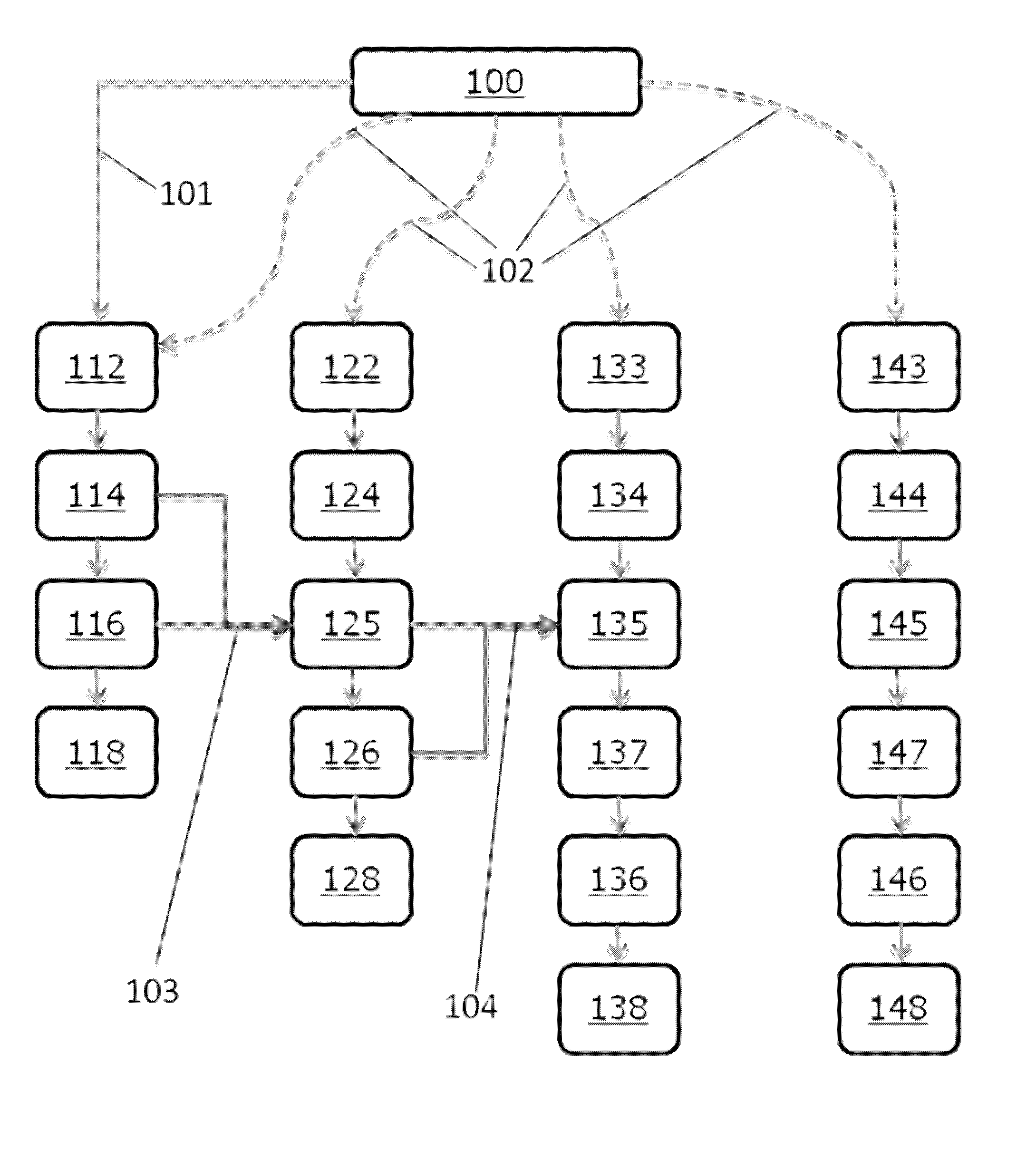
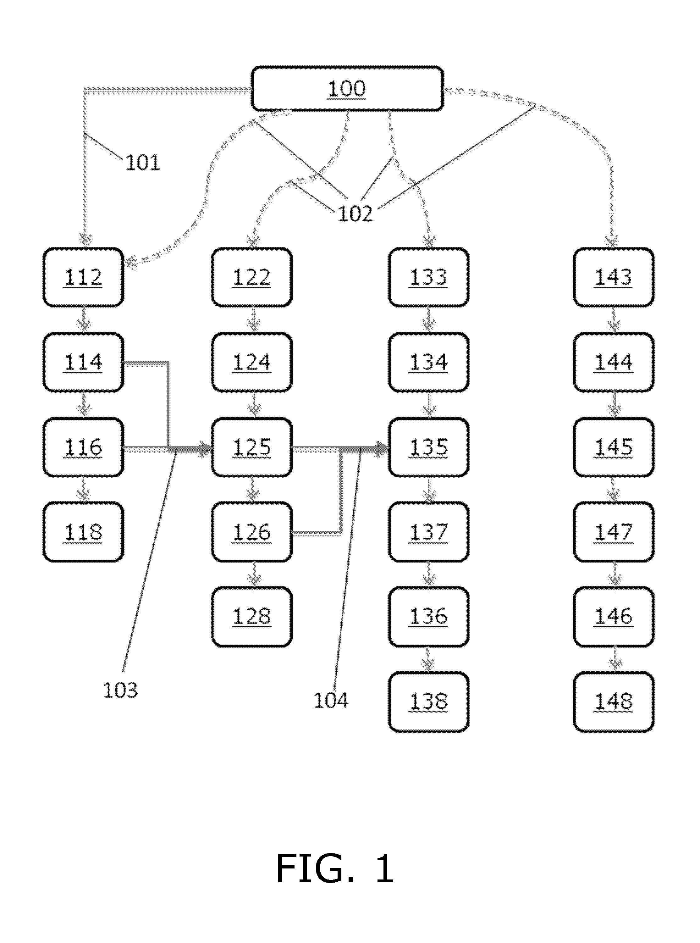
Image

Examples
example 1
Segmentation of an Image Comprising TTP Data
Materials & Methods
Morphological Grayscale Reconstruction
[0137]Finding a voxel within perfusion lesions, so-called seed point, appear easily discernible by eye, however, searching for the maximum of the image intensities often leads to a false positive seed point e.g. high intensities in regions of the image corresponding to irrelevant regions, such as eyes, cerebrospinal fluid (CSF), intensities outside the brain etc. To automatically detect a seed point, we use morphological grayscale reconstruction to truncate small spikes while preserving the edges of TTP lesion and homogenize the background of TTP maps. Compared with conventionally smoothing e.g. with a Gaussian kernel, morphological grayscale reconstruction is less sensitive to spikes and for that reason more suitable for seed point detection. In the present context, spikes are understood to be areas which are relatively small compared to a lesion, and which comprise data points with...
example 2
Segmentation of an Image of Type MTT Data
Materials and Methods
Level Sets
[0162]The approach emerges from the assumption that the average MTT value, Mhypo, in a hypoperfused region is higher than the average normal MTT value, Mnorm. In the hypoperfused area, MTT values will vary around Mhypo, while in normal tissue they are close to Mnorm. This corresponds to low mean squared errors between image MTT values, MTT(x,y), and the respective averages. Identifying the lesion, such as the ischemic lesion, can then be formulated as finding a smooth, closed curve C, which minimizes the total variation (Eq. (1))
E=∫Inside C(MTT(x,y)−Mhypo)2dxdy+∫Outside C(MTT(x,y)−Mnorm)2dxdy
[0163]Since ischemic regions are not necessarily coherent, for instance in cases with occlusions causing lesions in the watershed areas, it is important that C can represent multiple curves. This can be ensured by defining C implicitly as the zero-level set of a function φ which assigns a value to each point the plane, C={φ...
example 3
Segmentation of an Image Obtained by DWI
Materials & Methods
Morphological Grayscale Reconstruction
[0187]DWI lesion appears easily discernible by eye as a hyperintense region, although the diffusion lesion contains artefactual hypointensities and hyperintensities are present throughout the image due to anatomy and noise artifacts. Therefore simple thresholding of the image intensities leads to both high false positive and false negative rates as indicated in FIG. 11B. Typical image blurring, such as convolution by Gaussian kernel, remedies artifacts to some extent, although at the potential cost of ‘moving’ the lesion boundary. Morphological grayscale reconstruction uniquely truncates high peaks in the image while tending to preserve original edges of coherent areas, as determined by the kernel size. We use morphological grayscale reconstruction to enhance the contrast between the DWI lesion and the background. Compared with conventional blurring filter, morphological grayscale recons...
PUM
 Abstract
Description
Claims
Application Information