Specific a1at monoclonal antibodies for detection of endometriosis
a monoclonal antibody and endometriosis technology, applied in the field of diagnostics and/or detection of endometriosis, can solve the problems of limited sensitivity to certain types of endometriosis, and low acceptance rate by patients
- Summary
- Abstract
- Description
- Claims
- Application Information
AI Technical Summary
Benefits of technology
Problems solved by technology
Method used
Image
Examples
example 1
Preparation of Antigens
[0059]Serum samples were obtained with informed consent from women having endometriosis. The serum samples were precipitated by adding 4 times volume of cold acetone containing 10% (w / v) trichloracetic acid (TCA). The mixture was kept at −20° C. for 90 min, then centrifuged at 4° C. with a speed of 15,000×g for 20 min. The pellet was collected and washed with ice-cold acetone, followed by another centrifugation at a speed of 15,000×g for 20 min. The supernatant was discarded, and the pellet was dissolved in rehydration buffer (7M urea, 4% CHAPS ([3-(3-cholamidopropyl)dimethylammonio]-1-propanesulfonate), 2M thiourea, 0.002% bromophenol blue, and 65 mM dithioerythritol (DTE)).
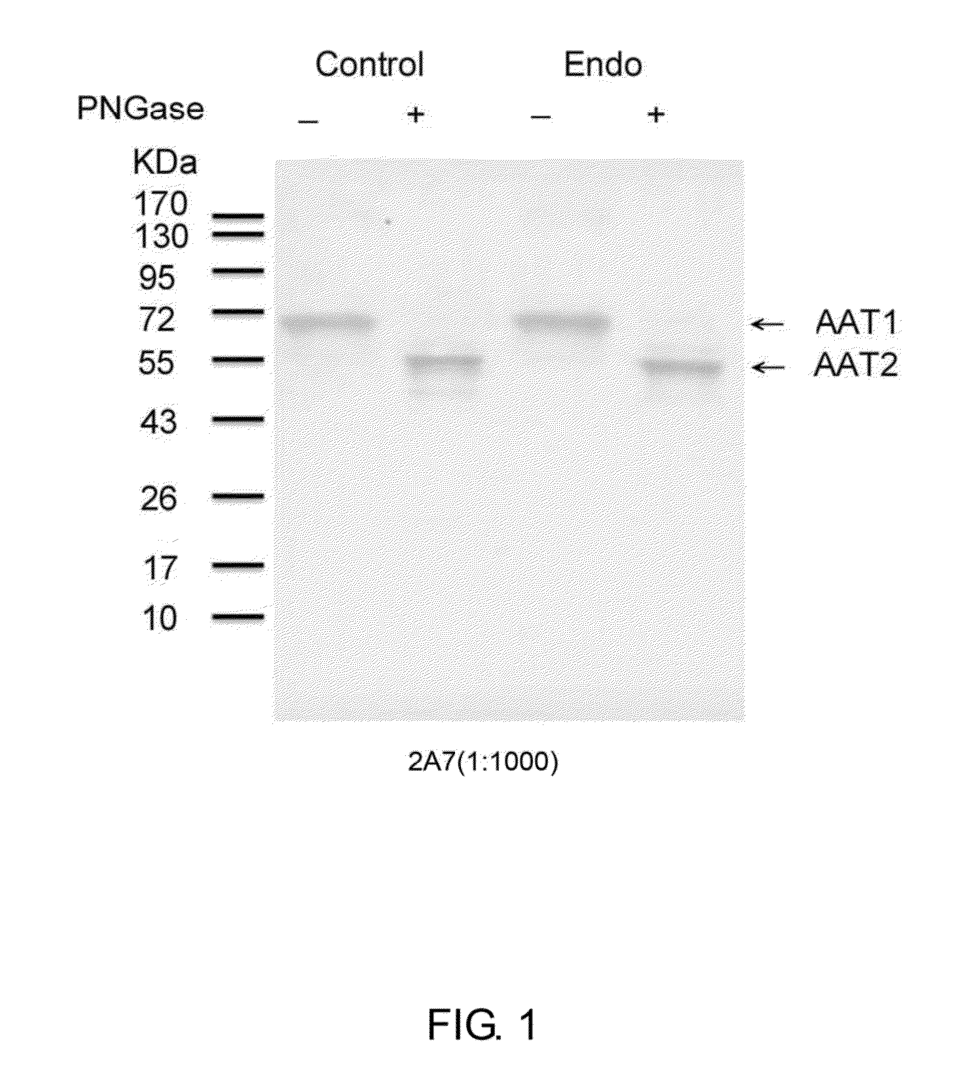
[0060]The proteins in the rehydration buffer were subsequently treated with PNGase F to remove any N-glycan chains of the proteins. The digested proteins were then identified by Western blot. Briefly, equal amount of protein samples were electrophoresized with 10% SDS-PAGE and then transfe...
example 2
Production of Monoclonal Antibodies
[0062]2.1 Immunization the Animals with Alpha 1-Antitrypsin (A1AT) and Measurement of Antibody Titers
[0063]Mice were immunized with alpha 1-antitrypsin (A1AT, purchased from Sigma Inc) at a dose of 30 μg / animal, 2 to 6 times, in 4 week intervals. After 2 boosting, the blood samples were taken every week, and were immediately centrifuged to separate sera. The resultant sera were subjected to serial dilution, followed by measurement of antibody titers by Western blot
2.2 Preparation of Antibody-Producing Cells
[0064]Animals with desired titers in example 2.1 (i.e., when a 1:5,000 dilution of the sera were positive in western blot) were selected for a fusion. Antibody-producing cells were prepared from splenic cells, regional lymph nodes of the immunized animals, and hybridomas were generated by fusion the antibody-producing cells with a myeloma FO cell line in accordance with the procedures described by Hong et al (J Immunol Methods 120: 151-157 (1989)...
example 3
The Detection of Endometriosis Using Monoclonal Antibodies of Example 2
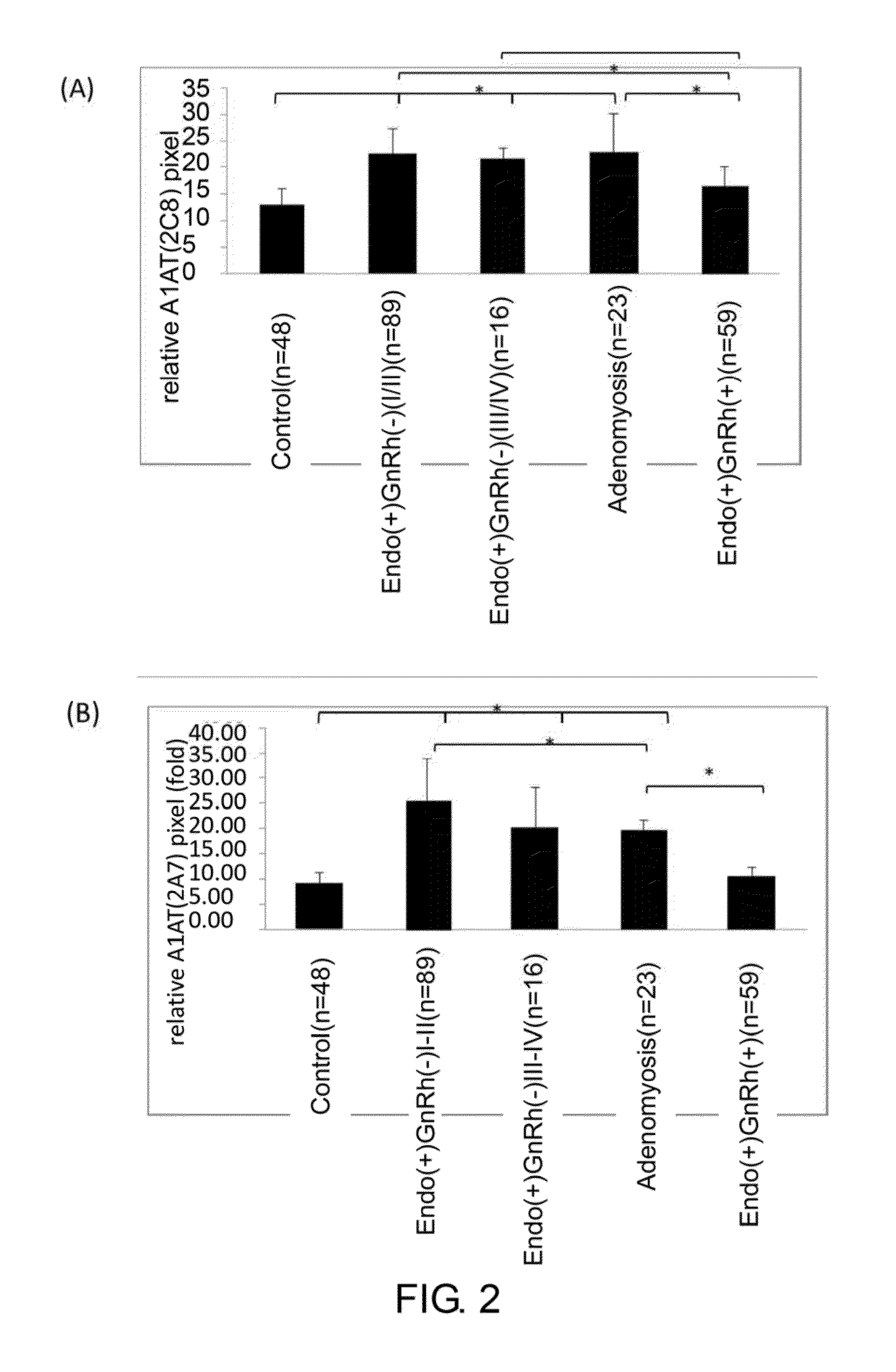
[0069]The specificity of the monoclonal antibodies 2A7 and 2C8 of Example 2 was analyzed using immune-dot blot analysis.
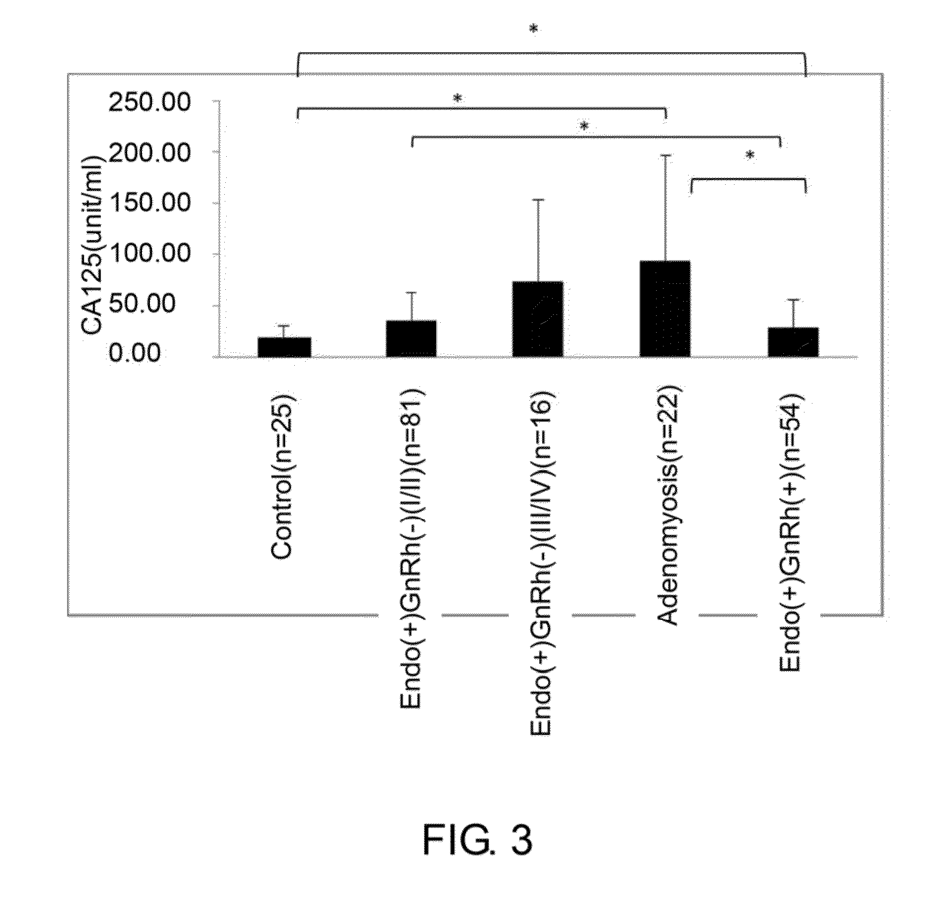
[0070]In this example, serum samples from total of 235 women were obtained with informed consent, and were classified into 5 groups. The control group was composed of 48 women without endometriosis at reproducing age. 89 women with mild (or early) stage pelvic endometriosis and whom had not received gonadotropin-releasing hormone (GnRh) treatment were classified as Ecdo(+)GnRh(−) I / II group. 16 women with severe stage of pelvic endometriosis and whom had not received GnRh treatment were classified as Ecdo(+)GnRh(−) III / IV group. 23 women with adenomyosis were classified as Adenomyosis group. Finally, 59 women with pelvic endometriosis and whom have received GnRh treatment were classified as Endo(+)GnRh(+) group.
[0071]To perform dot-blot analysis; 5 ng of serum collected from women described abov...
PUM
| Property | Measurement | Unit |
|---|---|---|
| Atomic weight | aaaaa | aaaaa |
| Atomic weight | aaaaa | aaaaa |
| Molecular weight | aaaaa | aaaaa |
Abstract
Description
Claims
Application Information
 - R&D
- Intellectual Property
- Life Sciences
- Materials
- Tech Scout
- Unparalleled Data Quality
- Higher Quality Content
- 60% Fewer Hallucinations
Browse by: Latest US Patents, China's latest patents, Technical Efficacy Thesaurus, Application Domain, Technology Topic, Popular Technical Reports.
© 2025 PatSnap. All rights reserved.Legal|Privacy policy|Modern Slavery Act Transparency Statement|Sitemap|About US| Contact US: help@patsnap.com


