Assessment of coronary heart disease with carbon dioxide

Inactive Publication Date: 2014-06-19
CEDARS SINAI MEDICAL CENT
View PDF18 Cites 6 Cited by
  • Summary
  • Abstract
  • Description
  • Claims
  • Application Information

AI Technical Summary

Benefits of technology

[0101]The methods described herein to functionally assess the oxygen status of the myocardium include administering an effective amount of CO2 to the subject in need thereof. In an embodiment, the O2 level is held constant while the CO2 level is altered so as to induce hyperemia. Applicants herein show the vascular reactivity in subjects in response to changes in PaCO2. The existing methods use adenosine, dipyridamole, or regadenoson which have significant side-effects described above. As described in the Examples below, CO2 is at least just as effective as the existing methods (which use, for example, adenosine) but without the side effects.
[0102]The use of CO2 provides distinct advantages over the use of, for example, adenosine. Administering CO2 is truly non-invasive because it merely involves inhaling physiologically sound levels of CO2. The instant methods are simple, repeatable and fast and most likely have the best chance for reproducibility. Not even breath-holding is necessary during acquisition of images using the methods described herein. The instant methods are also highly cost-effective as no pharmacological stress agents are required, cardiologists may not need to be present during imaging and rapid imaging reduces scan times and costs.
[0103]Further, the improved BOLD MRI technique described above provides a non-invasive and reliable determination of ischemic volume (no radiation, contrast-media, or adenosine) and other value-added imaging biomarkers from the same acquisition (Ejection Fraction, Wall Thickening). Additionally, the subject does not experience adenosine-related adverse side effects and thus greater patient tolerance for repeat

Problems solved by technology

As the coronary arteries narrow, blood flow to the heart can slow down or stop, causing, amongst other symptoms, chest pain (stable angina), shortness of breath and/or myocardial infarction.
Since nuclear methods are hampered by the need for radioactive tracers combined with limited imaging resolution, other imaging methods, such as ultrasound (using adenosine along with microbubble contrast) and MRI (also using adenosine and various conjugates of gadolinium (Gd) (first-pass perfusion) or alterations in oxygen saturation in response to hyperemia, also known as the Blood-Oxygen-Level-De

Method used


Image

  • Assessment of coronary heart disease with carbon dioxide
  • Assessment of coronary heart disease with carbon dioxide
  • Assessment of coronary heart disease with carbon dioxide

Examples


example 1

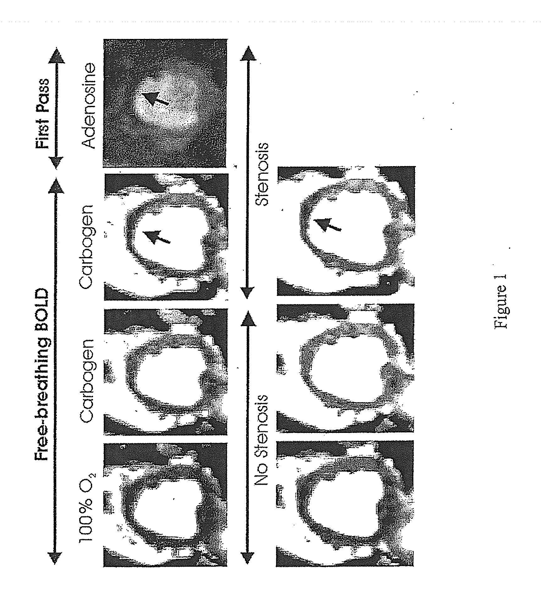
[0105]The inventor has shown that CO2 can increase myocardial perfusion by a similar amount, as does adenosine in canine models. The inventor has also shown that in the setting of coronary artery narrowing, it is possible to detect regional variations in hyperemic response with the use of MRI by detecting signal changes in the myocardium due to changes in oxygen saturation (also known as the BOLD effect) using a free-breathing BOLD MRI approach.

[0106]As show in FIG. 1, the inventor found a 20% BOLD signal increase (hyperemic response) with medical-grade carbogen breathing in the absence of stenosis in dogs. With a severe stenosis, the hyperemic response was significantly reduced in the LAD (left anterior descending) territory but the other regions showed an increase in signal intensity (as observed with adenosine). First-pass perfusion images acquired with adenosine under severe stenosis (in the same slice position and trigger time) is also shown for comparison. Heart rate increase ...

example 2

Co-Relation Between Inhaled CO2 and Oxygen Saturation

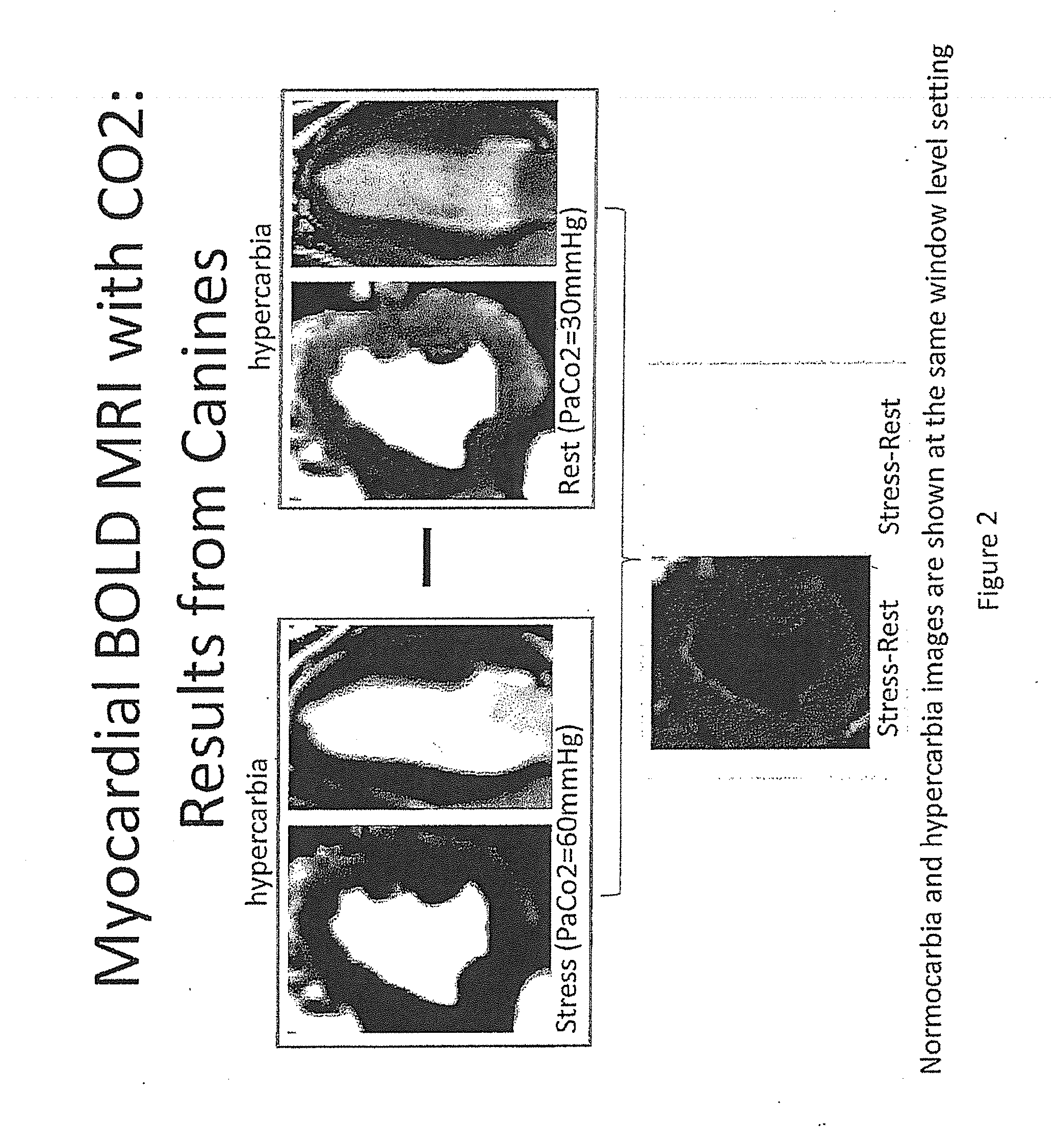
[0108]Applicants assessed the difference between myocardial blood-oxygen-level dependent (BOLD) response under hypercarbia and normocarbia conditions in canines. The BOLD signal intensity is proportional to oxygen saturation.

[0109]Top panels of FIG. 2 depict the myocardial response under hypercarbia (60 mm Hg) and normocarbia (30 mmHg) conditions and show an increase in BOLD signal intensity under hypercarbia condition. The lower panel depicts the difference as obtained by subtracting the signal under rest from that under stress. The myocardial BOLD signal difference between the two is depicted in grey and shows the responsiveness of canines to hypercarbia conditions.

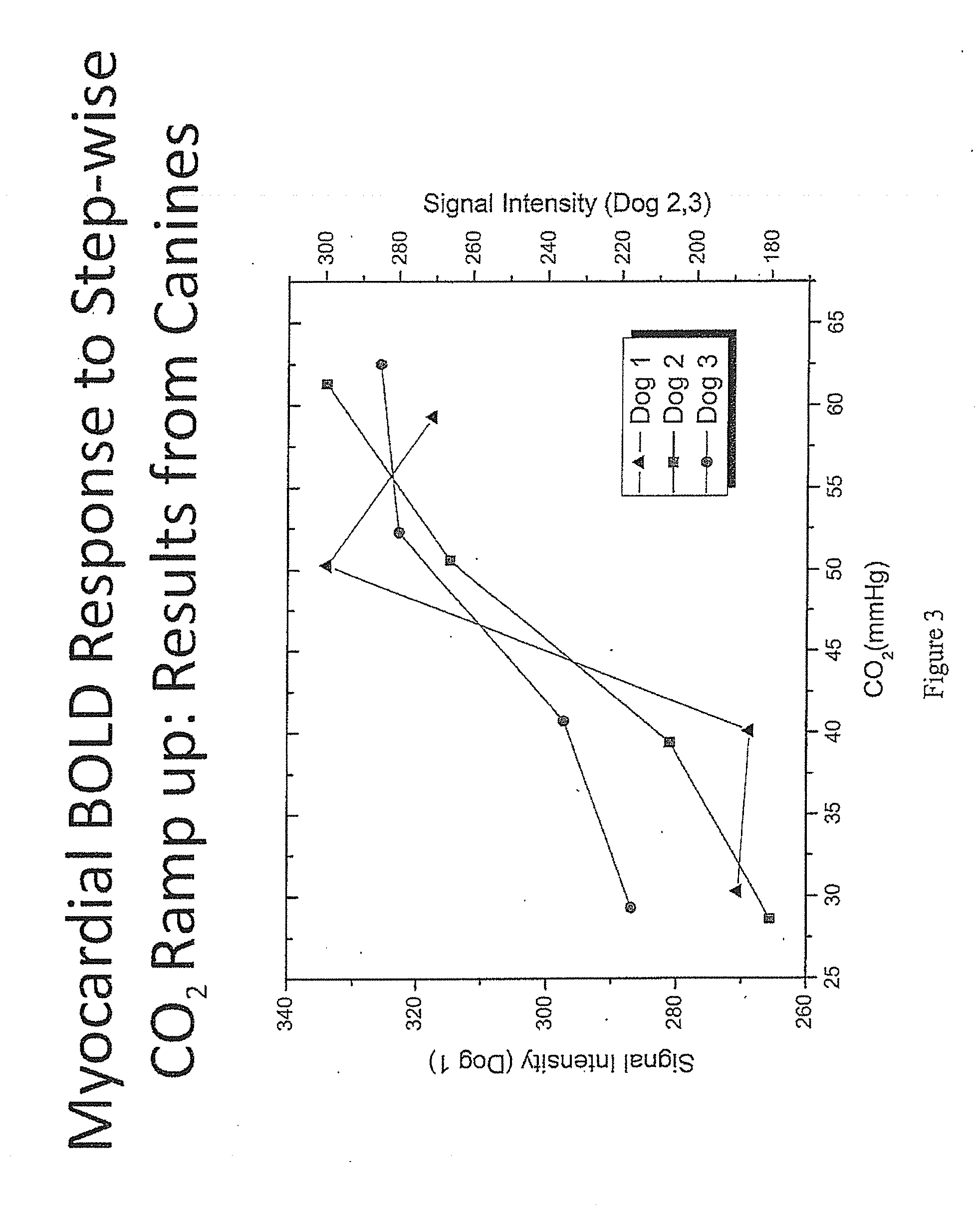
[0110]Applicants further assessed the myocardial BOLD response to stepwise CO2 increase (ramp-up) in canines. As shown in FIG. 3, as the amount of CO2 administered increases, the BOLD signal intensity increases which is indicative of an increase in hyperemic response ...

example 3

[0112]Co-Relation Between the Amount of CO2 Inhaled and Doppler Flow

[0113]Doppler flow, an ultrasound-based approach which uses sound waves to measure blood flow, was used to show that administration of CO2 leads to vasodilation which results in greater blood flow, while PaO2 is held constant. The Doppler flow was measured in the left anterior descending (LAD) artery. As shown in FIG. 5, as the amount of administered CO2 increases the Doppler flow increases. The relative change in coronary flow velocity is directly proportional to the amount of CO2 administered.


PUM


Abstract

The invention provides methods for diagnosing coronary heart disease in a subject in need thereof comprising administering an admixture comprising CO2 to a subject to reach a predetermined PaCO2 in the subject to induce hyperemia, monitoring vascular reactivity in the subject and diagnosing the presence or absence of coronary heart disease in the subject, wherein decreased vascular reactivity in the subject compared to a control subject is indicative of coronary heart disease. The invention also provides methods for increasing sensitivity and specificity of BOLD MRI.

Description

GOVERNMENT RIGHTS[0001]The invention was made with government support under Grant No. HL091989 awarded by the National Institutes of Health. The government has certain rights to the invention.FIELD OF INVENTION[0002]The invention is directed to methods for detecting coronary heart disease using carbon dioxide (CO2) to induce hyperemia and monitor vascular reactivity.BACKGROUND[0003]All publications herein are incorporated by reference to the same extent as if each individual publication or patent application was specifically and individually indicated to be incorporated by reference. The following description includes information that may be useful in understanding the present invention. It is not an admission that any of the information provided herein is prior art or relevant to the presently claimed invention, or that any publication specifically or implicitly referenced is prior art.[0004]Coronary artery disease (CAD) leads to narrowing of the small blood vessels that supply blo...

Claims


Application Information

Patent Timeline
IPC IPC(8): A61K49/00
CPCA61K33/00A61K31/7076A61K49/08A61K2300/00
Inventor DHARMAKUMAR, ROHANLI, DEBIAOTSAFTARIS, SOTIRIOS A.
Owner CEDARS SINAI MEDICAL CENT
Why Eureka
Try Eureka