Method for preparing induced pluripotent stem cells using microvesicles derived from embryonic stem cells

a technology of embryonic stem cells and microvesicles, which is applied in the field of preparation of induced pluripotent stem cells, can solve the problems of undesired modifications, easy degradation of mrna, and no successful case of dedifferentiation having similar characteristics, and achieves the effects of reducing damage, reducing the number of cells, and effectively protecting the content of the delivery

Inactive Publication Date: 2014-06-19
POSTECH ACAD IND FOUND
View PDF2 Cites 2 Cited by
  • Summary
  • Abstract
  • Description
  • Claims
  • Application Information

AI Technical Summary

Benefits of technology

[0013]According to the present invention, a cytoplasm delivery method using fusion of microvesicles does not need polyethyleneglycol (PEG) needed in cell fusion or a cytotoxic material such as streptolysin used in cytoplasm injection, and thus cells are damaged less. In addition, during the cytoplasm delivery, the microvesicle has high delivery efficiency, and more effectively protects a delivered content, and thus it is possible to perform specific delivery of the cytoplasm. According to conventional research on cytoplasm delivery, cytoplasm delivery efficiency is decreased according to an amount of proteins to be expressed in a nucleus, and thus perfect dedifferentiation does not occur. However, the microvesicles of the present invention can freely control a concentration of the cytoplasm and prevent loss of intracellular materials in fusion depending on a preparation method, so that the efficiency of dedifferentiation of somatic cells using the cytoplasm can be increased.
[0014]In addition, according to the method using microvesicles of the present invention, targeting to induce in-vivo dedifferentiation is possible. Particularly, since a signal generated in a wounded organ of a patient is a major factor of targeting, the dedifferentiation performed in vitro is expected to become a new means capable of being performed in vivo.

Problems solved by technology

In addition, the method of examining cell fusion (cell, cytoplasm) has a disadvantage in that many cells die due to induction of fusion using polyethylene glycol (PEG) that can damage the cells to help fusion of large-sized cells.
However, in the case of the dedifferentiation using cytoplasm, there has been no successful case of dedifferentiation having similar characteristics to embryonic stem cells as of 2010.
However, due to the characteristics of a virus, the genes are integrated in several sites, thereby forming undesired modifications.
However, it was seen that mRNA is easily degraded and has a difficult synthesis process, an immune response of RNA is induced, and the delivery of only a protein cannot achieve perfect dedifferentiation.

Method used


Image

  • Method for preparing induced pluripotent stem cells using microvesicles derived from embryonic stem cells
  • Method for preparing induced pluripotent stem cells using microvesicles derived from embryonic stem cells
  • Method for preparing induced pluripotent stem cells using microvesicles derived from embryonic stem cells

Examples


example 1

Preparation of Embryonic Stem Cell-Derived Microvesicles

[0042]Mouse embryonic stem cells were resuspended in 3 ml of a phosphate buffered saline (PBS) solution at a concentration of 5×10⁶ cells / ml. The resuspension was passed through a membrane filter having a pore size of 10 μm 10 times, and through a membrane filter having a pore size of 5 μm 10 times. 1 ml of 50% OptiPrep™, 1 ml of 5% OptiPrep™, and 3 ml of a cell suspension passed through the membrane filter were each put in a 5 ml ultracentrifuge tube. Afterward, ultracentrifugation was performed at 100,000×g for 2 hours. A microvesicle was obtained from a layer between 50% OptiPrep™ and 5% OptiPrep™.

example 2

Analysis of Characteristics of Embryonic Stem Cell-Derived Microvesicles

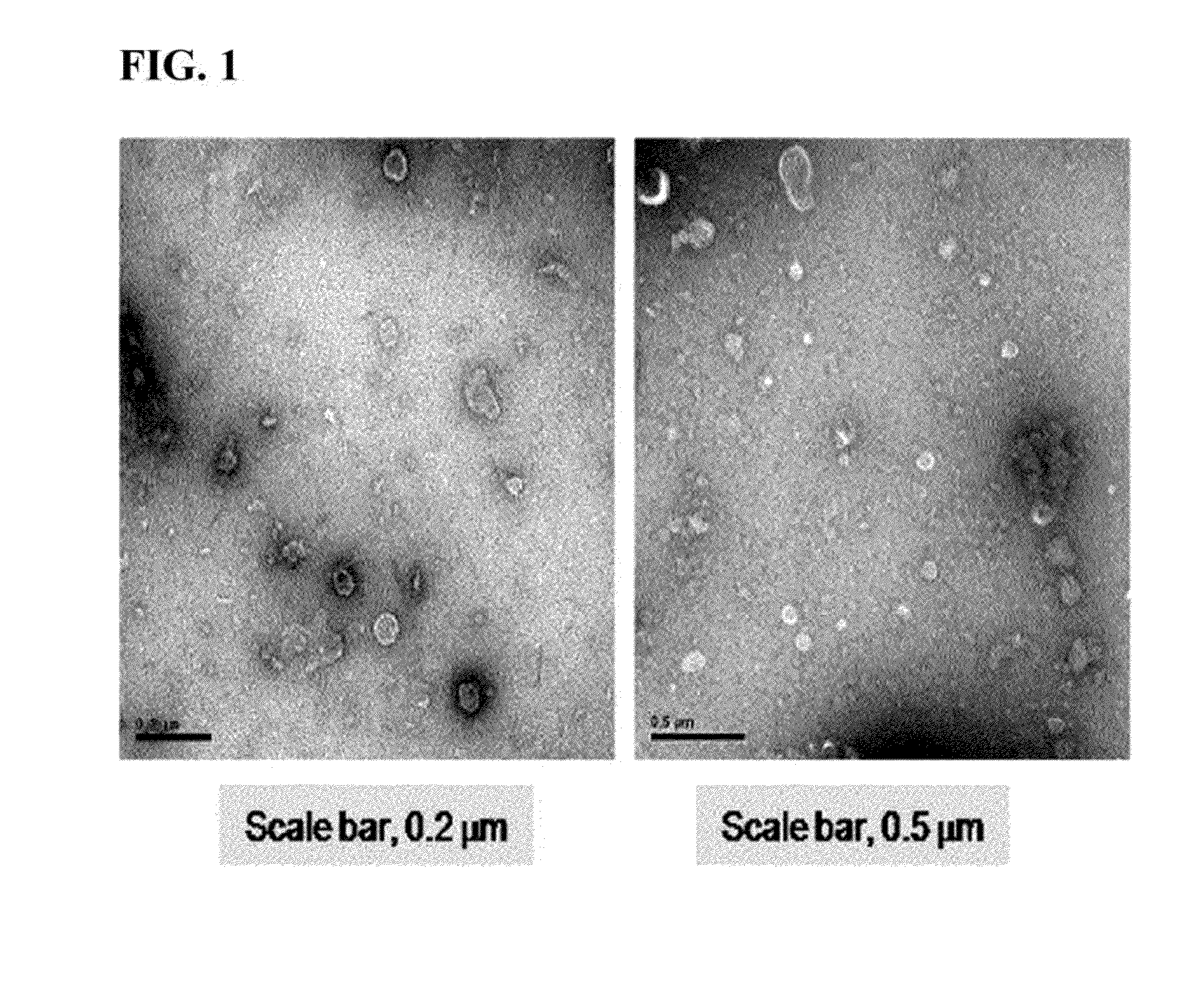
[0043]The microvesicles prepared in the embryonic stem cells according to the method described in Example 1 were adsorbed on a glow-discharged carbon-coated copper grid for 3 minutes. The grid was washed with distilled water and stained with 2% uranylacetate for 1 minute, and results observed using a transmission electron microscope, JEM101 (Jeol, Japan), are shown in FIG. 1.

[0044]As shown in the TEM images of FIG. 1, it can be seen that the microvesicle prepared by extrusion from the embryonic stem cells was composed of a lipid bilayer, and usually formed in a sphere having a size of 100 to 200 nm.

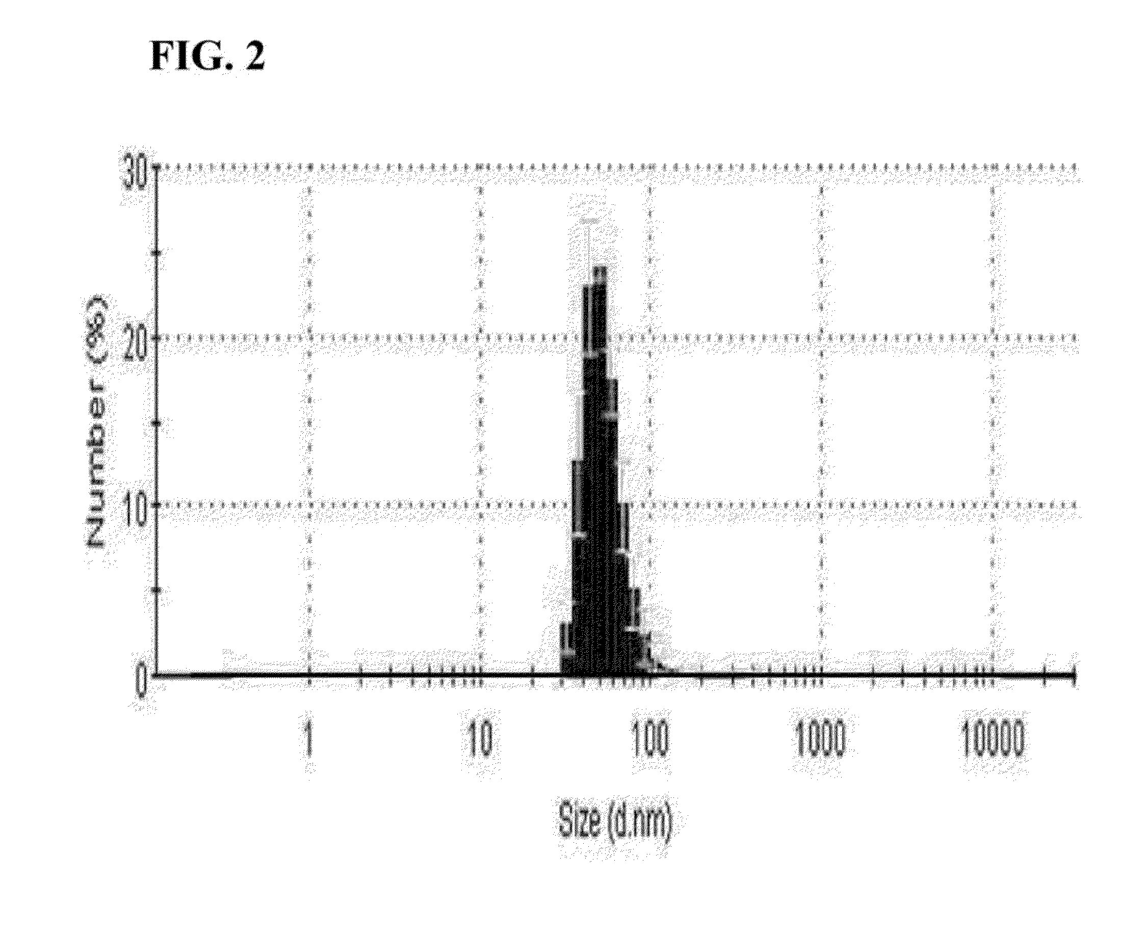
[0045]The microvesicle prepared from the embryonic stem cells described in Example 1 was diluted in 1 ml of PBS at a concentration of 5 μg / ml. 1 ml of PBS containing the microvesicles was put into a cuvette and analyzed using a dynamic light scattering particle size analyzer, and results are shown in FIG. 2.

[0046]As s...

example 3

Dedifferentiation of Somatic Cells using Embryonic Stem Cell-Derived Microvesicles

[0051]0.1% gelatin was coated on a 6-well plate and inoculated with 8×10⁴ of NIH3T3 cells, and the cells were incubated for 24 hours. Afterward, each well was washed with PBS, 2 ml of the microvesicles prepared in the embryonic stem cells according to the method described in Example 1 were diluted in a fibroblast medium (DMEM, 10% FBS, 100 U / ml penicillin-streptomycin) at a concentration of 100 μg / ml, and then treated to the incubated NIH3T3 cells. After 48 hours, approximately 2 to 3 colonies per well were identified, and each colony had a size of approximately 10 to 100 μm. The colonies were observed using an electron microscope, and results are shown in FIG. 5.

[0052]As shown in FIG. 5, it is confirmed that the NIH3T3 cells were dedifferentiated using the embryonic stem cell-derived microvesicles, thereby inducing colonies. Each well was washed with PBS, and 400 μl of 0.1× TE (Typsin-EDTA) was added....


PUM

PropertyMeasurementUnit
sizeaaaaaaaaaa
concentrationaaaaaaaaaa
sizeaaaaaaaaaa

Abstract

Provided is a method of dedifferentiating somatic cells using embryonic stem cell-derived microvesicles. Particularly, a method of preparing induced pluripotent stem cells by treating a composition including embryonic stem cell-derived microvesicles to the somatic cells. According to the method of preparing induced pluripotent stem cells, the dedifferentiation of the somatic cells may be efficiently performed without side effects using the embryonic stem cell-derived microvesicles, and moreover, the method is expected to be very useful in developing a cell therapy product having immunocompatibilities by individuals.

Description

TECHNICAL FIELD[0001]The present invention relates to a method of preparing induced pluripotent stem cells by dedifferentiating somatic cells using embryonic stem cell-derived microvesicles.BACKGROUND ART[0002]Dedifferentiation is a process in which mature somatic cells revert to younger stem cells, and relates to regeneration in vivo, which occurs in insects, amphibians, plants. It does not occur naturally in mammals such as humans, but only through artificial methods. Cellular dedifferentiation methods began with methods using cell fusion. The first method of dedifferentiating somatic cells to be discovered was a method using a characteristic in which, when embryonic stem cells (ESCs) are fused with somatic cells, the embryonic stem cells become more dominant. Afterward, research on inducing dedifferentiation of the somatic cells has progressed through fusion of somatic cells with stem cells having similar capabilities to the ESCs, for example, embryonic germinal cells (EGCs), or ...

Claims


Application Information

Patent Timeline
Patent Type & Authority Applications(United States)
IPC IPC(8): C12N5/074
CPCC12N2502/02A61K35/545C12N2506/13C12N5/0696A61P43/00
Inventor PARK, JAE-SUNGGHO, YONG SONGKIM, YOON KEUNKIM, JUN HOJANG, SU CHULYI, NAMWOOJEONG, DAYEONGCHOI, EUN-JEONG
Owner POSTECH ACAD IND FOUND
Why Eureka
Try Eureka