Nanoparticle-based scaffolds and implants, methods for making the same, and applications thereof

a nanoparticle and scaffold technology, applied in the field of devices, can solve the problems of significant healthcare and economic burden, increased morbidity and substantially worse outcomes, and insufficient methods for treating such tumors, so as to reduce or eliminate microbial infections, eliminate or reduce a source of microbial infections

Active Publication Date: 2014-09-25
RGT UNIV OF CALIFORNIA
View PDF4 Cites 8 Cited by
  • Summary
  • Abstract
  • Description
  • Claims
  • Application Information

AI Technical Summary

Benefits of technology

[0026]Further provided herein are and methods for using the devices in reducing or eliminating microbial infections. For example, the devices can be used to eliminate or reduce a source of microbial infection that can be introduced during a surgical procedure when the device (e.g., a scaffold, a fixture or an implant) is introduced into a recipient (e.g., a mammal). In some embodiments, the source of microbial infection can be an additional bioactive agent that the device can be used to mask.

Problems solved by technology

Implant-associated microbial infections are one of the most serious complications in orthopedic surgery because they are extremely difficult to treat and result in increased morbidity and substantially worse outcomes.
Despite a recent focus on aseptic surgical and procedural techniques, catheter- and surgical implant-associated infections account for nearly half of the 2 million cases of nosocomial infections in the United States per year, representing a significant healthcare and economic burden.
Consequently, methods for treating such tumors are also lacking due to the inability in combining high specific and sensitive imaging with highly conformal radiation.’
However, these additional surgical procedures and medical therapies not only increase the healthcare costs, but also result in an increased rate of recurrence, particularly because it is difficult to clear the infection from devascularized bone and other necrotic tissues.
Once a biofilm forms, it is extremely difficult to treat these infections because the biofilm blocks the penetration of both host immune cells (such as macrophages) and systemic antibiotics, promoting further bacterial survival.
Although this local antibiotic therapy may be effective, they are limited to certain bacterial species and these infections can be caused by a spectrum of bacteria, including Gram-positive Staphylococcus aureus, Staphylococcus epidermidis and Streptococci species, and Gram-negative Pseudomonas and Enterobacter species.

Method used


Image

  • Nanoparticle-based scaffolds and implants, methods for making the same, and applications thereof
  • Nanoparticle-based scaffolds and implants, methods for making the same, and applications thereof
  • Nanoparticle-based scaffolds and implants, methods for making the same, and applications thereof

Examples


example 1

Stainless Steel Alloy Coating

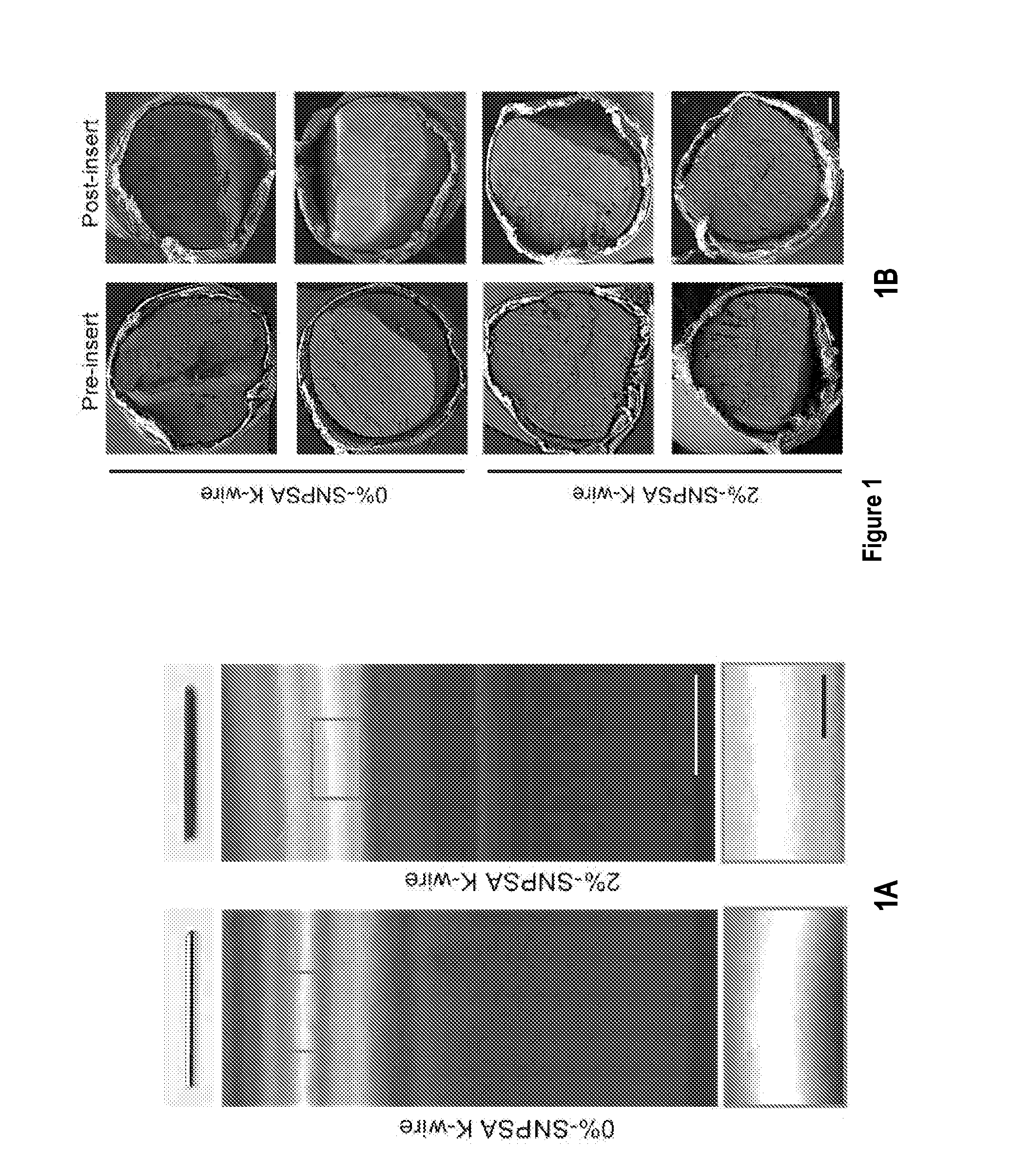
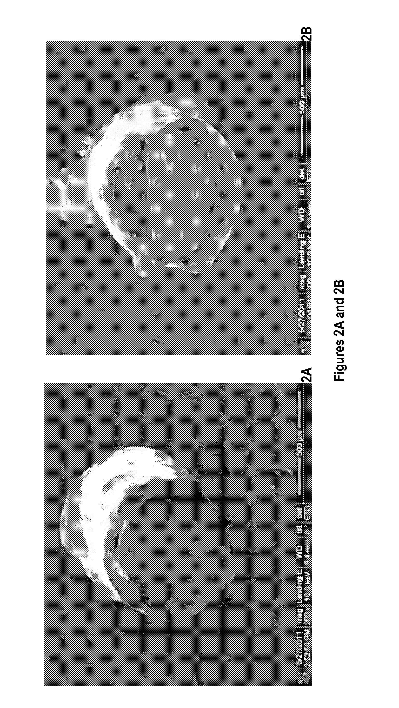
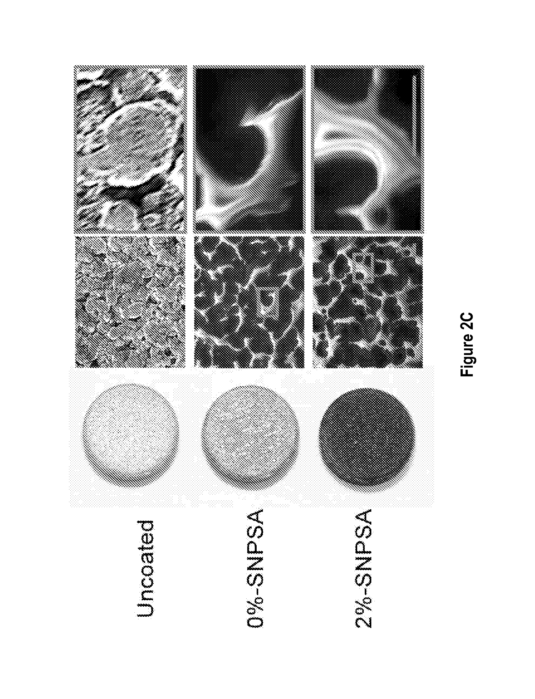
[0121]20-40 nm-diameter spherical silver nanoparticles (QSI-Nano® Silver, QuantumSphere, Inc., Santa Ana, Calif.) were thoroughly mixed with 17.5% (w / v) PLGA (lactic:glycolic=85:15, inherent viscosity: 0.64 dl / g in chloroform; Durect Co., Pelham, Ala.) solution. The proportion of silver nanoparticles refers to the weight ratio of silver nanoparticles to PLGA. 316L stainless steel alloy Kirschner (K)-wire (length: 1 cm, diameter: 0.6 mm; Synthes. Monument, Colo.) and discs (thickness: 1.59 mm, diameter: 6.35 mm; Applied Porous Technologies, Inc., Tariffville, Conn.) were soaked in the silver nanoparticle / PLGA-chloroform solution for 30 s and air-dried completely. The soak-dry process was repeated three times for each SNPSA implant. After incubating for 12 h at 37° C. to ensure a uniform coating, SNPSAs were stored at −20oC until use. Morphology of the SNPSA was evaluated by scanning electron microscopy (SEM; NovaNano SEM 230-D9064, FEI Company, Hillsboro,...

example 2

Surface Free Energy

[0122]Surface free energy of SNPSAs was obtained from contact angle measurements. Contact angles of multiple standard liquids on the tested SNPSAs were measured using a contact angle analyzer (FTA125; First Ten Ångestroms, Portsmouth, Va.). In order to obtain an accurate description of the wetting behavior of various SNPSAs, the surface free energy of the solid (γs), was considered to be the sum of separate dispersion (yd) and non-dispersion (γsnd) contributions. From this two-component model, the following relationship was derived from the dispersion γd and non-dispersion (also known as ‘polar’) γnd interactions between liquids and solids.

γL×(cosθ+1)=2×(γLd×γsd)12+(γLnd×γsnd)12(1)

[0123]Eq. (1), known as the geometric mean model, allows the calculation of the solid surface free energy using the contact angle (θ) and the surface tension components of the standard liquids, where γL, γLd, and γLd represent the surface tension and its dispersion and non-dispersion com...

example 3

In Vitro Antimicrobial Activity

[0124]The Gram-positive vancomycin-intermediate S. aureus (VISA / MRSA) strain Mu50 (ATCC 700699) was cultured in brain heart infusion broth (BHIB; BD, Sparks, Md.) at 37° C.; while biofilm-forming, Gram-negative opportunistic pathogen P. aeruginosa PAO-1 (ATCC 15692) was cultured in Luria Bertani broth (LB; Fisher Scientific, Hampton, N.H.) at 30° C. 103, 104, and 105 colony forming units (CFU) of bacteria were suspended in 1 ml culture broth and incubated with the SNPSA K-wires at 225 rpm on a shaker for 1, 2, 6, and 24 hours.

[0125]At the end of the incubation, Mu50 and PAO-1 bacteria attached to the surface were collected by 0.9% saline solution and plated onto 10-cm BHIB or LB culture medium plates overnight, respectively.

[0126]After 18 h incubation, the number of colonies on each plate was counted and the total viable CFU load was determined.


PUM

PropertyMeasurementUnit
mean sizeaaaaaaaaaa
mean sizeaaaaaaaaaa
mean sizeaaaaaaaaaa

Abstract

Implant-associated bacterial infections are one of the most serious complications in orthopedic surgery. Treatment of these infections often requires multiple operations, device removal, long-term systemic antibiotics, and extended rehabilitation, and is frequently ineffective, leading to worse clinical outcomes and increased financial costs. Silver nanoparticle/poly(DL-lactic-co-glycolic acid) (PLGA)-coated stainless steel alloy (SNPSA) was evaluated as a potential antimicrobial implant material. It was found that SNPSA exhibited strong antibacterial activity in vitro and ex vivo, and promoted MC3T3-E1 pre-osteoblasts proliferation and maturation in vitro. Furthermore, SNPSA implants induced osteogenesis while suppressing bacterial survival in contaminated rat femoral canals. The results indicate that SNPSA has simultaneous antimicrobial and osteoinductive properties that make it a promising therapeutic material in orthopedic surgery.

Description

CROSS-REFERENCE TO RELATED APPLICATIONS[0001]This application claims priority to International Application No. PCT / US2012 / 061217, filed on Oct. 19, 2012, which in turn claims priority from U.S. Provisional Patent Application No. 61 / 550,349, filed on Oct. 21, 2011, each of which is incorporated herein by reference in its entirety.STATEMENT REGARDING FEDERALLY-SPONSORED RESEARCH[0002]This invention was made with Government support of Grant No. W81XWH-09-1-0090, awarded by the United States Army Medical Research and Materiel Command. The Government has certain rights in the invention.FIELD OF THE INVENTION[0003]The invention disclosed herein generally relates to devices (e.g., scaffolds, fixture and / or implants) for osteoconductive applications. In particular, the invention relates to nanoparticle-based scaffolds and / or implants and methods for making the same. The invention further relates to applications of these devices.BACKGROUND[0004]Implant-associated microbial infections are one...

Claims


Application Information

Patent Timeline
Patent Type & Authority Applications(United States)
IPC IPC(8): A61L27/34A61L31/16A61L31/06A61L27/54
CPCA61C8/0016A61L31/06A61F2002/3084A61F2310/00359A61F2310/00365A61F2310/0052A61F2310/0097A61F2310/00976A61L27/34A61L27/54A61L31/10A61L31/16A61L2300/104A61L2300/404A61L2400/12A61F2/4455
Inventor SOO, B. CHIATING, KANGZHENG, ZHONG
Owner RGT UNIV OF CALIFORNIA
Why Eureka
Try Eureka