Methods for obtaining single cells and applications of single cell omics

Inactive Publication Date: 2014-10-16
THE SCRIPPS RES INST
View PDF2 Cites 20 Cited by
  • Summary
  • Abstract
  • Description
  • Claims
  • Application Information

AI Technical Summary

Benefits of technology

[0025]In some embodiments, said one or more cellular or molecular analysis comprise whole-genome analysis of the CTCs.
[0026]Some embodiments provide a method for assessing response of a patient suffering from cancer to a treatment, the method comprises: providing a circulating tumor cell (CTC) or a substantially pure population of CTCs from the pati

Problems solved by technology

Cancer is a difficult disease to treat and manage for several reasons.
First, tumor biology changes over the course of the disease.
Second, heterogeneity is a characteristic trait of cancer.
For a particular cancer treatment, some patients may

Method used


Image

  • Methods for obtaining single cells and applications of single cell omics
  • Methods for obtaining single cells and applications of single cell omics
  • Methods for obtaining single cells and applications of single cell omics

Examples


example 1

Identification of CTCs in a Blood Sample

[0087]Peripheral blood is collected from primary lung cancer patients in a cell-free DNA blood collection tube (Streck, Omaha, Nebr.). A white blood cell count is taken from the blood sample using a hemocytometer, and the cellular concentration of the sample is titrated so that it is about 3 million cells per slide when the titrated sample is disposed on a glass slide. After lysing red blood cells using ammonium chloride, the nucleated cells are distributed in a monolayer onto the glass slide. After paraformaldehyde fixation and methanol permeabilization, cells are incubated with anti-Cytokeratin cocktail and anti-CD45 antibodies followed by Alexa 555-conjugated secondary antibody and DAPI as a nuclear stain.

[0088]The glass slide is imaged (custom high speed scanning microscope, Epic Sciences at 10×) and “candidate” CTCs are identified as being Cytokeratin positive (CK+), CD45 negative (CD45−) with an intact nucleus using proprietary computer ...

example 2

Capture of Individual CTCs in a Blood Sample

[0089]CTCs are identified in a blood sample according to the general procedure described in Example 1. The CTCs on the glass slide are relocated on a fluorescence microscope. The glass slide is soaked in PBS buffer for 30 minutes to let the coverslip float off, and then soaked in methanol for about 1 hour to dissolve the glycerol-based mounting media. To perform CTC picking, the slide is covered with BSA solution which can help loosen the adhesion of CTCs on the glass slide and significantly reduce the stiction of CTCs to glass capillaries used for picking. A micromanipulator mounted on the microscope stage is used to pick CTCs from the slide one CTC at a time. The isolated CTC is put into a tube, either separately or with other isolated CTCs, for downstream analysis.

example 3

Capture of Single CTCs in a Blood Sample

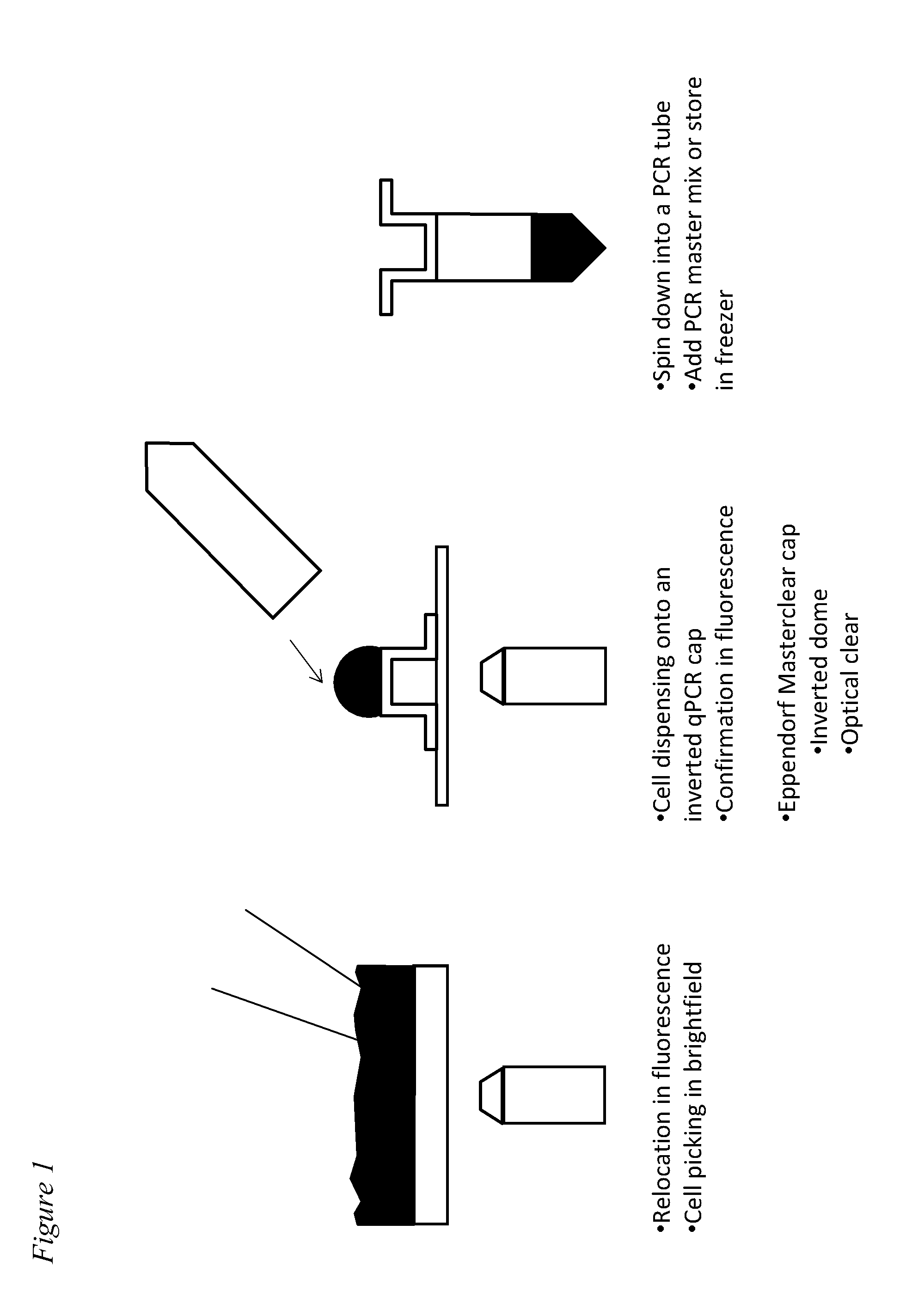
[0090]An exemplary embodiment of the method for capturing single CTCs is illustrated in FIG. 1. In this example, transparent qPCR tube cap that allows the detection of fluorescence detection through the cap for real-time PCR is laid upside down on top of glass slide. A small (for example, 1 to 5 μl) droplet of PBS buffer is put into the cap and the aspirated CTCs is dispensed into the PBS droplet. Then, fluorescence detection is performed to allow detection and confirmation of the number of CTCs and the purity of CTCs in the droplet. Finally, the cap is closed with a PCR tube. With a quick spin, the droplet will be at the bottom of the PCR tube.


PUM

PropertyMeasurementUnit
Fractionaaaaaaaaaa
Fractionaaaaaaaaaa
Fractionaaaaaaaaaa

Abstract

The present application provides methods for obtaining single cells from a sample. Methods for isolating and analyzing molecular features obtained from a single cell are also disclosed herein. For example, individual circulating tumor cells (CTCs) from a sample such as a patient's blood sample can be identified and obtained using methods disclosed herein, and picked for further analysis.

Description

RELATED APPLICATIONS[0001]The present application claims priority under 35 U.S.C. §119(e) to U.S. Provisional Application Nos. 61 / 435704, filed Jan. 24, 2011; 61 / 435724, filed on Jan. 24, 2011; and 61 / 435721, filed on Jan. 24, 2011. The contents of each of these related applications are herein expressly incorporated by reference in their entirety.STATEMENT REGARDING FEDERALLY SPONSORED R&D[0002]This invention, in part, was made with government support under grant U54CA143906 from the National Cancer Institute.PARTIES OF JOINT RESEARCH AGREEMENT[0003]This invention, in part, was made under a Research Funding and Option Agreement dated Jun. 25, 2009 with The Scripps Research Institute.BACKGROUND[0004]1. Field[0005]The present application relates to the field of cell biology and medicine. More particularly, disclosed herein are methods of obtaining and analyzing single cells from a sample. Also disclosed herein are methods for evaluating the condition of a patient, predicting treatment...

Claims


Application Information

Patent Timeline
IPC IPC(8): C12Q1/68G01N33/574
CPCG01N33/57426C12Q1/6886G01N33/56966G01N33/574
Inventor YANG, XINGNELSON, DAVID M.KUHN, PETERLAZAR, DANIEL CHESNAYE
Owner THE SCRIPPS RES INST
Why Eureka
Try Eureka