Flowcell, sheath fluid, and autofocus systems and methods for particle analysis in urine samples

a flowcell and autofocus technology, applied in the field of system, analyzer, composition and method of particle analysis, can solve the problems of difficult to distinguish from renal epithelial cells, yeast may be seen in urine, and infections are a problem

Active Publication Date: 2014-11-06
IRIS INT
View PDF0 Cites 30 Cited by
  • Summary
  • Abstract
  • Description
  • Claims
  • Application Information

AI Technical Summary

Benefits of technology

[0026]In another aspect, embodiments of the present invention encompass methods for imaging a plurality of particles using a particle analysis system configured for combined viscosity and geometric hydrofocusing. The particles can be included in a body fluid sample having a sample fluid viscosity. Exemplary methods include flowing a sheath fluid along a flowpath of a flowcell, the sheath fluid having a sheath fluid viscosity that differs from the sample fluid viscosity by a viscosity difference in a predetermined viscosity difference range, and injecting the body fluid sample into the flowing sheath fluid within the flowcell so as to provide a sample fluid stream enveloped by the sheath fluid. Methods can also include flowing the sample fluid stream and the sheath fluid through a reduction in flowpath size toward an imaging site, such that a viscosity hydrofocusing effect induced by an interaction between the sheath fluid and the sample fluid stream associated with the viscosity difference, in combination with a geometric hydrofocusing effect induced by an interaction between the sheath fluid and the sample fluid stream associated with the reduction in flowpath size, is effective to provide a target imaging state in at least some of the plurality of particles at the imaging site while a viscosity agent in the sheath fluid retains viability of cells in the sample fluid stream leaving structure and content of the cells intact when the cells extend from the sample fluid stream into the flowing sheath fluid. Further, methods can include imaging the plurality of particles at the imaging site. In some embodiments, the body fluid sample is a urine sample.
[0027]In yet another aspect, embodiments of the present invention enc

Problems solved by technology

They may be difficult to distinguish from renal epithelial cells, but they are generally larger and more spherical.
In addition, yeast may be seen in urine, especially as the result of vaginal contamination such as contamination from female patients with yeast infections.
Yeast is a common contaminant, from skin and the environment, and infections are a problem in debilitated and immunosuppressed or immunocompromised patients.
However, many of these earlier developed urine analysis systems generally lacked the throug

Method used


Image

  • Flowcell, sheath fluid, and autofocus systems and methods for particle analysis in urine samples
  • Flowcell, sheath fluid, and autofocus systems and methods for particle analysis in urine samples
  • Flowcell, sheath fluid, and autofocus systems and methods for particle analysis in urine samples

Examples


examples

[0192]Any of a variety of urinalysis or urine particle analysis techniques can be performed using images of sample fluid flowing through the flowcell. Often, image analysis can involve determining certain cell or particle parameters, or measuring, detecting, or evaluating certain cell or particle features. For example, image analysis can involve evaluating cell or particle size, cell nucleus features, cell cytoplasm features, intracellular organelle features, and the like. Relatedly, analysis techniques can encompass certain counting or classification methods or diagnostic tests. Relatedly, with reference to FIG. 4, the processor 440 can include or be in operative association with a storage medium having a computer application that, when executed by the processor, is configured to cause the system 400 to differentiate different types of cells or particles based on images obtained from the image capture device. For example, diagnostic or testing techniques can be used to differentiat...


PUM


Abstract

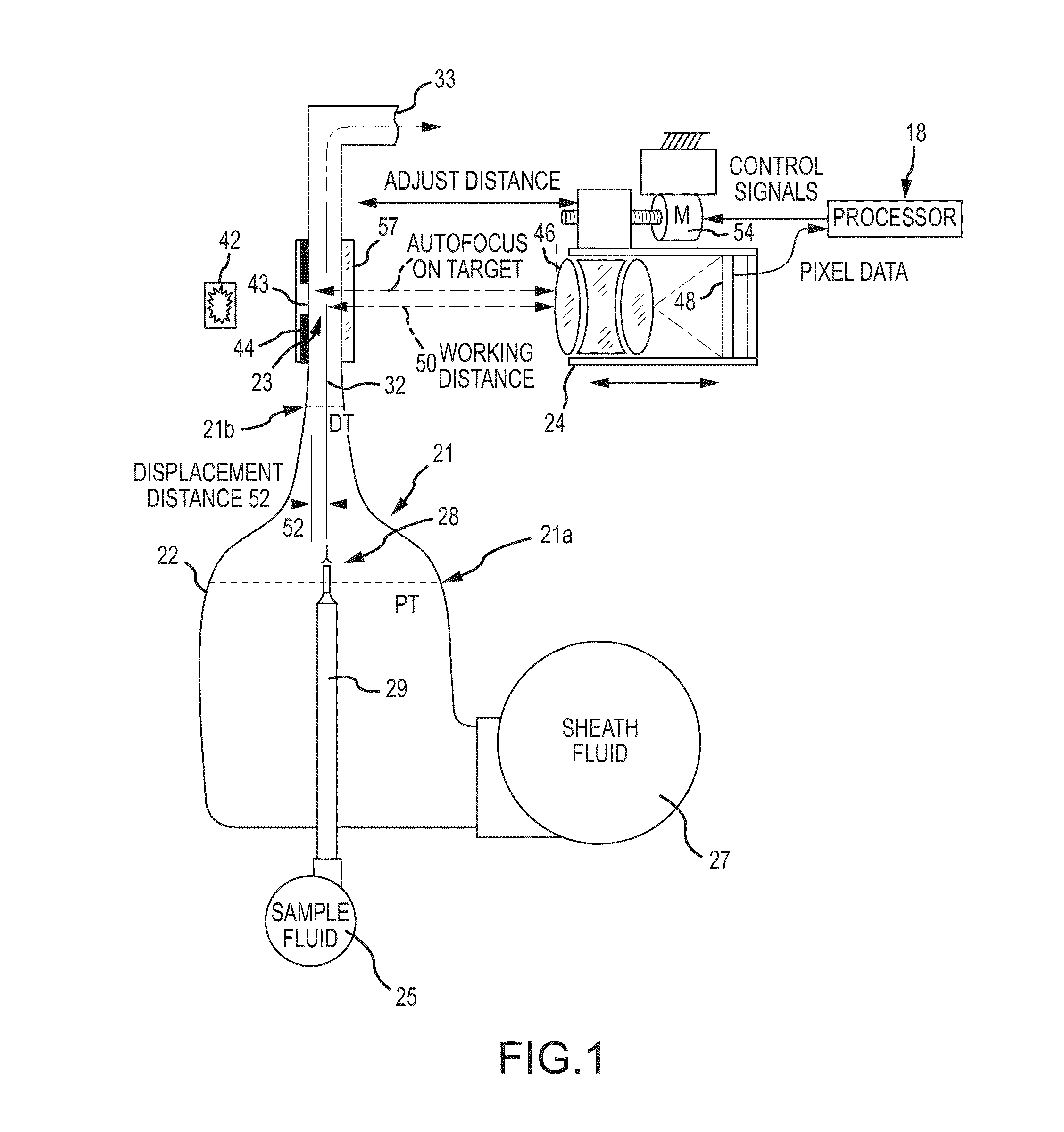
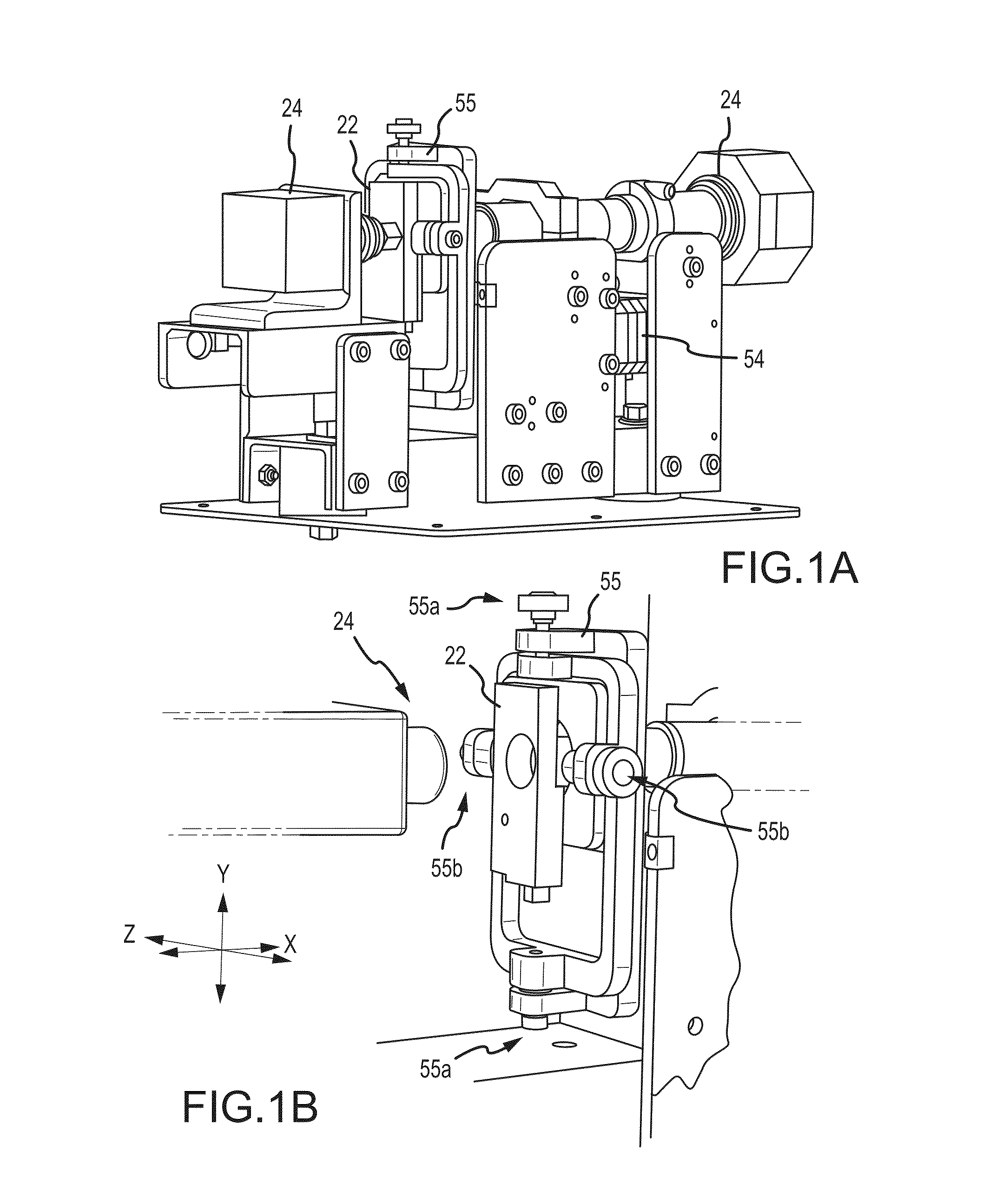
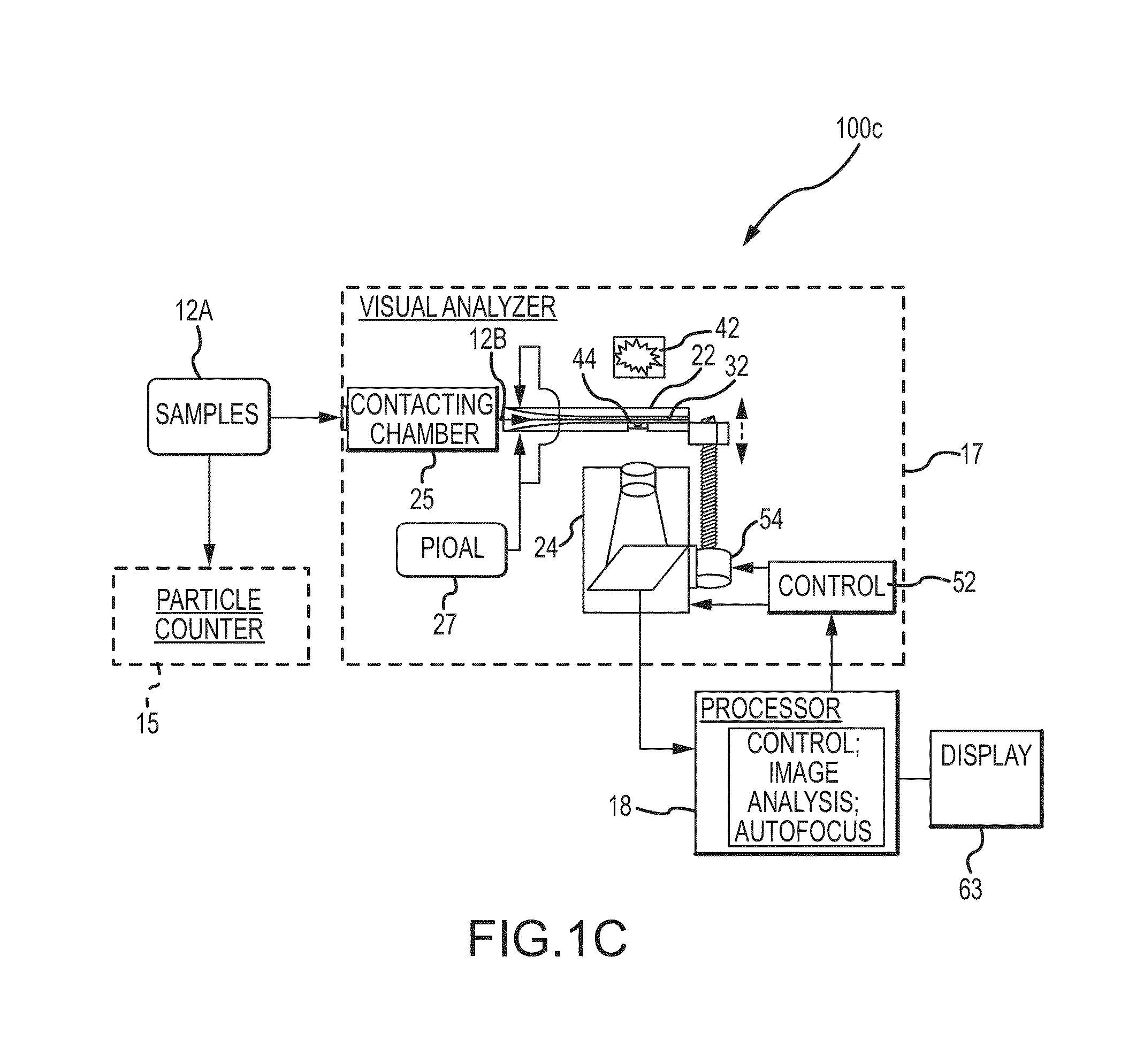
The present disclosure relates to apparatus, systems, compositions, and methods for analyzing a sample containing particles. A particle imaging system or analyzer can include a flowcell through which a urine sample containing particles is caused to flow, and a high optical resolution imaging device which captures images for image analysis. A contrast pattern for autofocusing is provided on the flowcell. The image processor assesses focus accuracy from pixel data contrast. A positioning motor moves the microscope and/or flowcell along the optical axis for autofocusing on the contrast pattern target. The processor then displaces microscope and flowcell by a known distance between the contrast pattern and the sample stream, thus focusing on the sample stream. Cell or particle images are collected from that position until autofocus is reinitiated, periodically, by input signal, or when detecting temperature changes or focus inaccuracy in the image data.

Description

CROSS-REFERENCES TO RELATED APPLICATIONS[0001]This application is a non-provisional of, and claims the benefit of priority to, U.S. Provisional Patent Application No. 61 / 799,014 filed Mar. 15, 2013, the content of which is incorporated herein by reference. This application is also related to U.S. patent application Ser. No. 14 / 216,562 and International Patent Application No. PCT / US14 / 30856, both filed Mar. 17, 2014. The content of each of these filings is incorporated herein by reference.BACKGROUND OF THE INVENTION[0002]This disclosure relates to the field of systems, analyzers, compositions, and methods for analysis of particles, including imaging of particles in fluid samples, such as urine samples, using wholly or partly automated devices to discriminate and quantify particles in the sample. The present disclosure also relates to a particle and / or intracellular organelle alignment liquid (PIOAL) useful for analyzing particles in a urine sample from a subject, methods for producin...

Claims


Application Information

Patent Timeline
IPC IPC(8): G01N33/50
CPCG01N33/5091G01N2015/1452G01N15/1468G01N2015/0065G01N15/1404G01N15/147G01N2021/8557
Inventor WANDERS, BART J.CHAPOULAUD, ERICJORDAN, BRETT
Owner IRIS INT
Why Eureka
Try Eureka