Cell suspension medium and cell suspension medium additive for the three dimensional growth of cells
- Summary
- Abstract
- Description
- Claims
- Application Information
AI Technical Summary
Benefits of technology
Problems solved by technology
Method used
Image
Examples
example 1
Spheroid Formation in Cell Culture Media Containing the Additive
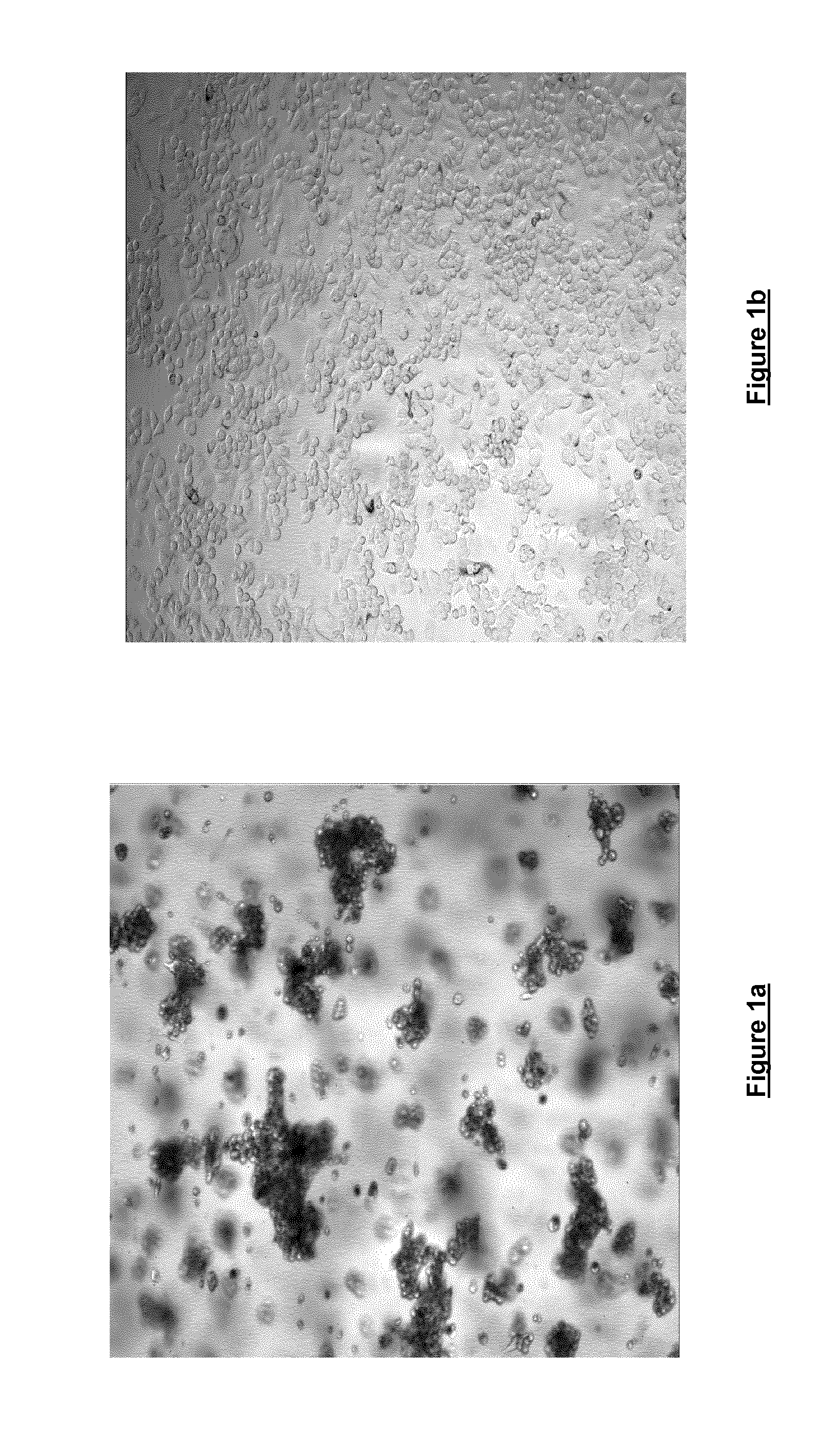
[0055]Freshly trypsinised SKBR3 breast cancer cells were seeded at a density of 0.9×10⁴ cells / 100 μl in DMEM either in the presence (FIG. 1a) or the absence (FIG. 1b) of 0.06% w / v cell culture medium additive [blend of xanthan and enzyme-modified guar (50:50 w / w; guar enzymatically modified by α-galactosidase to give a mannose:galactose ratio of approx. 80:20 w / w)]. Cells were seeded in Nunc F96 Microwell plates and were incubated for 120 hours at 37° C. / 5% CO2 and 95% humidity. Cells were then imaged at 4× magnification using an IN Cell Analyser High Content Analysis imaging system. As can be seen, the cells grown in the presence of the cell culture medium additive (FIG. 1a) show notable spheroid formation, whereas the cells grown in the absence of the cell culture medium additive show little spheroid formation.
example 2
Imaging of Spheroids in Cell Culture Media Containing the Additive
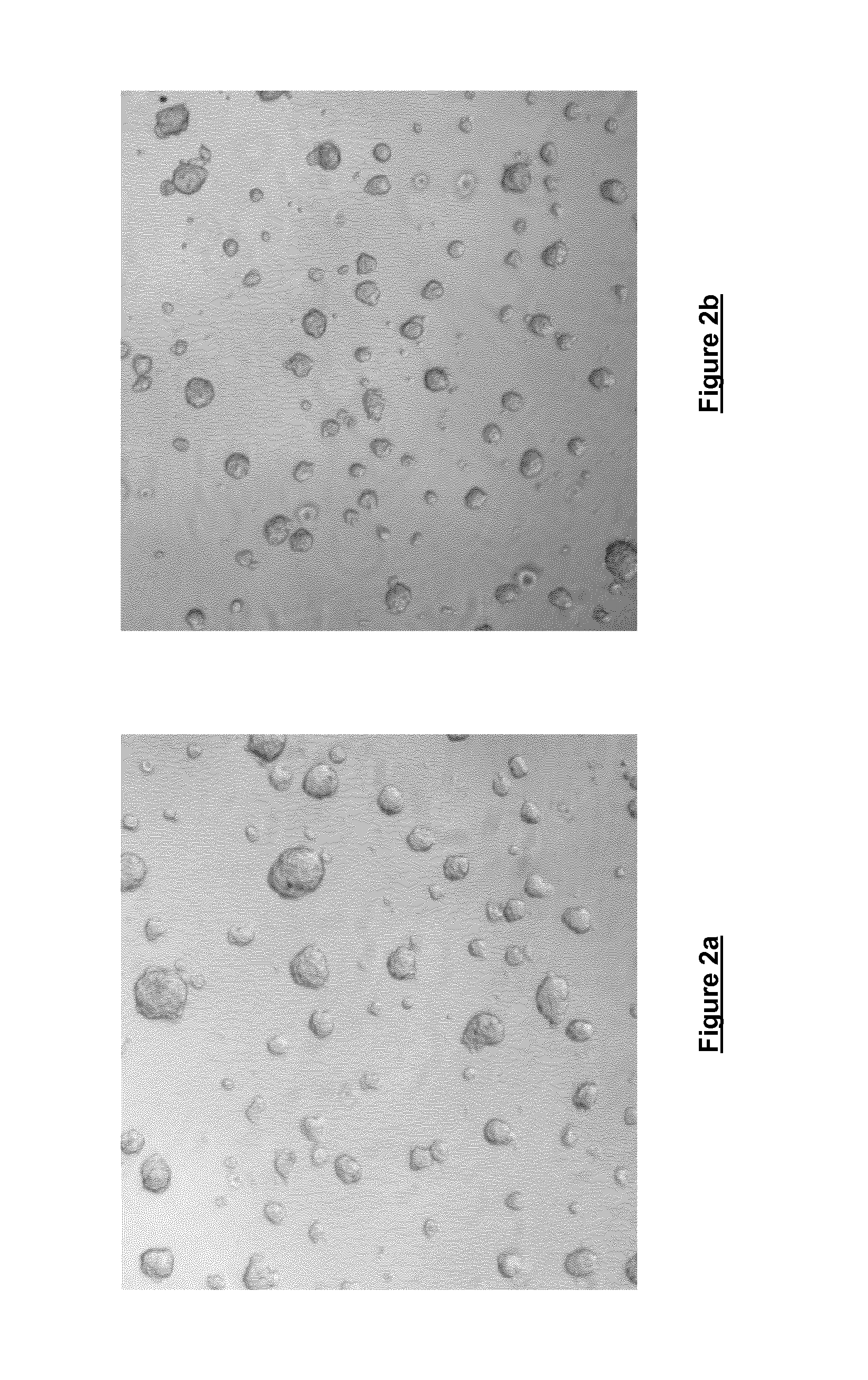
[0056]Freshly trypsinised Prostate cancer cells were seeded at a density of 0.9×10⁴ cells / 100 μl in DMEM containing 0.06% w / v cell culture medium additive [blend of xanthan and enzyme-modified guar (50:50 w / w; guar enzymatically modified by α-galactosidase to give a mannose:galactose ratio of approx. 80:20 w / w)] in a ‘Hydrocell’ low binding 96-well plate (Nunc) and incubated for 72 hours at 37° C. / 5% CO2 and 95% humidity. Cells were then imaged at 4× magnification using an IN Cell Analyser 1000 High Content Analysis imaging system. Both FIGS. 2a and 2b illustrate the results of this imaging. The images produced clearly illustrate that it is possible to conduct High Content Analysis on cells grown in the cell culture containing the cell culture medium additive.
[0057]Subsequent experiments performed under similar conditions for five days (cells grown in RPMI containing 0.06% w / v cell culture medium additive [blend of xa...
example 3
Characterisation of Spheroids in Cell Culture Media Containing the Additive
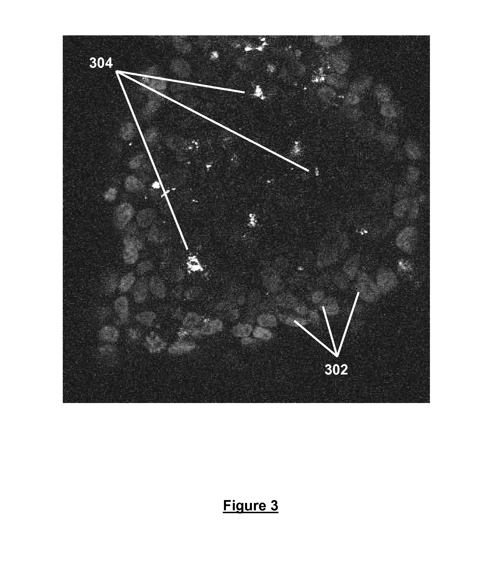
[0058]Freshly trypsinised Prostate cancer cells were seeded in DMEM containing 0.06% w / v cell culture medium additive [blend of xanthan and enzyme-modified guar (50:50 w / w; guar enzymatically modified by α-galactosidase to give a mannose:galactose ratio of approx. 80:20 w / w)] at a density of 1×10⁵ cells / 500 in a Lab-Tek chamber slide (Thermo Scientific) and incubated for 48 hours at 37° C. / 5% CO2 and 95% humidity. Spheroids produced were then subsequently stained with Hoechst (blue fluorescent DNA dye) and propidium iodide, and imaged at 40× magnification using a Zeiss Meta Confocal microscope (FIG. 3). The imaged spheroid comprises a viable outer mantle of cells 302 stained in blue (Hoechst) surrounding a region of dead and / or necrotic cells 304 stained in red (propidium iodide). Spheroids of this type are considered to be more tumour-like than cells grown in 2D.
PUM
 Abstract
Description
Claims
Application Information
 - R&D
- Intellectual Property
- Life Sciences
- Materials
- Tech Scout
- Unparalleled Data Quality
- Higher Quality Content
- 60% Fewer Hallucinations
Browse by: Latest US Patents, China's latest patents, Technical Efficacy Thesaurus, Application Domain, Technology Topic, Popular Technical Reports.
© 2025 PatSnap. All rights reserved.Legal|Privacy policy|Modern Slavery Act Transparency Statement|Sitemap|About US| Contact US: help@patsnap.com


