Preparation of thin tissue sections for imaging mass spectrometry

a mass spectrometry and thin tissue technology, applied in material analysis, instruments, electric discharge tubes, etc., can solve the problems of unfavorable signal-to-noise ratio at the ion detector and limit the spatial resolution of the imag

Active Publication Date: 2015-04-02
BRUKER DALTONIK GMBH & CO KG
View PDF2 Cites 12 Cited by
  • Summary
  • Abstract
  • Description
  • Claims
  • Application Information

AI Technical Summary

Benefits of technology

[0014]In order to measure peptides and proteins efficiently, it is helpful to rinse the lipids out of the thin tissue section before the preparation using methods that are basically known.

Problems solved by technology

Unfortunately, the liquid of each droplet spreads radially in all directions, so the analyte substances are also transported laterally within the thin tissue section; this limits the spatial resolving power of the image because the spatial information of the analyte molecules is smeared out.
The consequence of this is that only a small number of analyte molecules are formed during laser bombardment in the course of an ionization with matrix-assisted laser desorption, and the signal-to-noise ratio is unfavorably low at the ion detector.
However, this slow diffusion of the analyte molecules to the matrix layer in the swollen thin tissue section is also associated with an undesirable lateral diffusion of the analyte molecules in the same order of magnitude, which in turn smears out the spatial information of the analyte molecules.

Method used


Image

  • Preparation of thin tissue sections for imaging mass spectrometry
  • Preparation of thin tissue sections for imaging mass spectrometry

Examples


first embodiment

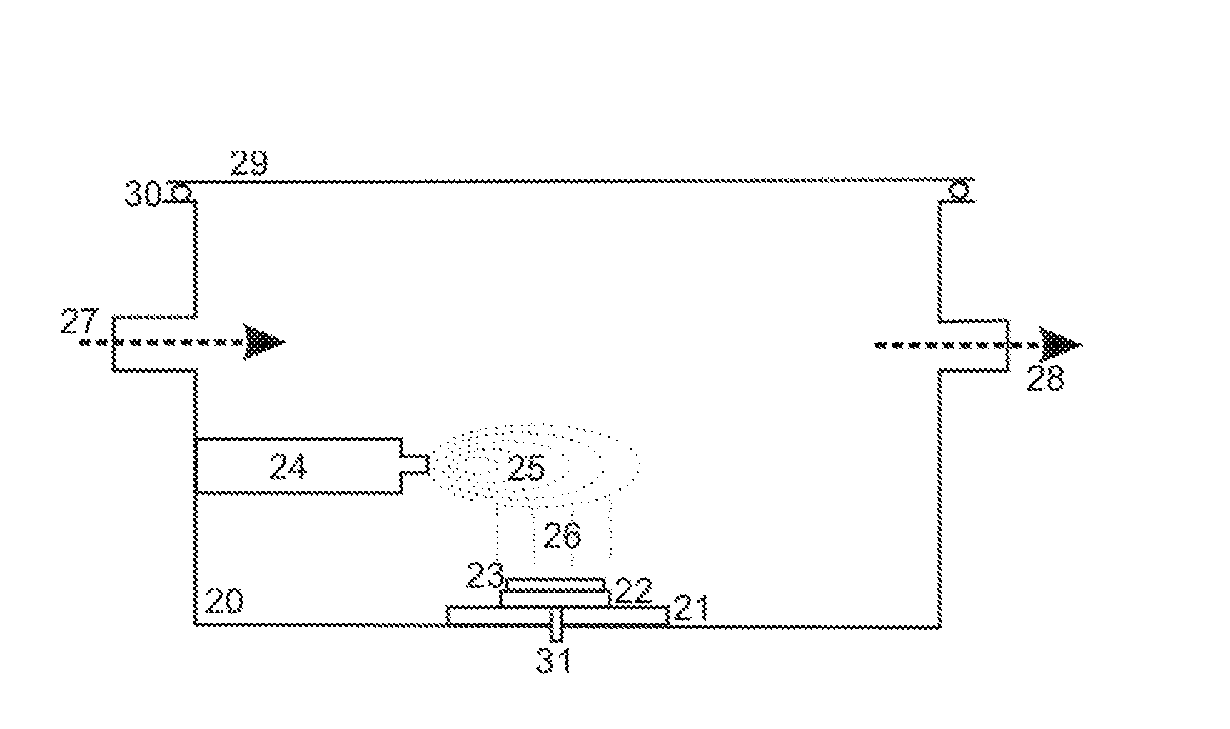
[0036]In the apparatus, as depicted in FIG. 1, the matrix substance is sublimed in a heated pan (3) on the floor of a chamber (1), while the thin tissue section (6) is located on a support (7) hanging from the lid at the top. The walls of the chamber (1) can be heated in such a way that no resublimation takes place there. The thin tissue section (6) on its support (7) can be cooled or heated to a specific temperature, by a Peltier element (8), for example. The matrix crystal layer is fed with matrix vapor from below. The chamber (1) can be charged with the vapors of the solvents for the swelling via an inlet (11), and can also be evacuated, if required, via the outlet (12).

[0037]In this first embodiment apparatus, a special preparation method may be performed. This special method uses a solvent which at the same time can swell the thin tissue section, transport peptides, and solve a part of the matrix layer for a recrystallization with embedding the peptides into the crystals. After...

second embodiment

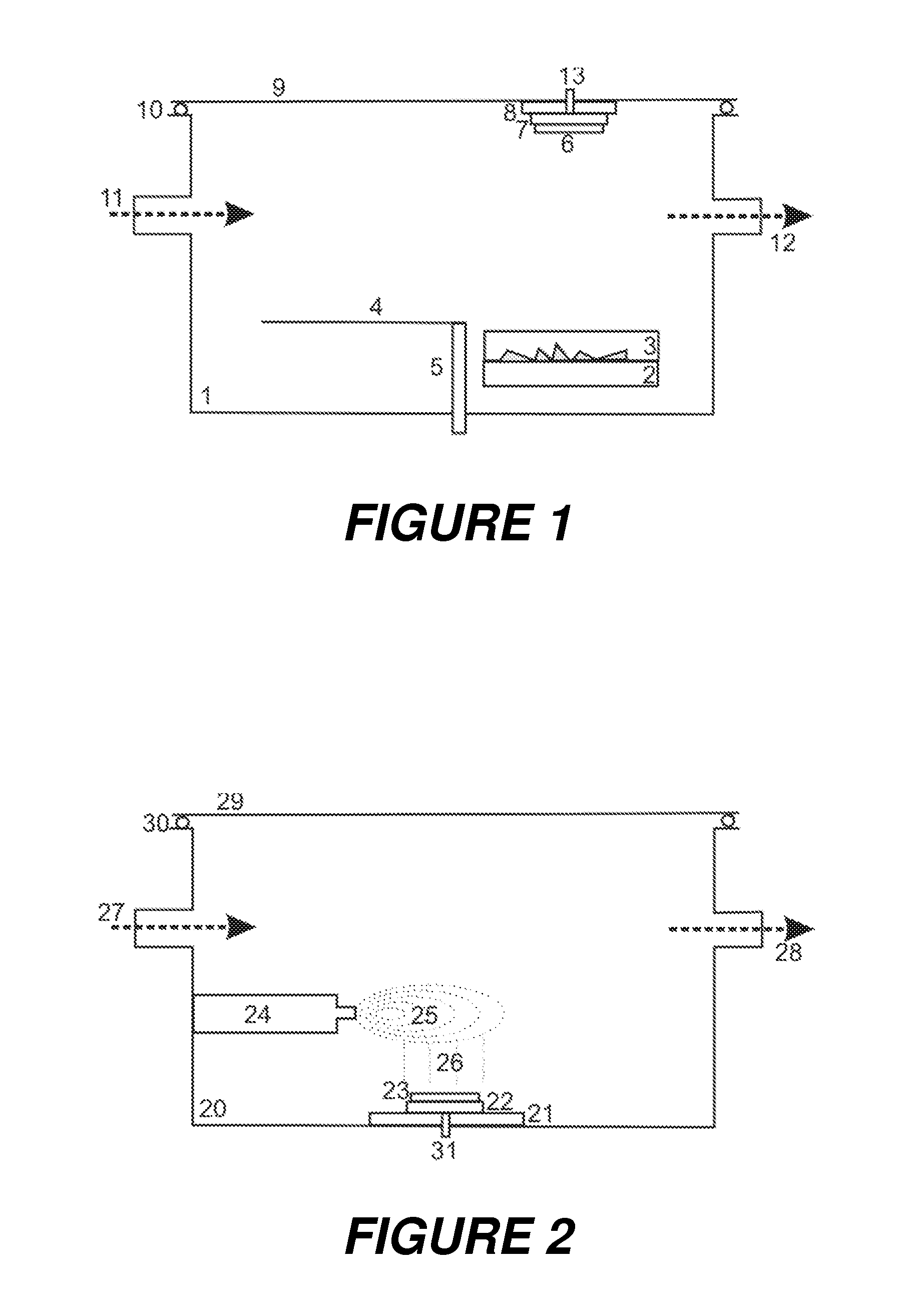
[0040]In the apparatus, which is schematically depicted in FIG. 2, the thin tissue section (23) on its support (22) is located near the floor of a chamber (20). In this position, the matrix snow (26) can be applied by a sprayer (24), for example. To ensure the snow is applied uniformly, it must be possible to move the support (22) for the thin tissue section (23) in a variety of ways. Here again, the thin tissue section (23) on its support (22) should be able to be heated and cooled, by the Peltier element, for example. It is also possible here to feed in vapors of solvents or other working gases, such as inert nitrogen N2, via the inlet (27); the pressure can be regulated via the outlet (28). For example, a solvent can be fed in which forms a floor-level mist above the cold thin tissue section on the support (22). This mist slightly dissolves the matrix crystals of the snow and thus incorporates the analyte molecules into the matrix crystals during the subsequent recrystallization....


PUM

PropertyMeasurementUnit
pressureaaaaaaaaaa
pressureaaaaaaaaaa
thickaaaaaaaaaa

Abstract

The invention relates to the preparation of thin tissue sections for mass spectrometric (MALDI) imaging, and proposes a method wherein a microcrystalline layer of the matrix material is produced on the surface of the thin tissue sections, and soluble analyte molecules are transported orthogonally through the thin tissue section to the matrix layer, without substantial lateral diffusion, by means of a solvent flow with alternating or constant direction, and are then deposited in the matrix layer and, if possible, embedded in the matrix crystals. A solvent flow which alternates in direction can be produced by successive, alternating phases of swelling and drying, brought about by periodic changes to the thin tissue section temperature and/or the partial pressures of the solvents. A continuous solvent flow can be generated by applying the thin tissue section onto a porous support which supplies solvent to the rear surface of the thin tissue section.

Description

BACKGROUND OF THE INVENTION[0001]1. Field of the Invention[0002]The invention relates to the preparation of thin tissue sections for mass spectrometric imaging with ionization by matrix-assisted laser desorption (MALDI), particularly for high-resolution and high-sensitivity imaging of soluble peptides and proteins.[0003]2. Description of the Related Art[0004]Imaging mass spectrometry (imaging MS or IMS) acquires images of thin tissue sections which shows for every pixel the mass spectrum of the ionizable components, corresponding to the color spectrum in every pixel of an optical image. The ionization is usually carried out by matrix-assisted laser desorption (MALDI), not least because MALDI primarily produces singly charged molecular ions, which means that the resulting mass spectra are very easy to interpret.[0005]Several methods are known for the preparation of thin tissue sections in imaging mass spectrometry with MALDI, particularly spray methods for matrix solutions. For examp...

Claims


Application Information

Patent Timeline
Patent Type & Authority Applications(United States)
IPC IPC(8): G01N1/30
CPCG01N1/30G01N1/31G01N1/4055H01J49/0418G01N1/28G01N33/6851
Inventor SCHURENBERG, MARTINFRANZEN, JOCHENRUCKRIEM, KAIBELAU, ECKHARD
Owner BRUKER DALTONIK GMBH & CO KG
Why Eureka
Try Eureka