
Method for Identification, Selection and Analysis of Tumour Cells

a tumour cell and selection technology, applied in the field of tumour cell identification, selection and analysis, can solve the problems of low sensitivity and specificity, widespread dosage of psa, poor sensitivity and specificity, and non-negligible costs for the health system, etc., to achieve high diagnostic and predictive reliability, high purity, and high accuracy.

Active Publication Date: 2015-05-07
MENARINI SILICON BIOSYSTEMS SPA
View PDF3 Cites 8 Cited by
  • Summary
  • Abstract
  • Description
  • Claims
  • Application Information

AI Technical Summary

Benefits of technology

The present invention is a method for identifying, isolating, and analyzing tumor cells. The method uses a silicon microchip with millions of electrodes to isolate individual cells with high purity. The method is automated and highly reliable for molecular analysis. It can be used for non-invasive early diagnosis, sensitive and accurate follow-up in treatment, and post-operative stage monitoring. Overall, the method can improve oncological treatment and monitoring of diseases progression and treatment response.

Problems solved by technology

Consequently, their sensitivity and specificity are not particularly high.
In the case, considered by way of example, of prostate cancer, one of the most widespread forms of cancer in the male population (approximately one quarter of all the cases of cancer, 300,000 new cases a year in Europe), screening on the basis of serological parameters (dosages of PSA) is widespread, but has a poor sensitivity and specificity.
More invasive procedures, such as digital rectal examination can at times complete the clinical picture, but more frequently it is necessary to carry out a prostatic biopsy to have a more accurate diagnosis, in an escalation of invasiveness that results in a growing discomfort for the patient and in non-negligible costs for the health system.
Since they are difficult to identify and eliminate, it is frequently impossible to treat all the metastases successfully.
Consequently, since there has been demonstrated, in the case of breast cancer, cancer of the colon, and cancer of the prostate, a correlation between the number of CTCs and the increased risk of unfavourable outcome of the illness, it appears evident that the possibility of isolating them and, by means of molecular analysis, obtaining a complete informative picture therefrom having clinical importance, would have very significant diagnostic repercussions.
13(5), 441-53 (2008)) that the over-expression of HER2, which can be detected in a single DTC but not in the primary tumour, proves predictive of a very high risk of death.
At the current state of the art, for isolation of circulating and disseminated tumour cells there are, however, available approaches of a chiefly analytical nature that are very complex and laborious, which, however, have limited yields and generate specimens distinguished by a low degree of purity.
Consequently, said specimens of CTCs or DTCs are not compatible with some of the most refined and reliable diagnostic procedures, such as sequencing.
The limited purity, in fact, determines a high risk of false positives and false negatives caused by wrong calls in the bases of the sequence.
However, said methods have led to levels of purity on average of 50%-60%, which do not prove sufficient for carrying out, downstream, analyses for which the purity of the specimen is crucial, such as, for example, the evaluation of copy-number variations, microsatellite instability, loss of heterozygosity (LoH), gene expression in which a better signal-to-noise (S / N) ratio is fundamental for diagnostic applications, or identification of new drugs, or again in the sector of research into cancer-initiating cells.
The problems encountered with molecular analyses on low purity samples of CTCs are the same that arise when only small samples of solid tumours are available.
Preparing samples enriched with tumor cells for molecular analysis can be challenging.
For many tumors, finding suitable tissue sections with sufficient tumor cells, or tissue not impaired by the presence of necrotic or damaged cells, can be difficult.
Such composition can result in equivocal or inconclusive test results that may ultimately disqualify patients for certain therapies or confound biomarker analyses in clinical studies.
The drawbacks of this method are however that, very often, cells recovered from FFPE samples are too little in number to perform a FACS analysis, and that separations are incomplete, producing partially enriched rather than pure, separate population of tumour and normal cells.
Similar considerations can be extended to other methods the enormous diagnostic potential of which has been highlighted, which, however, cannot be concretely implemented since the known procedures of enrichment of biological specimens and isolation of a population of rare cells therein do not enable sufficient levels of purity to be obtained, if not following upon a heavy, complex and costly analytical workload that cannot, consequently, find application in medical practice.
Also in this perspective, however, the methodologies of enrichment, identification and isolation of rare cells in the blood, in other biological fluids, in small biopsies and in FFPE tissue samples, do not enable a sufficient purity to be obtained to guarantee that the subsequent analysis will be reliable (exclusion of false positives, false negatives, etc.) and, in some cases, that they can even materially be conducted (for example, for the cases in which, since the purity is low, the signal-to-noise ratio is too low to obtain a valid reading).

Method used


Image

  • Method for Identification, Selection and Analysis of Tumour Cells
  • Method for Identification, Selection and Analysis of Tumour Cells
  • Method for Identification, Selection and Analysis of Tumour Cells

Examples


first embodiment

Circulating Tumour Cells or Disseminated Tumour Cells

Sampling

[0086]The samples can be taken from the peripheral circulation of the patient, or else from bone marrow through various techniques known in the sector.

Pre-Enrichment

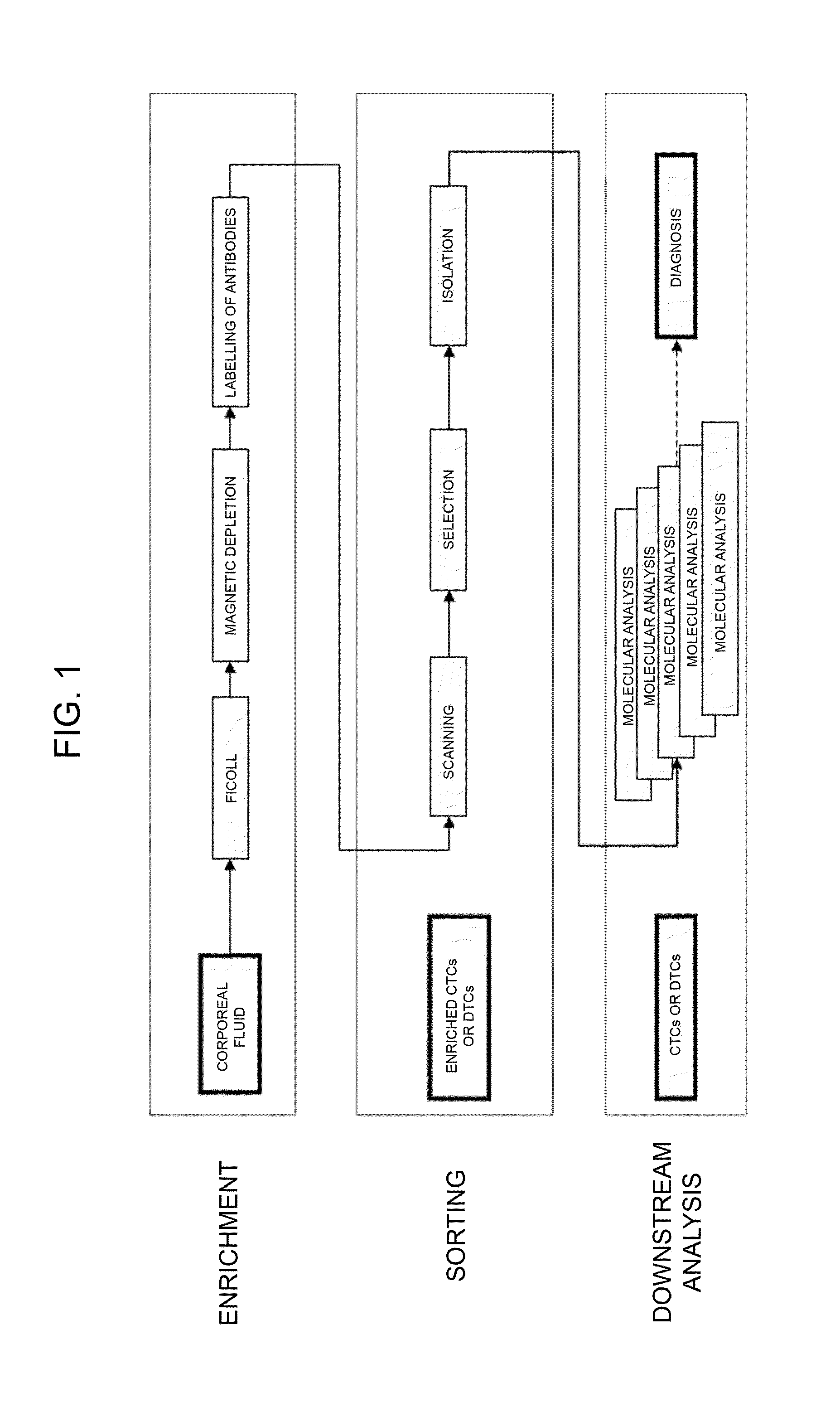
[0087]The proportion of tumour cells in the sample taken can be enriched using various methods, such as for example: centrifugation on density gradient, constituted by solutions such as Ficoll or Percoll; mechanical enrichment, such as filters of various types; enrichment by means of separation by dielectrophoresis via a purposely provided device—dielectrophoretic-activated cell sorter (DACS); selective lysis, such as for example, selective lysis of erythrocytes not of interest; immunomagnetic separation, via immunomagnetic beads with positive selection (using beads bound to specific antibodies for the population to be recovered) or with negative selection (depletion of cell populations that are not of interest), and in which the two types of selection can be c...

second embodiment

Formalin-Fixed-paraffin Embedded Samples

Sampling

[0109]The samples are formalin-fixed and paraffin-embedded samples routinely used and prepared by known methodologies.

Sample Processing

[0110]The samples are deparaffinised and cells are dissociated with a 1% dispase / 1% collagenase solution so as to obtain a cell suspension in a fluid which includes tumour cells and stromal cells. The methodology employed is known from Corver W E et al. (J Pathol, 206, 233-241 (2005).

[0111]Isolation of single tumour cells (common to both embodiments)

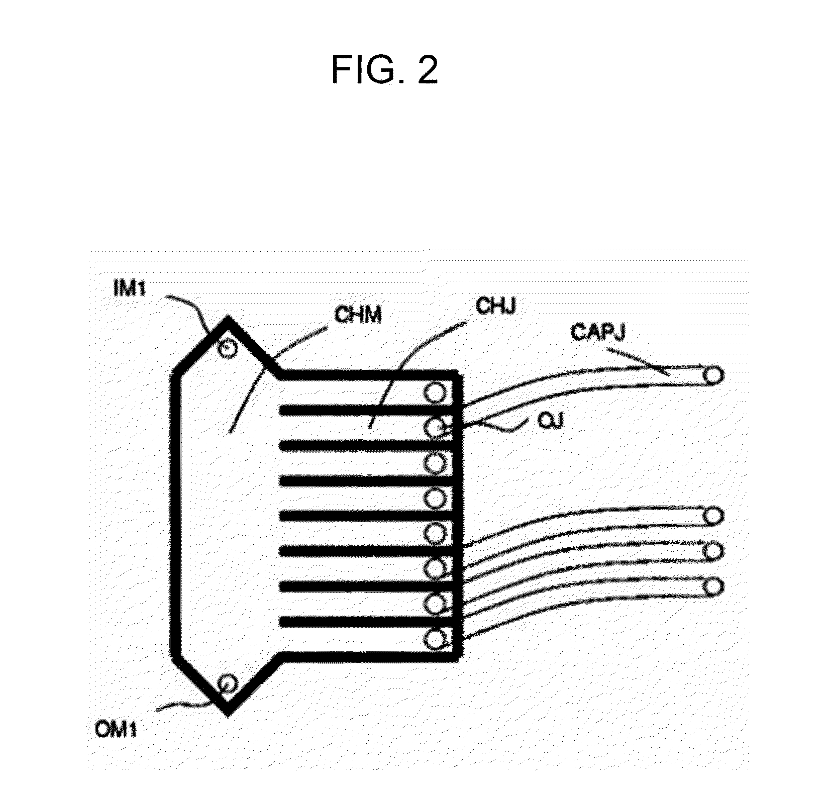
[0112]Next, the sample containing the tumour cells is inserted in a microfluidic device that is able to select individually single cells and isolate them in an automatic or semi-automatic non-manual way. For this purpose, it is possible to use a dielectrophoretic isolation (DEPArray, using for example, the techniques described in PCT / IB2007 / 000963 or in PCT / IB2007 / 000751, or again in Manaresi et al., IEEE J. Solid-State Circuits, 38, 2297-2305 (2003) and in ...

example 1

[0182]Non-invasive evaluation of mutations (for example, in the K-RAS gene) in cancer patients.

[0183]Described in what follows is the case of isolation of CTCs from peripheral bloodflow of patients affected by metastatic Colon-Rectal Cancer (mCRC).

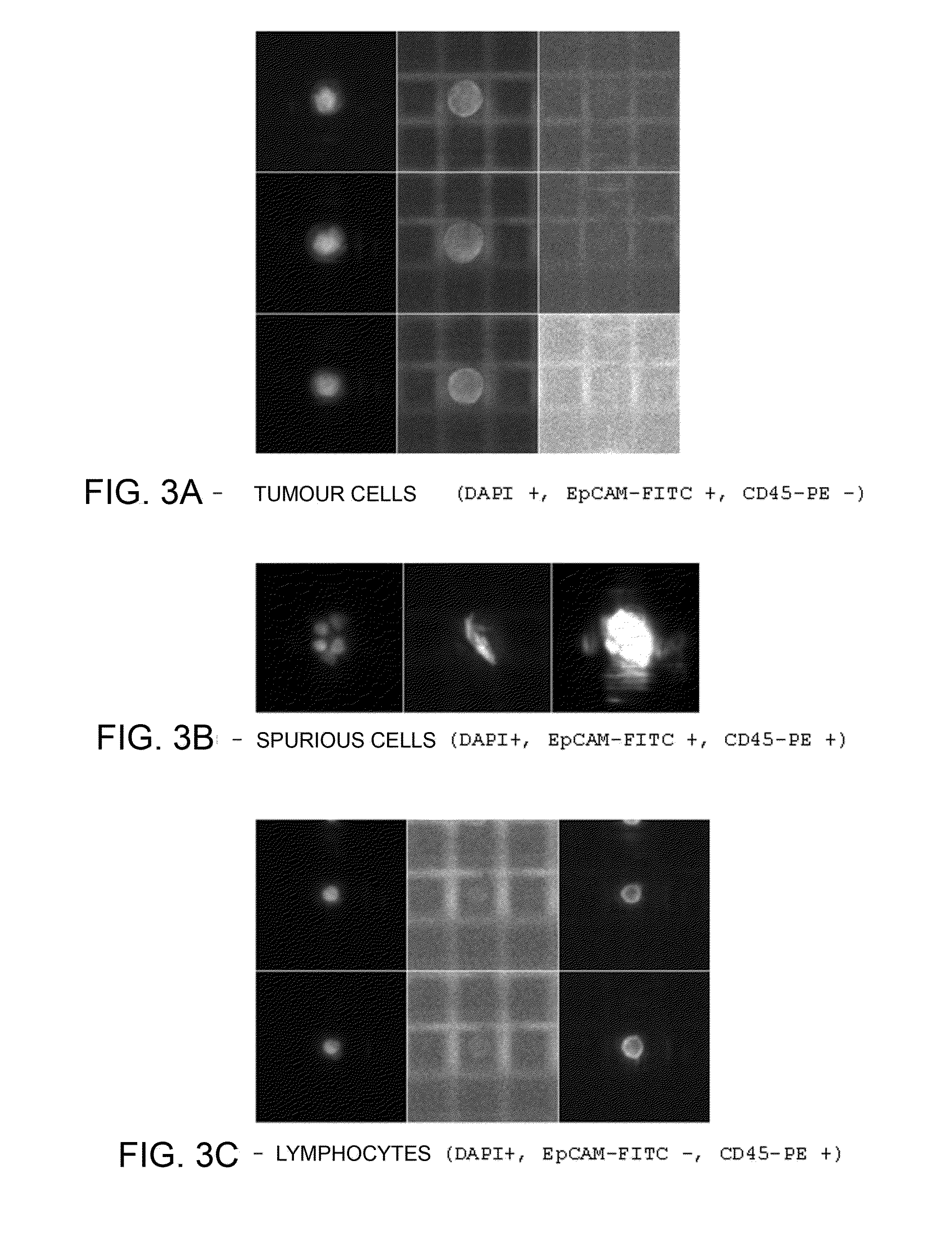
[0184]A specimen of 7.5 ml of peripheral bloodflow was drawn from the patient in Vacutainer tubes with EDTA anticoagulant (Beckton Dickinson). The specimen was analysed with Coulter Counter (Beckman Coulter) and presented 42×106 leukocytes (WBCs) and 43.95×10⁹ erythrocytes (RBCs). The PBMCs were then isolated via centrifugation (in Ficoll 1077). The PBMCs recovered (16.5×10⁶ on the basis of the count with Coulter Counter) were washed in PBS with BSA and EDTA (Running Buffer, Miltenyi) and were selected via depletion with magnetic microbeads conjugated to anti-CD45 and anti-GPA antibodies (Miltenyi) according to the instructions of the manufacturer. The resulting cells (negative fraction for CD45 and GPA) were fixated with 2% PFA in PBS for...


PUM

PropertyMeasurementUnit
volumeaaaaaaaaaa
purityaaaaaaaaaa
fluorescence imagesaaaaaaaaaa

Abstract

The present invention relates to a method for the diagnosis of tumoural conditions and / or of the corresponding state of advance, wherein a sample from a patient comprising at least one tumour cell is obtained. According to the invention, a purified specimen of the at least one tumour cell is obtained by individually selecting and isolating single cells in a microfluidic device the purified specimen having a purity of at least 90%. On the purified specimen thus obtained there is subsequently performed a molecular analysis such as to highlight a characteristic thereof suited to enabling diagnosis.

Description

CROSS-REFERENCE TO RELATED APPLICATION[0001]This is a continuation-in-part of U.S. patent application Ser. No. 13 / 127,640, which is a U.S. national stage application of International Patent Application No. PCT / IB2009 / 007316 filed on Nov. 4, 2009, which in turn claims the benefit of Italian Patent Application No. TO2008A000814, filed Nov. 4, 2008, the respective disclosures of which are each incorporated herein by reference in their entireties.BACKGROUND[0002]1. Field of the Art[0003]The present invention relates, in general, to diagnostic methods based upon identification, isolation, and subsequent analysis of tumour cells. In particular, the invention relates to diagnostic methods that find application in the oncology sector, and that hence involve identification and isolation of circulating tumour cells (CTCs) or disseminated tumour cells (DTCs) or tumour cells from very small samples.[0004]2. State of the Art[0005]The diagnosis of numerous pathological conditions, in particular t...

Claims


Application Information

Patent Timeline
Patent Type & Authority Applications(United States)
IPC IPC(8): C12Q1/68G01N33/574
CPCC12Q1/6886G01N33/57419C12Q2600/156C12Q2600/158G01N33/57423G01N33/574
Inventor MANARESI, NICOLMEDORO, GIANNIGIORGINI, GIUSEPPE
Owner MENARINI SILICON BIOSYSTEMS SPA
Learn More