Engineered antibody fragments for targeting and imaging cd8 expression in vivo

a technology of cd8 and antibody fragments, applied in the field of mouse cd8 imaging in vivo, can solve the problems of inability to provide dynamic information, prone to error in invasive tissue sampling methods, and difficulty in non-invasive monitoring of immune cells in the fields of infection, cancer and autoimmunity, and achieve the effect of improving the accuracy of immunotherapy protocol evaluation, and improving the quality of immunotherapy

Inactive Publication Date: 2015-07-09
RGT UNIV OF CALIFORNIA
View PDF1 Cites 25 Cited by
  • Summary
  • Abstract
  • Description
  • Claims
  • Application Information

AI Technical Summary

Benefits of technology

[0047]In certain embodiments of any of the second or third embodiments, the in vivo expression of CD8 is imaged less than four hours post-administration. In certain embodiments of any of the second or third embodiments, the in vivo expression of CD8 is imaged between 3 and 5 hours post-administration. In certain embodiments of any of the second or third embodiments, the in vivo expression of CD8 is imaged between 5 and 7 hours post-administration. In certain embodiments of any of the second or third embodiments, the in vivo expression of CD8 is imaged between 7 and 10 hours post-administratio...

Problems solved by technology

Nov. 1 2012; 30(31):3884-3892), the non-invasive monitoring of immune cells in the fields of infection, cancer, and autoimmunity remains challenging.
However, the invasive tissue sampling methods are prone to error and do not provide dynamic information that reflect the number, location and movement of lymphoid cells.
Therefore, problems still exist for the evaluation of immunotherapy protocols due to the lack of effective methods to monitor the extent and duration of the therapy.
However, this method has inherent limitations such as radioisotope half-life and cell division in vivo that leads to probe dilution.
Accordingly, reporter gene imaging allows for longitudinal tracking of cell...

Method used


Image

  • Engineered antibody fragments for targeting and imaging cd8 expression in vivo
  • Engineered antibody fragments for targeting and imaging cd8 expression in vivo
  • Engineered antibody fragments for targeting and imaging cd8 expression in vivo

Examples


example 1

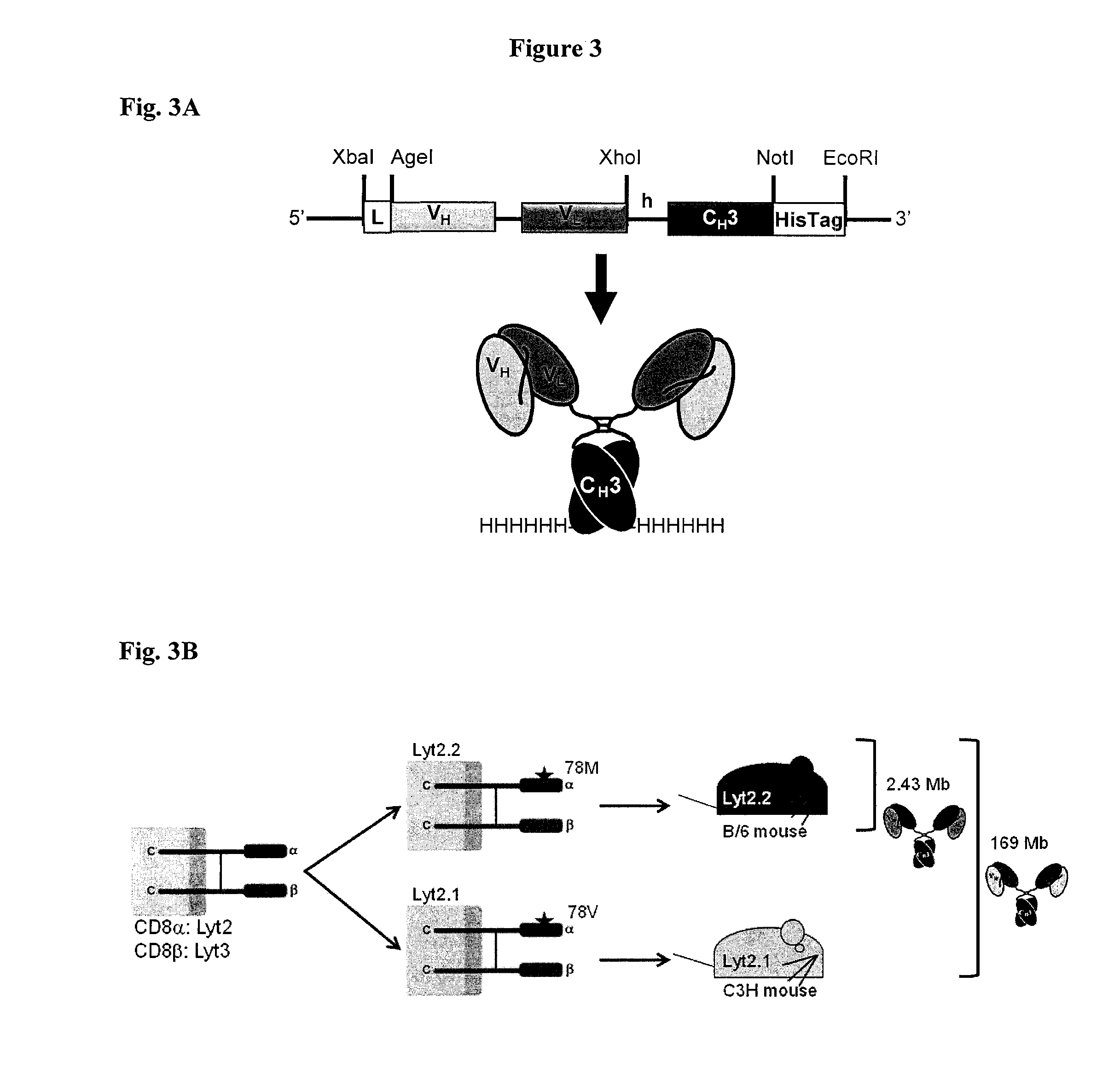
Design and Production of the Anti-CD8 Minibodies

[0323]Determination of VH and VL Sequences from Parental Hybridomas.

[0324]The YTS169 hybridoma was obtained from the Therapeutic Immunology Group at Oxford University, UK and cultured in Iscove's Modified Dulbecco's Medium (IMDM) plus 10% FBS and Pen / Strep (21). The 2.43 hybridoma was obtained from ATCC (TIB-210) and cultured in DMEM plus 10% FBS and Pen / Strep. VH and VL sequences were obtained by RT-PCR using primers published by Dubel, S., et al. (Dubel S. et al., Isolation of IgG antibody Fv-DNA from various mouse and rat hybridoma cell lines using the polymerase chain reaction with a simple set of primers. J Immunol Methods. Sep. 30 1994; 175(1):89-95). For the hybridoma 2.43, the obtained VH and VL sequences were confirmed with trypsin digest-mass spectrometry of the purified parental antibody performed at the UCLA core facility. The YTS169 hybridoma was engineered to antibody fragments without further VH and VL validation.

[0325]S...

example 2

In Vivo Preparation and Administration of Anti-CD8 Minibodies

[0348]CD8 depletion. Mice were treated for three consecutive days with 330 μg of anti-CD8 depleting antibody (Clone 53-6.7 purchased from UCSF Monoclonal Antibody Core) injected intraperitoneally (165 μL in saline) or 250 μg of 2.43 minibody injected intravenously (125 μL in saline). Two to three days post-treatment, single cell suspensions from the spleen, peripheral blood, thymus and lymph nodes were isolated and stained as described above for CD8 depletion analysis.

[0349]SCN-NOTA Conjugation.

[0350]All solutions were made metal-free (MF) using Chelex 100 (1.2 g / L; BioRad). The 2.43 and YTS169 Mbs were dialyzed against MF-PBS overnight using Slide-A-Lyzer MINI dialysis units (Thermo Scientific). Proteins at 1-2 mg / mL were then incubated with an 80-fold molar excess of S-2-(4-Isothiocyanatobenzyl)-1,4,7-triazacyclononane-1,4,7-triacetic acid (p-SCN-Bn-NOTA; Macrocyclics) for 4 hours at 4° C. pH was adjusted to 8.5 using 1 ...

example 3

Production of Anti-CD8 Antibodies

[0360]Sequencing Variable Regions of Parental Rat Anti-Murine CD8 Antibodies.

[0361]RT-PCR was repeated until at least 2 individual experiments produced three replicates of the same sequence for the VH and VL domains for each hybridoma. For sequence validation of hybridoma 2.43, the VH and VL from RT-PCR sequences were confirmed with tryptic digest-mass spectrometry of the parental antibody. VH amino acid coverage was 35% (41 / 117) including the complete CDR1 and half of CDR2 and VL amino acid coverage was 62% (66 / 107) including both CDR2 and CDR3. For YTS169, no sequence verification was performed.

[0362]For the hybridomas YTS 105 and YTS 156, the sequences were compared to the crystal structures of the Fab fragment in complex with either sCD8a or sCD8β deposited with PDB ID codes 2ARJ and 3B9K, respectively. RT-PCR sequences for YTS 105 were identical to the published crystal structure except for a few mutations at the 5′ and 3′ terminal ends due to t...


PUM

PropertyMeasurementUnit
Timeaaaaaaaaaa
Lengthaaaaaaaaaa
Volumeaaaaaaaaaa

Abstract

Disclosed herein, the parental antibodies from the hybridomas YTS 169.4.2.1 (YTS169) and 2.43 were engineered into minibody and diabody fragments. Both the YTS and 2.43 antibodies bind mCD8+. However, they differ in that the YTS 169 antibodies bind both Lyt2.1 and Lyt2.2 while the 2.43 antibodies bind an epitope that is Lyt2.2 specific. These novel minibodies and diabodies retained their antigen specificity as shown by flow cytometry and ImmunoPET imaging. Most importantly, both the 2.43 and YTS169 minibodies and diabodies produced high contrast ImmunoPET images of CD8+ lymphoid organs at only four hours post-injection.

Description

CROSS REFERENCE TO RELATED APPLICATIONS[0001]This application is a 371 National Phase of PCT / US2013 / 053862 filed Aug. 6, 2013 which claims the benefit under 35 U.S.C. §119(e) to U.S. Application No. 61 / 680,165 filed Aug. 6, 2012, the disclosures of which are incorporated by reference in their entireties.STATEMENT AS TO GOVERNMENT SUPPORT[0002]This invention was made with Government support under Grant Nos. CA016042, CA092131, and CA098010, awarded by the National Institutes of Health. The Government has certain rights in the invention.FIELD OF THE INVENTION[0003]This invention relates to novel antibody-based agents for imaging in vivo CD8 expression in mouse models of disease and novel, non-invasive and detection and quantification of CD8+ T cells in vivo.BACKGROUND[0004]Non-invasive detection of specific biomarkers of disease can provide crucial information for diagnosis, prognosis, response to therapy, dosage for radioimmunotherapy and targeted therapy selection. Specifically, the...

Claims


Application Information

Patent Timeline
IPC IPC(8): C07K16/28A61K51/10
CPCC07K16/2815A61K51/1027C07K2317/565A61K2039/505C07K2317/626C07K2317/24C07K2317/622C07K2317/33C07K2317/567C07K2317/64C07K2317/73C07K2317/92A61P35/00A61P37/00
Inventor WU, ANNA M.TAVARE, RICHARDOLAFSEN, TOVE
Owner RGT UNIV OF CALIFORNIA
Why Eureka
Try Eureka