Delivery tool and method for devices in the pericardial space

a delivery tool and pericardial space technology, applied in the field of cardiac rhythm therapy, can solve the problems of increased risk of reentry injury, increased risk of intrathoracic adhesion in patients undergoing sternotomy, and increased risk of venous occlusion, so as to achieve safe and accurate placement of cardiac medical devices and reduce the vasculature

Active Publication Date: 2015-08-20
CHILDRENS NAT MEDICAL CENT
View PDF9 Cites 83 Cited by
  • Summary
  • Abstract
  • Description
  • Claims
  • Application Information

AI Technical Summary

Benefits of technology

[0010]The present disclosure differs from U.S. Pat. Nos. 4,991,578 and 4,270,549, at least, in that navigation and visualization is achieved with the use of a camera embedded in the delivery tool as opposed to intermittent fluoroscopic images gathered throughout the procedure. As a result, pericardial access is achieved under direct visualization of the tissue, reducing the risk of myocardial puncture, and excess radiologic exposure.
[0016]A single cannula allows the camera to be positioned within the pericardial space itself, providing direct visualization for navigation and anchoring of the medical device. With this approach, a lead can be selectively placed on the right or left atrial epicardium for atrial pacing, or the right and / or left ventricular epicardium for ventricular pacing, cardiac resynchronization therapy, and / or defibrillation. Clear visualization of the pericardium, the beating heart, and critical structures such as coronary arteries is critical to position cardiac devices securely and safely while avoiding cardiac and coronary injury in the pericardial space.
[0020]A third group may be any patient, regardless of size and anatomy that may benefit from a subxiphoid pericardial approach versus standard epicardial approach due to less invasive procedure reducing surgical morbidity, and potentially shortening recovery time and total expense of procedure.
[0023]In some cases, the patient may not have a virgin pericardial sac to be accessed. Previous cardiac surgeries may result in the obliteration of the pericardial space all together. In this instance, a subxiphoid delivery tool may be beneficial to deliver multimodal imaging technology to the pericardial and epicardial surfaces of the heart. The multimodal imaging may include direct visualization, ultrasound transducers, multispectral light guide and camera, or any combination thereof. Use of multimodal imaging may help in the detection and identification of anatomical structures for safe and accurate placement of cardiac medical devices such as the coronary artery.
[0024]A subxiphoid approach may also be beneficial for the delivery of biological agents and therapeutic drugs to the pericardial space. In one application, the delivery of stem cells to the epicardium, pericardium, or pericardial space can be performed with a minimally invasive technique for selective placement under direct visualization. Pharmaceuticals can also be delivered directly to the epicardium, pericardium, or pericardial space with a minimally invasive technique for controlled, selective placement under direct visualization. In addition, procedures that require the access to the coronary arteries such as the placement of stents to remove obstructions will be possible with direct visualization in a subxiphoid approach. This approach will benefit patient groups such as infants and small children that are more susceptible to x-ray and have smaller vasculature, as well as the elderly who cannot tolerate an invasive open approach.

Problems solved by technology

Cardiac pacemaker implantation in small children and patients with congenital heart defects presents unique challenges to the cardiologist and surgeon.
Patients with congenital heart disease and device-dependent primary electrical diagnoses are likely to require multiple invasive procedures over the course of a lifetime, with attendant cumulative risk of venous occlusion.
The post-operative recovery typically entails multiple days in the intensive care unit with the commensurate costs and risks.
Patients undergoing sternotomy are also at increased risk of intrathoracic adhesions with heightened subsequent operative risk of reentry injury should the need for reoperation / exploration arise.
Re-operation can be difficult, as the fibrotic tissue must be fully dissected in order to reach viable cardiac tissue for acceptable pacing thresholds.
For pediatric, single ventricle, and abnormal vasculature patients, a transvenous approach is not suitable due to restriction in anatomy used for navigation.
First, the delivery tool only offers visualization parallel to the working channel, reducing the capability of a surgeon to accurately gauge depth during pericardial puncture.
Second, the delivery tool does not offer control of epicardial leads once inside in the pericardial space, limiting implantation to the ventricular surface.
There are currently no approved leads or delivery tools on the market for percutaneous pericardial pacing or defibrillator lead placement.
These patients may have contraindication to transvenous pacing due to intracardiac shunts or may have inaccessible endocardial surfaces due to abnormalities in venous or cardiac anatomy.
Therefore, they also would not be amenable to transvenous pacing and would require open-chest access to place epicardial pacing leads.
For defibrillation, there is not a suitable FDA-approved alternative, and they often undergo off-label indication use of current leads placed using an open-chest access to the pericardial space.
A third group may be any patient, regardless of size and anatomy that may benefit from a subxiphoid pericardial approach versus standard epicardial approach due to less invasive procedure reducing surgical morbidity, and potentially shortening recovery time and total expense of procedure.
Previous cardiac surgeries may result in the obliteration of the pericardial space all together.

Method used


Image

  • Delivery tool and method for devices in the pericardial space
  • Delivery tool and method for devices in the pericardial space
  • Delivery tool and method for devices in the pericardial space

Examples


Embodiment Construction

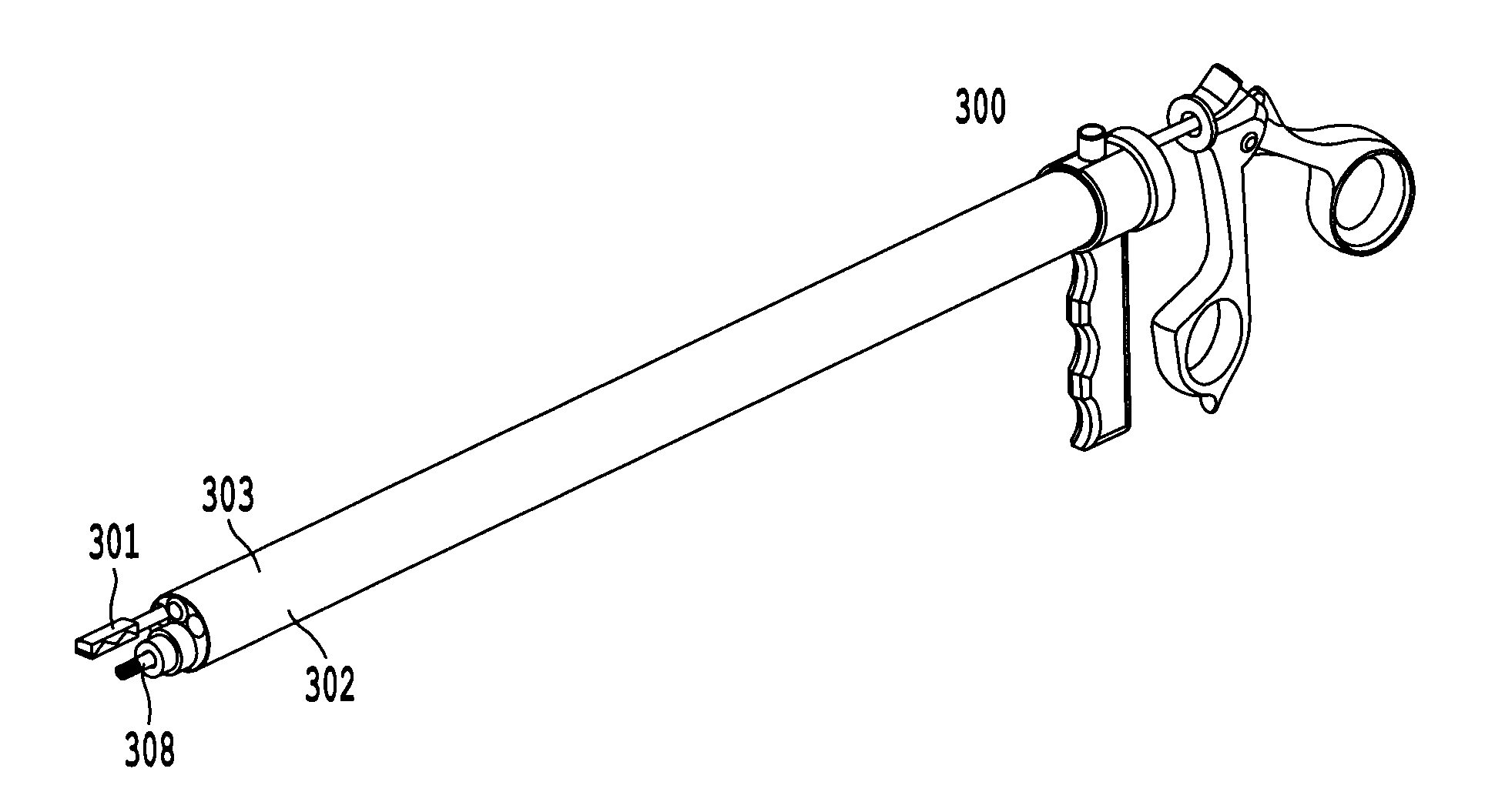
[0051]The present disclosure generally involves an apparatus and methods for delivering medical devices within the pericardial space. While several embodiments are disclosed, it is understood that the present disclosure is exemplary and can be embodied in many different forms. Therefore, the specific features and functionality of the tool disclosed are not to be interpreted as limiting, but to serve a basis for the claims, and to educate one skilled in the art as to the functionality of the tool with respect to the method of device delivery. For the purposes of teaching, the embodiments are direct towards the selective placement and implantation of cardiac leads and leadless pacemakers in the pericardial space, and should not be considered limiting.

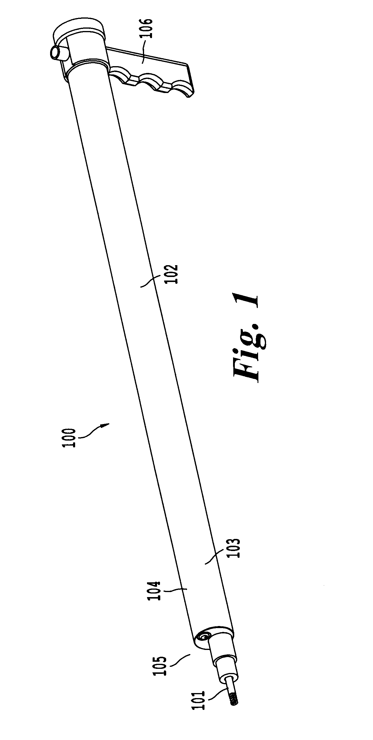
[0052]FIG. 1 illustrates one embodiment of the proposed delivery tool 100 configured or designed to selectively deliver a cardiac device 101 into the pericardial space and epicardial surface. The core 102 of the delivery tool includes of ...


PUM


Abstract

The present disclosure is a device and method associated with the delivery of medical devices in the pericardial space using a minimally invasive approach with direct visualization. More specifically, the device can be used to deliver permanent pacing, defibrillation and cardiac resynchronization leads, as well as leadless pacemakers for cardiac rhythm management to the epicardial surface of the heart. A subxiphoid procedure is proposed as a minimally invasive alternative to thoracotomy, while the delivery tool incorporates a camera for direct visualization of the procedure. The tool also incorporates a steerable catheter to provide selective control of the placement and orientation of the medical device in the pericardial space.

Description

CROSS-REFERENCE TO RELATED APPLICATIONS[0001]This application claims priority to U.S. Provisional Application No. 61 / 940,551, filed Feb. 17, 2014, which is hereby incorporated by reference in its entirety.BACKGROUND[0002]1. Field of the Disclosure[0003]The present disclosure is related to the field of cardiac rhythm therapy, and a device capable of delivering medical devices for cardiac pacing to the pericardial space under direct visualization and control via percutaneous approach.[0004]2. Description of the Related Art[0005]Cardiac pacing is utilized to stimulate the heart, and currently can be performed via two distinct approaches: transvenously to access the endocardium and direct surgical access to the epicardial surfaces. Cardiac pacemaker implantation in small children and patients with congenital heart defects presents unique challenges to the cardiologist and surgeon. These patients are often too small for insertion of pacemaker leads through a standard transvenous approach...

Claims


Application Information

Patent Timeline
Patent Type & Authority Applications(United States)
IPC IPC(8): A61B1/05A61B5/00A61N1/05A61B1/018
CPCA61B1/00154A61B1/3132A61B1/05A61B1/018A61N1/0592A61B5/0084
Inventor BERUL, CHARLESOPFERMANN, JUSTINKRIEGER, AXELKIM, PETER
Owner CHILDRENS NAT MEDICAL CENT
Why Eureka
Try Eureka