
Chemical bleaching of dyes using radical photoinitiators

Active Publication Date: 2015-10-08
LEICA MICROSYSTEMS CMS GMBH
View PDF4 Cites 0 Cited by
  • Summary
  • Abstract
  • Description
  • Claims
  • Application Information

AI Technical Summary

Benefits of technology

This patent describes a method for rapidly destroying signals in biological samples without damaging them. The method is also advantageous because it does not require high power light that may damage sample components. The technique allows for rapid signal cycling without significantly modifying the components of the sample that are different from the probe. Overall, this method provides improved signal-to-noise ratios and allows for the same sample to be used for multiple cycles without losing its biological integrity.

Problems solved by technology

In some embodiments, the disclosed methods also may be characterized by the absence of residual optical signals even in high expression targets resulting, e.g., in increased signal-to-noise ratio.

Method used


Image

  • Chemical bleaching of dyes using radical photoinitiators
  • Chemical bleaching of dyes using radical photoinitiators
  • Chemical bleaching of dyes using radical photoinitiators

Examples


example 1

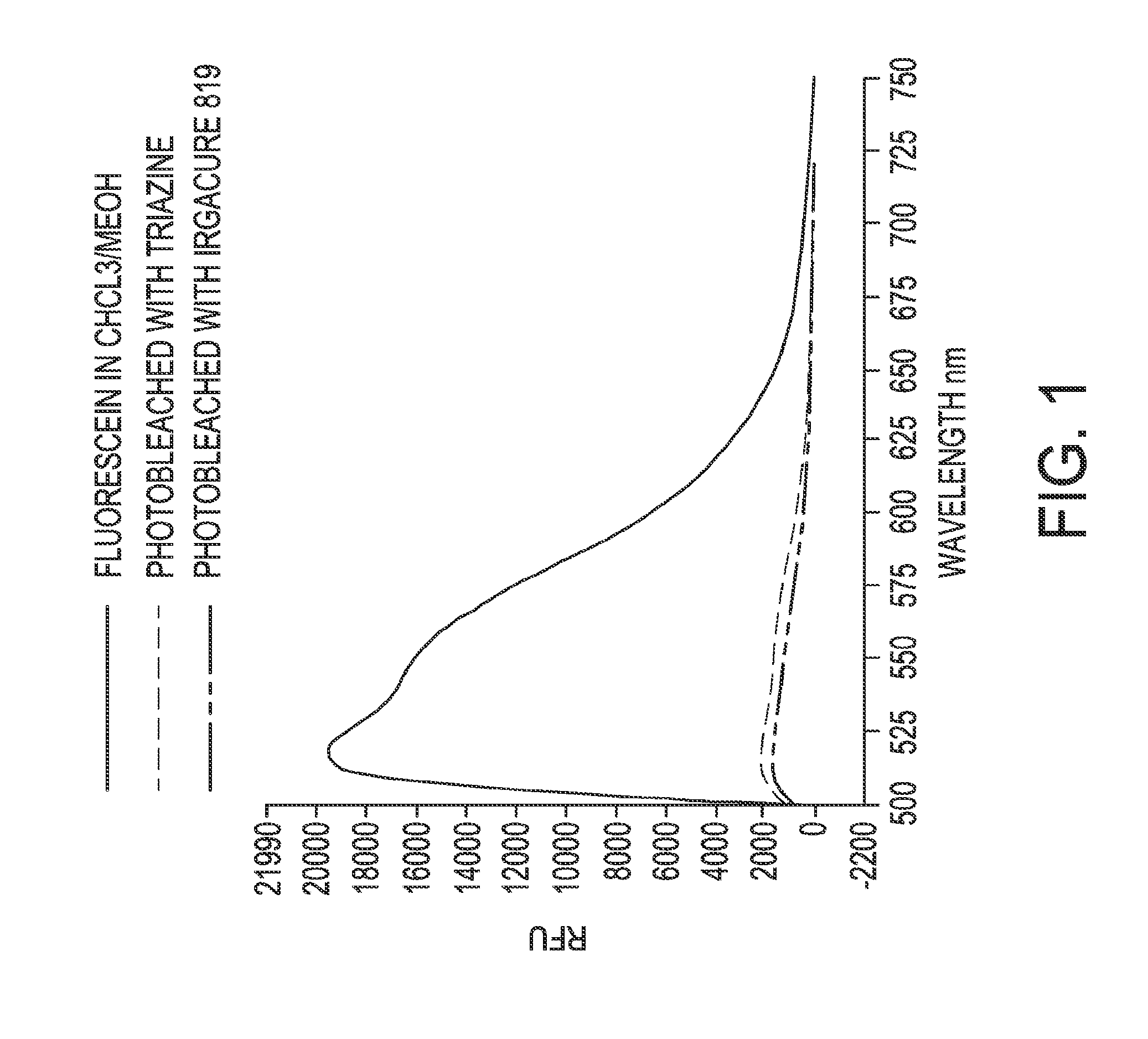
Chemical Bleaching of Fluorescein Using Photoinitiators

[0255]The bleaching reaction of fluorescein 0.75 mM was carried out in chloroform / methanol alone, with 2.2 mM solution of Methoxy styryl bis trichloro methyl triazine, or 2.4 mM solution of Irgacure 819. Irradiation of the samples was carried out for 5 minutes under 330 nm lamps rayonet reactor. FIG. 1 shows a fluorescence spectrum of fluorescein before and after bleaching, under each condition, respectively, measured using the Nanodrop Instrument (Thermo Scientific). The fluorescence spectrums demonstrate complete fluorescence quenching of fluorescein by chemical bleaching using both photoinitiators tested.

example 2

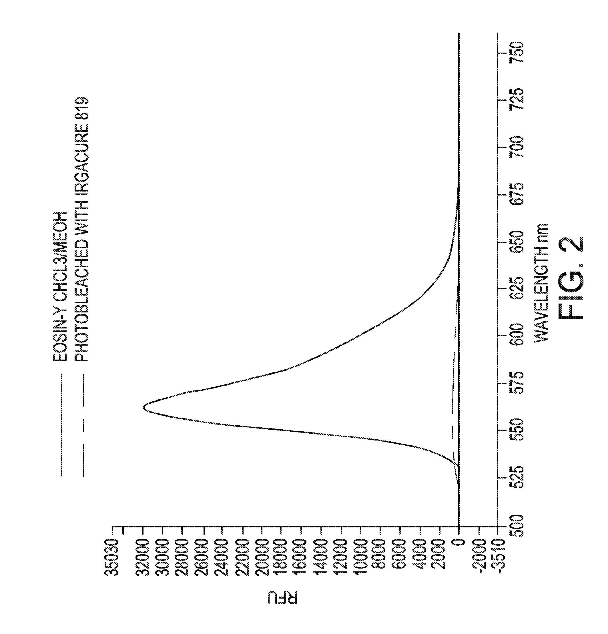
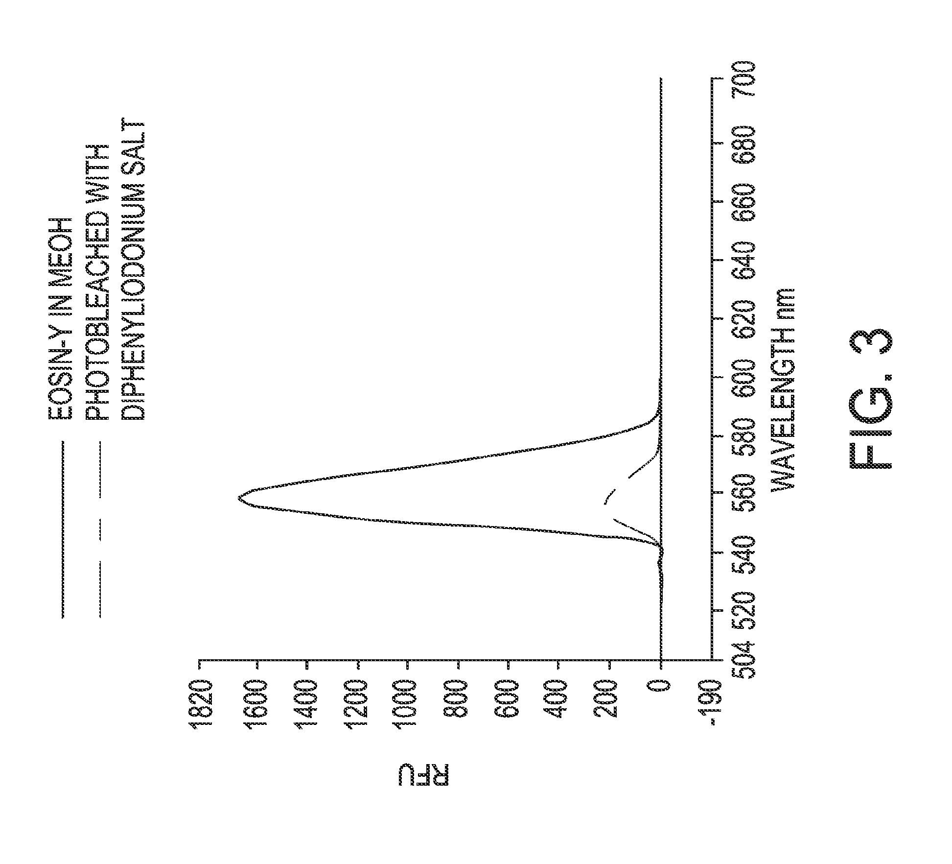
Chemical Bleaching of Eosin-Y Using Photoinitiators

[0256]In one experiment, the bleaching reaction of Eosin-Y 0.4 mM was carried out in chloroform / methanol or with 2.4 mM solution of Irgacure 819. In another experiment, the bleaching reaction of Eosin-Y 0.4 mM was carried out in methanol or with 2.32 mM solution of diphenyliodonium triflate. Irradiation of the samples was carried out for 5 minutes under 330 nm lamps rayonet reactor. FIGS. 2 and 3 show fluorescence spectrums of Eosin-Y before and after bleaching, under each condition, respectively, measured using the Nanodrop Instrument (Thermo Scientific). The fluorescence spectrums demonstrate complete fluorescence quenching of Eosin-Y by chemical bleaching using Irgacure 819, and over 85% fluorescence quenching of Eosin-Y by chemical bleaching using diphenyliodonium triflate.

example 3

Photoactivated Chemical Bleaching of Cy5 in Tissues

[0257]Preparation of Tissue Samples:

[0258]Human multi tissue array samples were obtained as tissue slides embedded in paraffin. These samples included microarray of normal, premalignant, and cancer tissues with progressive grades (Pantomics, MNT241).

[0259]Slide Clearing:

[0260]Three paraffin embedded slides were baked at 60° C. for one hour with tissue facing up and parallel to the oven rack. After baking, slides were deparaffinized by washing in xylene with gentle agitation for ten minutes. The samples were then rehydrated by washing in four solutions of ethanol with concentrations decreasing in the order of 100%, 95%, 70%, and 50% followed by a wash with 1× phosphate buffer saline (PBS, pH 7.4). After rehydration, the slides were washed with 1×PBS. A ten minute wash in 0.3% Triton X-100 in PBS was performed for membrane permeabilization of the tissue, followed by a wash with 1×PBS.

[0261]Antigen Retrieval:

[0262]After the slide clear...


PUM

PropertyMeasurementUnit
Temperatureaaaaaaaaaa
Timeaaaaaaaaaa
Wavelengthaaaaaaaaaa

Abstract

Methods comprising the use of photoactivated chemical bleaching for detecting multiple targets in a biological sample are provided. The methods include the steps of binding at least one probe to one or more target present in a biological sample including multiple targets, and detecting a signal from the probe. The method further includes the steps of contacting the sample comprising the bound probe with a photoinitiator and irradiating the sample, thereby initiating a photoactivated chemical bleaching process that substantially inactivates the probe. The method further includes the steps of binding at least one probe to one or more target present in the sample, and detecting a signal from the probe. The process of binding, detecting and bleaching may be iteratively repeated.

Description

FIELD OF THE INVENTION[0001]The present invention is directed to the detection of biomarkers on a biological sample. More specifically, the present invention is directed to the use of radical photoinitiators for bleaching of detectable signals in a method for detecting multiple targets in a biological sample. Also provided are a kit and a system for performing the novel method, as well as images of a biological sample generated using the novel method.BACKGROUND[0002]Various methods may be used in biology and in medicine to detect different targets in a biological sample. For example, analysis of proteins in histological sections and other cytological preparations may be performed using the techniques of histochemistry, immunohistochemistry (IHC), or immunofluorescence. Analysis of proteins in biological samples may also be performed using solid-state immunoassays, for example, using the techniques of western blots, or using cell-based assays that can be performed, for example, by us...

Claims


Application Information

Patent Timeline
IPC IPC(8): G01N33/58
CPCG01N33/582
Inventor SOOD, ANUPNATARAJAN, ARUNKUMARKAANUMALLE, LAKSHMI SIREESHALOWES, CHRISTINA
Owner LEICA MICROSYSTEMS CMS GMBH
Learn More