Method for inducing the formation of islet structures and improving beta cell function

a beta islet and cell culture technology, applied in the field of mammals, can solve the problems of reducing insulin production, reducing the phenotype of the islet, requiring extensive cell manipulation, and affecting the formation of pi, so as to avoid the need for immunosuppressive drugs, improve insulin production, and improve insulin sensing

Inactive Publication Date: 2016-01-21
AKIRAV EITAN MOSHE
View PDF1 Cites 2 Cited by
  • Summary
  • Abstract
  • Description
  • Claims
  • Application Information

AI Technical Summary

Benefits of technology

[0012]The islet cell cultures may be used in vitro to produce various hormones. The islets might also be transplanted in vivo as a therapy for islet dysfunction, e.g., diabetes. In some cases, an autotransplant may be employed, where β cells generated from stem cells from the afflicted individuals are co-cultured with primary islet derived ECs ex-vivo to expand the stock establish a supply of pseudoislets for re-implantation into the patient, e.g., on the pancreas, thus avoiding need for immunosuppressive drugs. The pseudoislets may also be used in research and therapies which comprise artificial organs, which may be extracorporeal or implanted in a patient.
[0013]A straightforward and rapid method for inducing free-floating PIs by co-culturing iEC and β-cell insulinoma lines is provided. Newly formed PIs are positive for ECM proteins produced by iECs and show improved insulin production, insulin sensing and glucose stimulated insulin secretion when compared with monolayer cells. iEC-induced PIs may serve as a useful tool for examining β-cell / iEC interactions and studying β-cell function in a native 3D configuration.

Problems solved by technology

Over time, however, cultured hIslets lose their 3D structure due to islet adhesion and flattening, resulting in a loss of islet phenotype (7, 8) and reduced insulin production (9).
Despite their usefulness, PI generation requires extensive cell manipulation and may take several weeks to form (7-14 d).

Method used


Image

  • Method for inducing the formation of islet structures and improving beta cell function
  • Method for inducing the formation of islet structures and improving beta cell function
  • Method for inducing the formation of islet structures and improving beta cell function

Examples


example 1

Generation of Pseudoislets (PIs) Using Islet Derived Murine Endothelial Cells and Insulinoma Cell Lines

[0024]The following procedure describes the formation of PIs using islet derived ECs (MS-1 cells).

[0025]This procedure allows for the formation of free floating islets which contain ECM components and show improved insulin production as described Spelios et al. (36 and 37).

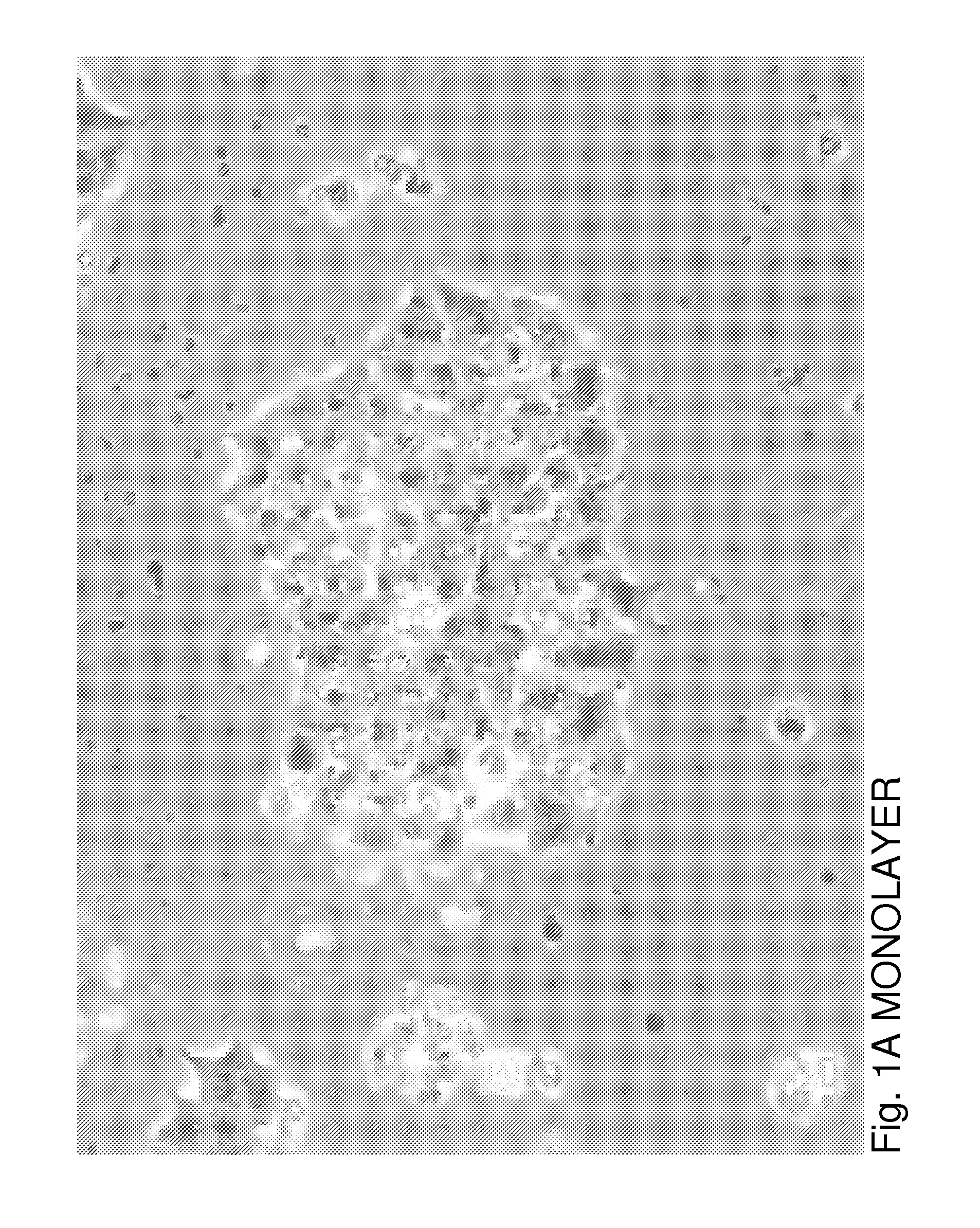
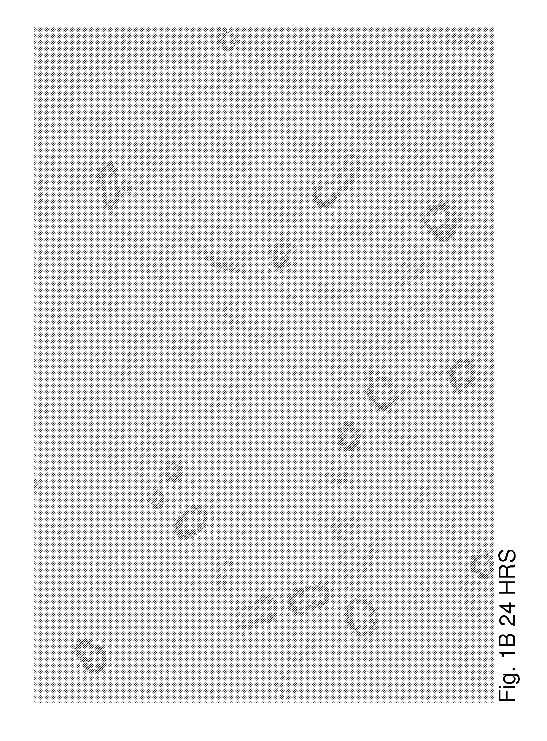
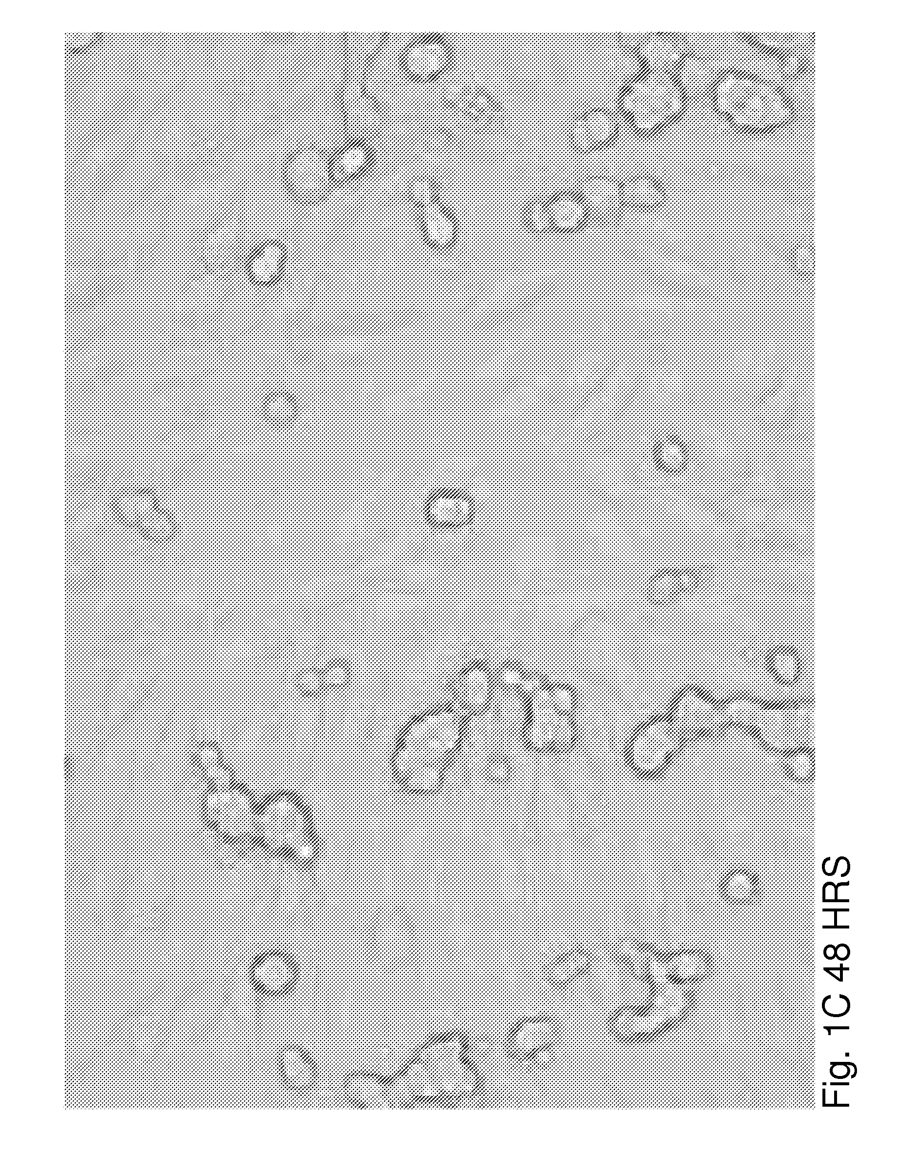
[0026]βTC3 is a murine-derived insulinoma cell line that does not produce ECM proteins. To evaluate the effects of ECM on βTC3 cells, we established a co-culture system of βTC3 cells and the islet-derived EC line, MS-1. RT-PCR and immunofluorescence (IF) staining showed expression of both laminin and collagen IV in MS-1 cells. Mixing of MS-1 and βTC3 cells resulted in the formation of free-floating islet-like structures as early as 48 h following co-culture, while βTC3 cultured alone remained attached to the surface as a monolayer. Confocal microscopy showed the deposition of laminin and collagen IV on the surfac...

example 2

Maintaining Insulin Production and ECM Proteins by Co-Cultures of Islet Derived Murine Endothelial Cells and Primary Human Islets

[0044]The following procedure describes the effects of MS-1 cells on insulin production and ECM deposition in primary human islets.

[0045]Procedures (See, FIGS. 4A-4B)

[0046]MS-1 cells are grown to ˜100% confluency in T-12.5 tissues culture treated flask. Primary hIslets (Primary human islets—1000 IEQ) are added to the flask containing the MS-1 cells. The flask is placed on an orbital shaker in a 37° C. 5% CO2 incubator running at 70 rpm. Media (DMEM, 5 mM glucose, 10% FCS, P / S / N) is replaced every 4 days.

[0047]FIGS. 4A and 4B show co-culturing of MS-1 and primary human islets increased ECM deposition and insulin staining in vitro. Human primary islets were cultured alone or in the presence of murine islet derived ECs (MS-1). FIG. 4A shows 8 day cultures of hIslet. White arrows point to high Col IV and laminin deposition in the hIslets. FIG. 4B shows pixel ...

example 3

Generating Human Islet Derived ECs to Increase the Viability and Function of Primary Human Islets

[0048]Production of primary islet derived human ECs (See FIGS. 5A, 5B, and 5C)

[0049]Previous reports show the ability of primary islet-derived ECs from rats to grow efficiently in vitro (17), expressly incorporated herein by reference. This previously reported protocol has been adapted for the enrichment and propagation of primary human islet derived-ECs. 100 islets are sufficient to produce a full 24 well plate by passage 3.

[0050]Purified human islets are cultured on collagen I coated plates for 2 wks. BS-1 positive ECs from mixed cultures are enriched using magnetic bead isolation (AutoMACS pro, Miltenyi). Passage 3 cells are tested for ECM components as well as other EC markers (i.e. PE-CAM / BS-1). Enriched human islet derived can be cryopreserved indefinitely.

Co-Cultures of Primary Islet Derived Human ECs and Primary Human Islets

[0051]Primary islet derived ECs are grown to ˜100% conf...


PUM

PropertyMeasurementUnit
adhesionaaaaaaaaaa
β cell functionaaaaaaaaaa
densityaaaaaaaaaa

Abstract

Insulin producing β cells are found in three dimensional (3D) structures, the Islet of Langerhans. The 3D structure is required for normal β cell function and survival. β cell pseudoislets (PIs) are useful for study of β cell physiology. Co-culturing of primary human islets and β cell lines together with islet-derived epithelial cells can improve β cell function and survival and maintain the cells' 3D structure, resulting a rapid and spontaneous formation of free-floating PIs. β cells in PIs were similar in size to native islets and showed increased percentage of pro-insulin-positive cells, increased insulin gene expression in response to glucose stimulation, improved glucose-stimulated insulin secretion, and reduced β cell death. Key ECM proteins, absent in monolayer β cells, are deposited by iECs in and round the PIs. iEC induced PIs are a useful tool for examining β-cell/iEC interactions and studying β-cell function in a native 3D configuration.

Description

BACKGROUND[0001]1. Field of the Invention[0002]The present invention relates to the field of mammalian cell culture, and more particularly to advancements to in vitro beta islet cell culture.[0003]2. Related Art[0004]The islets of Langerhans are 3-dimensional (3D) structures that contain insulin producing β cells. In vivo, islets contain key components of the extracellular matrix (ECM) which can be found in the islet capsule and around the islet microvessels. The ECM is made of defined proteins that include collagens and laminins (1). In vivo, islet endothelial cells (ECs) are a major source for ECM proteins, and previous studies demonstrated an active role for ECs in mediating β cell function, by promoting β-cell differentiation and increasing insulin production (2, 3). Following isolation, primary islets lose both ECs and ECM. This leads to impaired islet function in vitro (4-6) and results in apoptosis (7, 11). Previous reports show that culturing of human islets (hIslets) on dif...

Claims


Application Information

Patent Timeline
Patent Type & Authority Applications(United States)
IPC IPC(8): C12N5/071
CPCC12N5/0677C12N2506/22C12N2502/22
Inventor AKIRAV, EITAN, MOSHE
Owner AKIRAV EITAN MOSHE
Why Eureka
Try Eureka