
System and method for controlling depth of imaging in tissues using fluorescence microscopy under ultraviolet excitation following staining with fluorescing agents

a fluorescence microscopy and depth control technology, applied in the field of systems and methods for structural and molecular imaging of human and animal tissues using fluorescence microscopy, can solve the problems of difficulty, difficulty in limiting the use of intra-operative biopsy or surgical margin evaluation, and the quality of most frozen specimens may be less than optimal

Active Publication Date: 2016-03-17
RGT UNIV OF CALIFORNIA +1
View PDF12 Cites 25 Cited by
  • Summary
  • Abstract
  • Description
  • Claims
  • Application Information

AI Technical Summary

Benefits of technology

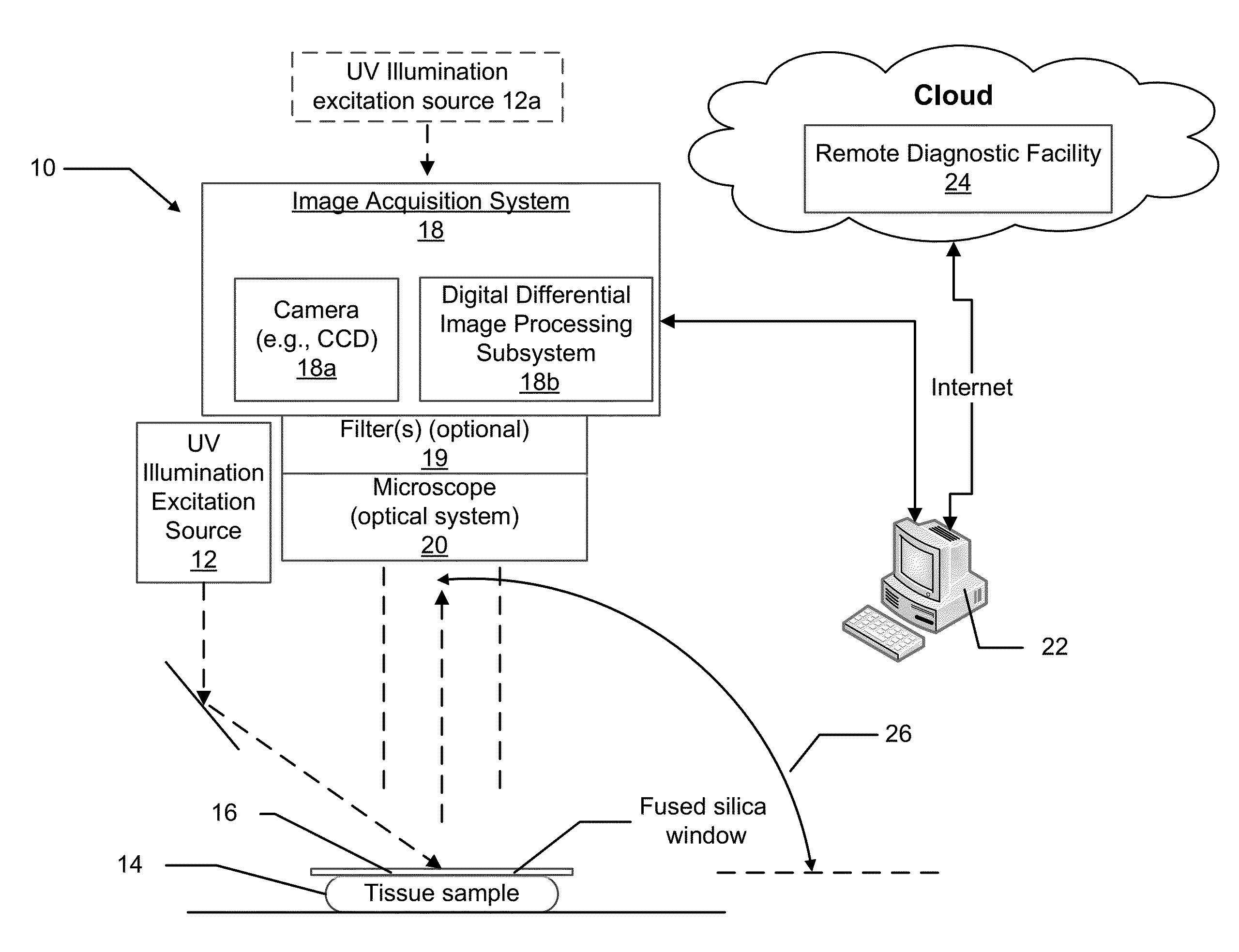
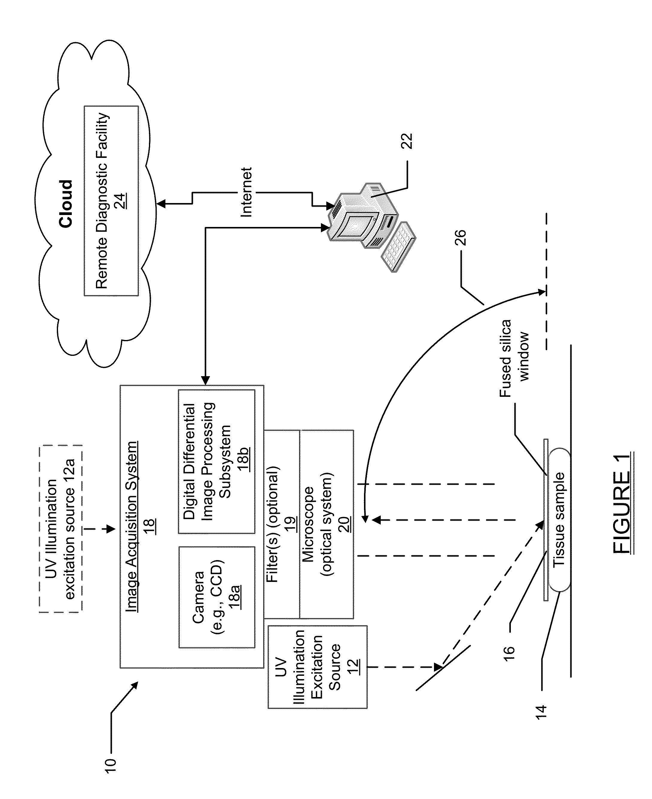
The present patent is related to a method and system for analyzing tissue. The method involves obtaining a tissue sample and exposing it to fluorophores that accumulate in tissue or cells. An illumination excitation source is used to illuminate the tissue sample with UV light that has a specific wavelength to limit its penetration into the sample. A microscope optical system is then used to image the tissue sample, and an image acquisition system records the optical information. The display system processes and displays the images for analysis. The technical effect of the patent is to provide a reliable and effective method for analyzing tissue samples by using UV light to excite the fluorophores and a microscope optical system to visualize the tissue sample. This method may have application in various fields such as in research, medical diagnosis, and treatment planning.

Problems solved by technology

Surgical margin evaluation: Frozen-section evaluation of biopsies obtained during surgery can be routine part of clinical care but is fraught with difficulties.
Moreover, the quality of most of the frozen specimens may be less than optimal, and often lower than that of formalin-fixed paraffin-embedded (“FFPE”) specimens.
The resulting delays and interpretation challenges limit the use of intra-operative biopsy or surgical margin evaluation.
Consequently, if margin assessment is not done intra-operatively, additional surgeries may be required.
Most tissues removed undergo conventional FFPE processing before definitive diagnoses are rendered, incurring delays of days or even weeks, depending on workflow.
Similarly, many molecular tests, such as immunohistochemistry or immunofluorescence for cancer markers or companion diagnostics are not ordered until after the initial tissue diagnosis, and incur additional days to weeks of delay.

Method used


Image

  • System and method for controlling depth of imaging in tissues using fluorescence microscopy under ultraviolet excitation following staining with fluorescing agents
  • System and method for controlling depth of imaging in tissues using fluorescence microscopy under ultraviolet excitation following staining with fluorescing agents
  • System and method for controlling depth of imaging in tissues using fluorescence microscopy under ultraviolet excitation following staining with fluorescing agents

Examples


Embodiment Construction

[0022]The following description is merely exemplary in nature and is not intended to limit the present disclosure, application, or uses. It should be understood that throughout the drawings, corresponding reference numerals indicate like or corresponding parts and features.

[0023]With traditional pathology methods, pathology specimens must be physically cut in order to present a thin slice of tissue to a standard microscope. If instead the tissue could be optically sectioned, then freezing, or fixation and paraffin embedding, followed by microtomy, would not be necessary. The previous methods discussed herein, as disclosed in U.S. Pat. Nos. 7,945,077 and 8,320,650 for imaging optically thick specimens inexpensively and efficiently, are centered around oblique wide-field fluorescent imaging of tissue using intrinsic-to-tissue fluorescing biomolecules with short UV light (typically 266 nm) excitation. The present disclosure expands on the teachings of U.S. Pat. No. 7,945,077 and U.S. P...


PUM

PropertyMeasurementUnit
wavelengthaaaaaaaaaa
incidence angleaaaaaaaaaa

Abstract

The present disclosure relates to a method for analyzing tissue specimens. In one implementation the method involves obtaining a tissue sample and exposing the sample to one or more fluorophores as contrast agents to enhance contrast of subcellular compartments of the tissue sample. The tissue sample is illuminated by an ultraviolet (UV) light having a wavelength between about 200 nm to about 400 nm, with the wavelength being selected to result in penetration to only a specified depth below a surface of the tissue sample. Inter-image operations between images acquired under different imaging parameters allow for improvement of the image quality via removal of unwanted image components. A microscope may be used to image the tissue sample and provide the image to an image acquisition system that makes use of a camera. The image acquisition system may create a corresponding image that is transmitted to a display system for processing and display.

Description

FIELD[0001]The present disclosure relates to systems and methods for structural and molecular imaging of human and animal tissues using fluorescence microscopy under short wavelength, such as ultraviolet light, excitation and more particularly to a system and method which is able to optically section thick tissue samples to obtain high-resolution images of the near-surface tissue microstructure without requiring formalin fixation and paraffin embedding (FFPE) followed by microtome sectioning, or freezing and sectioning of the tissue sample, and which further is able to be used with conventional or novel fluorescing stains and labels, to allow effective analysis of tissue for diagnosis, tissue composition, and / or surgical guidance, such as monitoring surgical margin areas of a biopsy specimen for the presence of tumor cells.BACKGROUND[0002]The statements in this section merely provide background information related to the present disclosure and may not constitute prior art.[0003]Tech...

Claims


Application Information

Patent Timeline
Patent Type & Authority Applications(United States)
IPC IPC(8): G01N21/64
CPCG01N21/6458G01N2201/062G01N2201/06113G01N21/6428G01N21/6486A61B5/0071G01N21/6445G01N2021/6419G01N2021/6421G01N2021/6441A61B5/0044
Inventor DEMOS, STAVROSLEVENSON, RICHARD
Owner RGT UNIV OF CALIFORNIA
Learn More