Diagnostic methods for infectious disease using endogenous gene expression

a technology of endogenous gene expression and diagnostic methods, applied in biocide, instruments, biochemistry apparatus and processes, etc., can solve the problems of limited application and accuracy, inadequate white blood cell count,

Inactive Publication Date: 2016-07-21
WASHINGTON UNIV IN SAINT LOUIS
View PDF1 Cites 4 Cited by
  • Summary
  • Abstract
  • Description
  • Claims
  • Application Information

AI Technical Summary

Problems solved by technology

Previous studies indicate that white blood cell count is an inadequate tool for distinguishing between viral and bacterial infection, a distinction often used to determine whether or not to treat the patient with antibiotics (Rudinsky, S. L. et al.
While some other previous studies provide clinical information in certain groups of subjects and for some particular diseases and / or infections, they can be limited in application and accuracy.

Method used


Image

  • Diagnostic methods for infectious disease using endogenous gene expression
  • Diagnostic methods for infectious disease using endogenous gene expression
  • Diagnostic methods for infectious disease using endogenous gene expression

Examples


example 1

[0138]This example illustrates detection of viruses in young children with Fever Without an Apparent Source (FWS).

[0139]Subjects were drawn from as study of children between 2 to 36 months of age with Fever Without an Apparent Source (Table 1) and afebrile children having ambulatory surgery who were recruited at St. Louis Children's Hospital as described previously (Colvin, J. M., et al. 2012, Pediatrics 130(6):e1455-1462). The febrile and afebrile groups were similar with respect to age, gender, and season of recruitment, but differed with respect to race, with more African-American children in the febrile group (57% vs. 13%). Patients were enrolled according to Institutional Review Board—approved protocol. The study was approved by the Washington University Human Research Protection Office. Each subject was tested for multiple viruses in blood and nasopharyngeal samples using panels of virus-specific PCR assays as described (Colvin, J. M., et al. 2012, Pediatrics 130(6):e1455-1462...

example 2

[0140]This example illustrates preparation of biological samples, including RNA from blood samples.

[0141]In these investigations, whole blood and nasopharyngeal samples were collected for virus-specific PCR and high-throughput sequencing. Additionally, a blood sample was collected in a Tempus™ Blood RNA Tube (Applied Biosystems, Carlsbad, Calif.) and stored at −80° C. for subsequent gene expression analysis.

[0142]Total RNA was isolated from whole blood collected in Tempus™ Blood RNA tubes (Applied Biosystems, Carlsbad, Calif.) according to the manufacturer's instructions. RNA quality was determined by gel-chip image (showing 28S, 18S and 5S bands) and RNA integrity number (RIN, generally a>7 RIN indicates good quality RNA) using an Agilent 2100 Bioanalyzer (Agilent, Palo Alto, Calif.). All but 3 of the RNA preparations had RIN scores≧7.0. Total RNA concentration was obtained from an absorbance ratio at 260 nm and 280 nm using a NanoDrop ND-100 spectrometry instrument (NanoDrop Inc.,...

example 3

[0143]This example illustrates gene expression microarray assays.

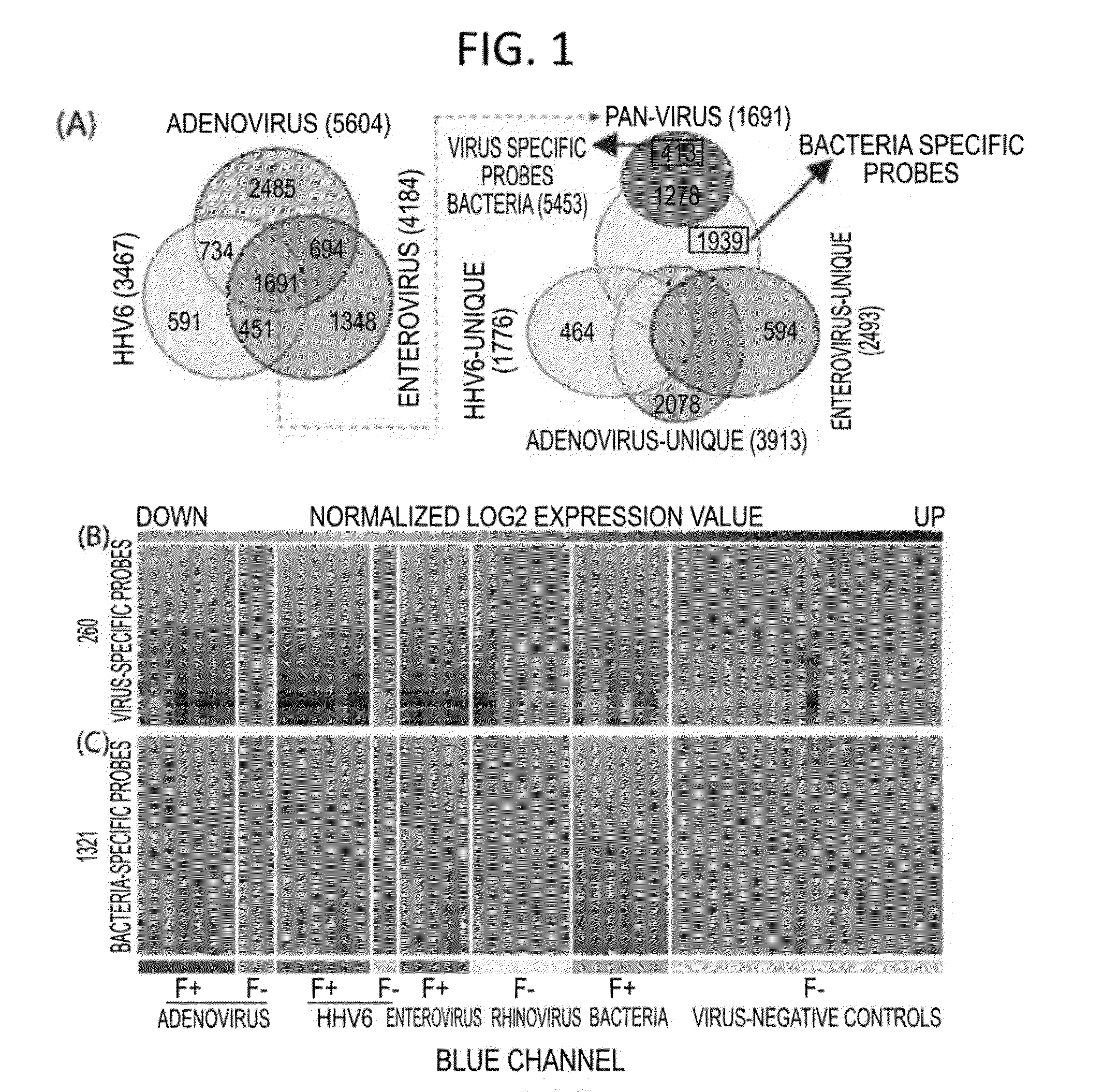
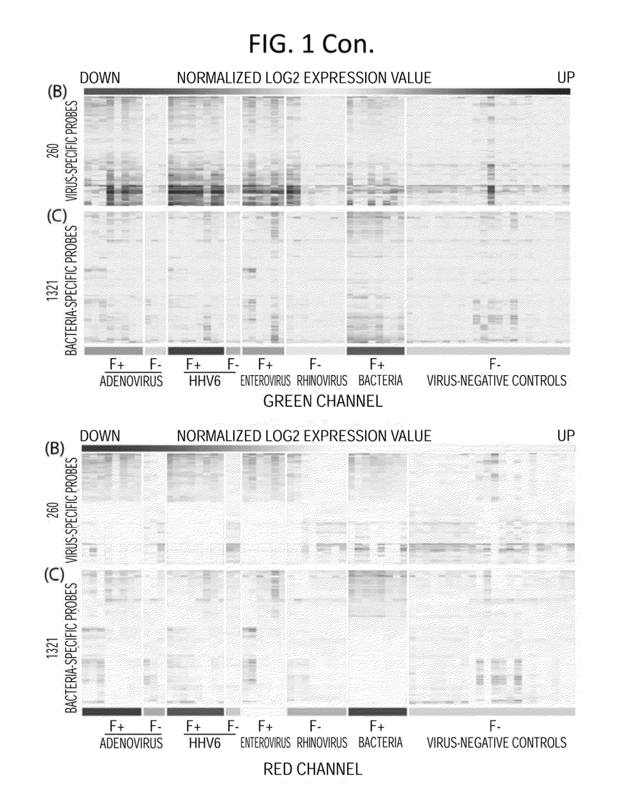
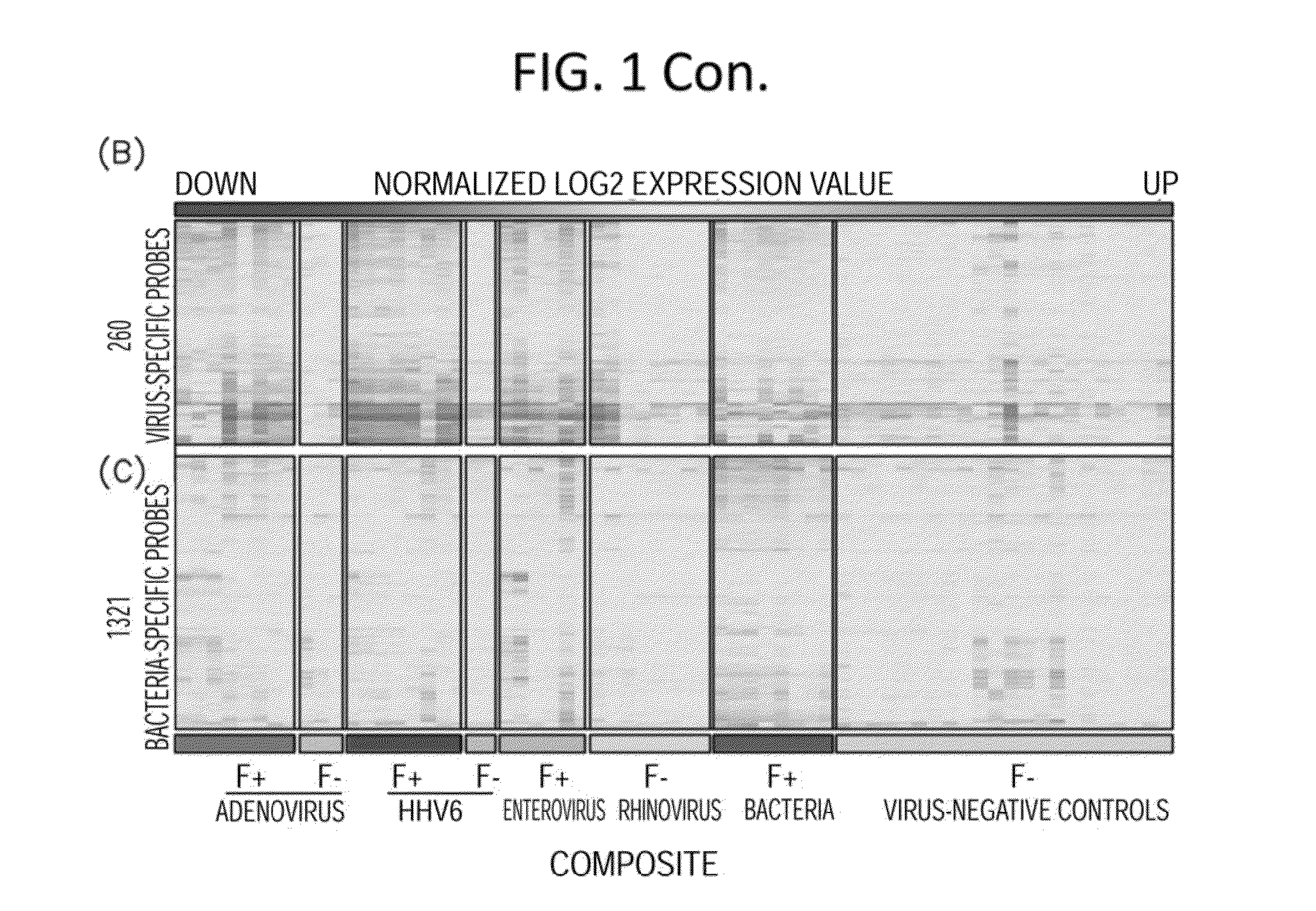
[0144]In these investigations, gene expression microarray analyses were conducted on blood samples from 35 febrile children positive for adenovirus, human herpesvirus 6 (HHV-6), or enterovirus infection or with acute bacterial infection, and 22 afebrile controls. Gene expression microarray assays were carried out at the Genome Technology Access Center in Washington University in St Louis. RNA transcripts were amplified by 17 linear amplification with the IIlumina 3TVT Direct Hybridization Assay Kit (Illumina Inc., San Diego, Calif.), and biotin-labeled cRNA targets were hybridized to the Illumina Human-HT12 v4 Expression BeadChips (>47,000 probes), which were scanned on an Illumina BeadArray Reader. Scanned images were quantitated by Illumina Beadscan software (v3). Quantified data were imported into Illumina GenomeStudio software (version 2011.1) to generate expression profiles and to make data quality assessments. Th...


PUM

PropertyMeasurementUnit
Tmaaaaaaaaaa
Northern blot assayaaaaaaaaaa
dual color fluorescentlyaaaaaaaaaa

Abstract

Disclosed are methods of diagnosis of a pathogen-associated disease. These methods comprise: providing a biological sample from a human subject; determining presence, absence and / or quantity of a bacterial pathogen, a viral pathogen, or a combination thereof, by a pathogen culture, a serum antibody detection test, a pathogen antigen detection test, a pathogen DNA and / or RNA detection test, or a combination thereof; determining in the sample, expression levels of at least one endogenous gene in which aberrant expression levels are associated with infection with a pathogen, by a microarray hybridization assay, RNA-seq assay, polymerase chain reaction assay, a LAMP assay, a ligase chain reaction assay, a Southern, Northern, or Western blot assay, an ELISA or a combination thereof. The subject can be diagnosed with the disease if the subject comprises the pathogen and an aberrant level of expression of an endogenous gene.

Description

CROSS REFERENCE TO RELATED APPLICATIONS[0001]This application claims the priority benefit of International Application PCT / US14 / 57164, filed on Sep. 24, 2014, which claims priority to U.S. Provisional Application Ser. No. 61 / 881,508 filed Sep. 24, 2013. Each of these applications is incorporated herein by reference, each in its entirety.STATEMENT REGARDING FEDERALLY SPONSORED RESEARCH[0002]This invention was made with government support under NIH Grants 1UAH2AI083266-01 from the NIAID, UL1 RR024992 from the NIH-National Center for Research Resources, and the Training Grant T32HD049338 from the NICHD. The government has certain rights in the invention.INTRODUCTION[0003]Despite absence of bacterial infection, many febrile children are treated with antibiotics. Virus and bacteria interact with different pattern recognition receptors in circulating blood leukocytes, triggering specific host transcriptional programs mediating immune response. Unique transcriptional signatures can be defi...

Claims


Application Information

Patent Timeline
Patent Type & Authority Applications(United States)
IPC IPC(8): C12Q1/68G01N33/569A61K45/00C12Q1/70
CPCC12Q1/6883C12Q1/689C12Q1/70G01N2469/10G01N33/56983A61K45/00C12Q2600/158G01N33/56911A61K31/00C12Q1/04C12Q2600/112G01N2469/20Y02A50/30
Inventor STORCH, GREGORY
Owner WASHINGTON UNIV IN SAINT LOUIS
Why Eureka
Try Eureka