System And Method For Motion-Robust 3D Magnetic Resonance Imaging Of Vessel Walls
a magnetic resonance imaging and motion-robust technology, applied in the field of magnetic resonance imaging system and method for the visualization of vascular structures, can solve the problems of poor slice resolution, long imaging time, and slow flow of turbulent blood, and achieve the effect of reducing artifacts and image degradation, and optimizing background suppression
- Summary
- Abstract
- Description
- Claims
- Application Information
AI Technical Summary
Benefits of technology
Problems solved by technology
Method used
Image

Examples
Embodiment Construction
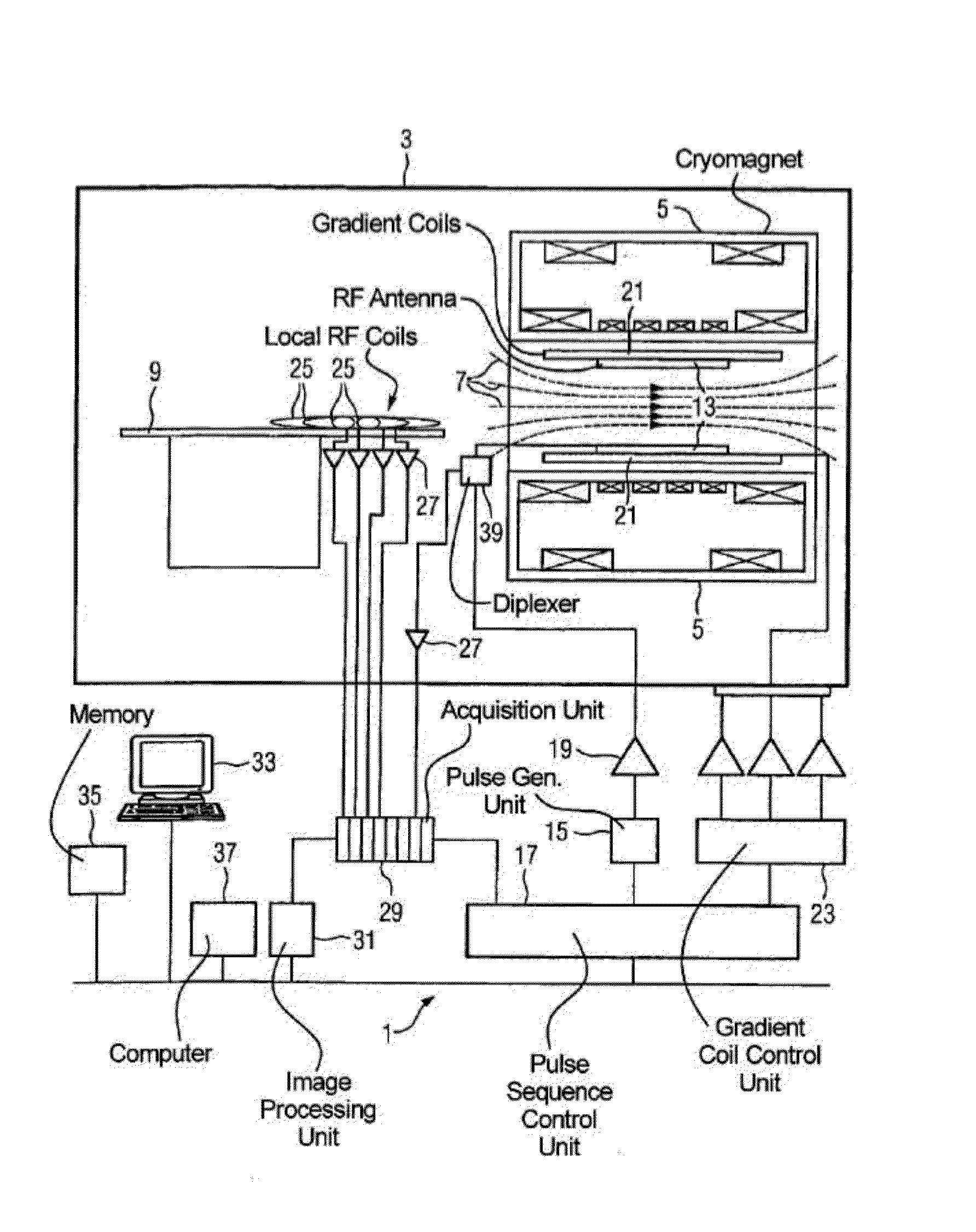
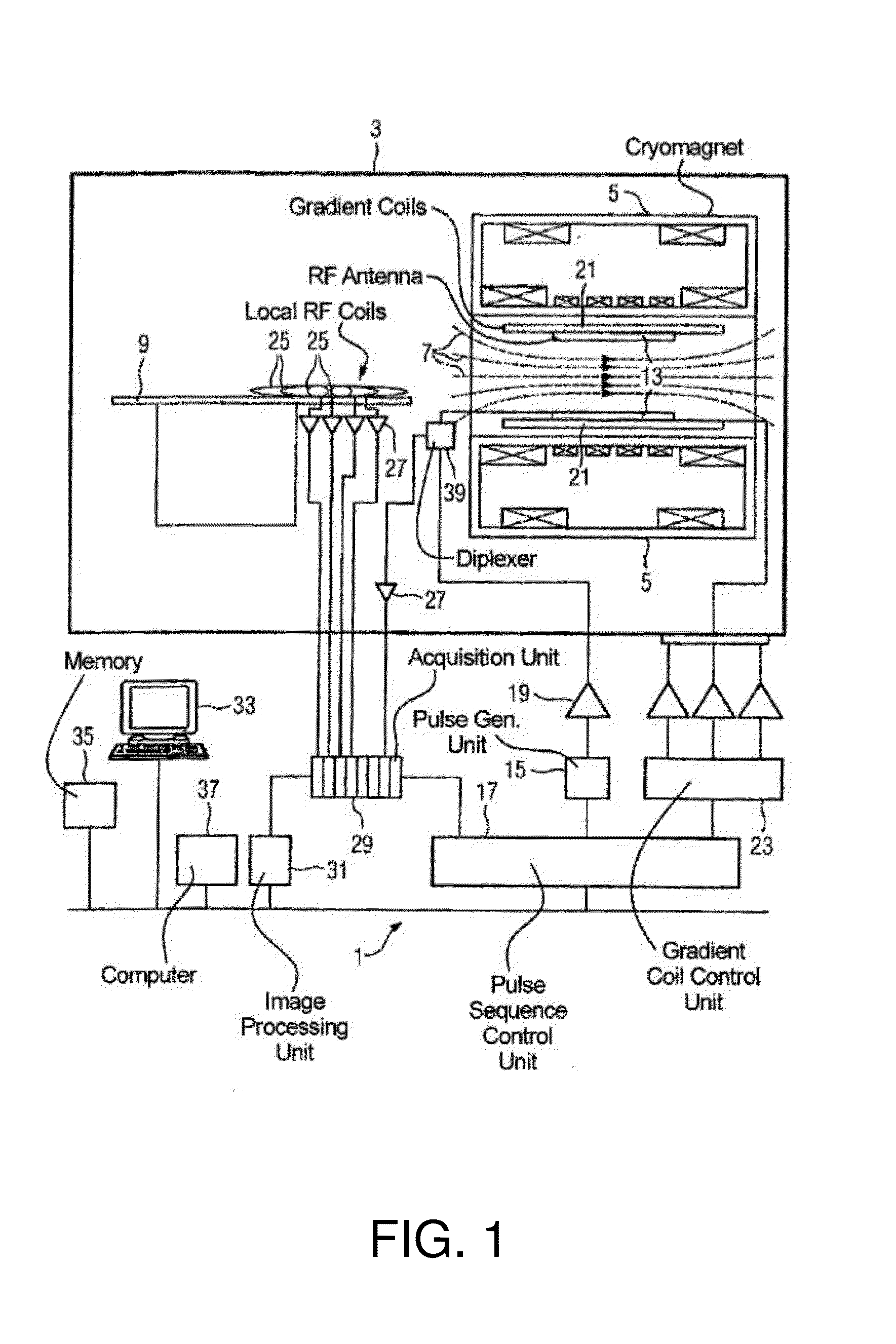
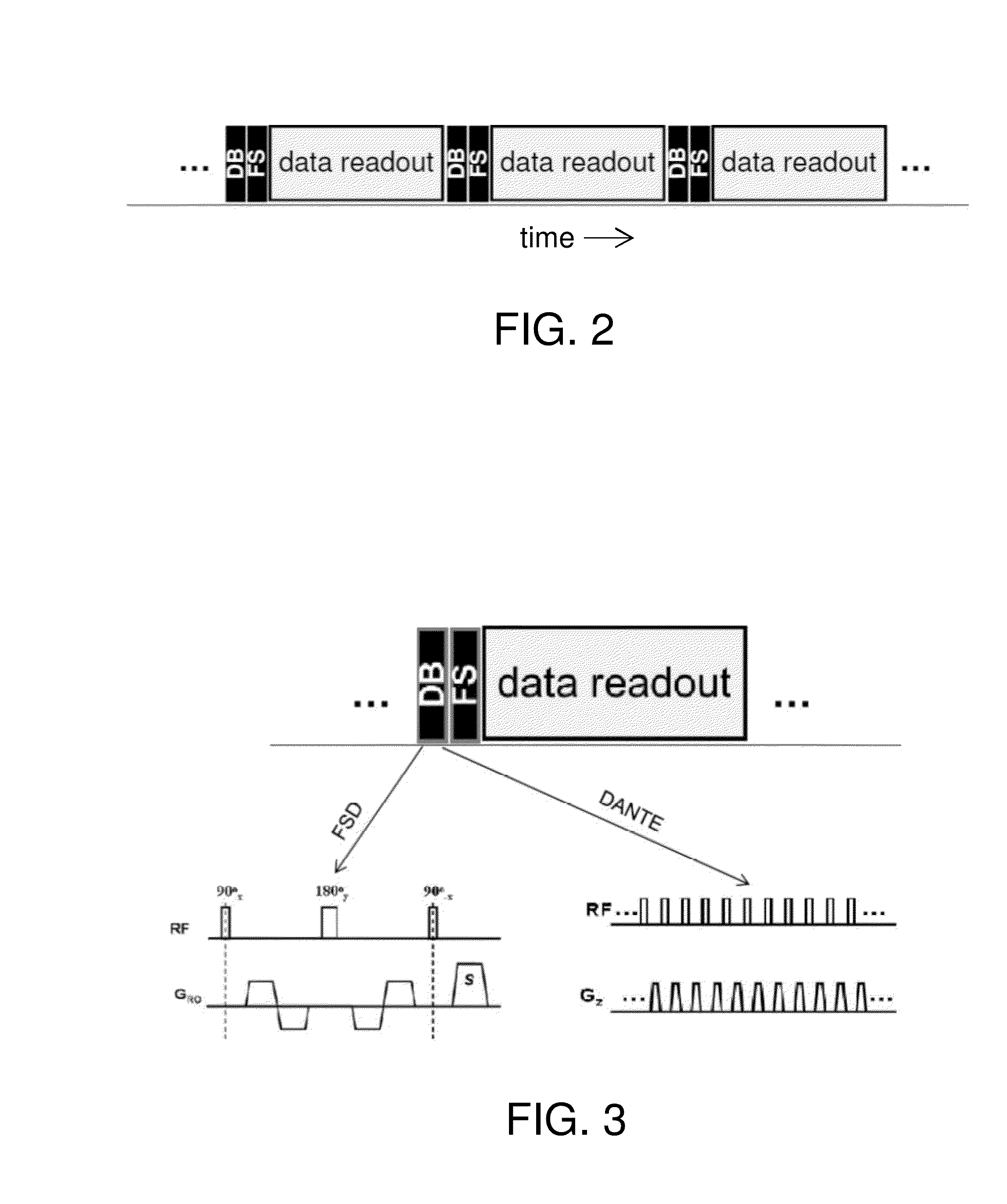
[0009]Exemplary embodiments of the present disclosure can provide systems and methods for 3D magnetic resonance (MR) imaging of vascular structures, e.g., a carotid wall or another vessel wall that is relatively insensitive to motion of the imaged volume. The imaging procedure described herein combines the advantages of both Cartesian and radial techniques, resulting in optimal background suppression (e.g., suppression of both blood and fat signals) while also reducing artifacts and image degradation arising from motion of the imaged volume.
[0010]In one embodiment, a magnetic resonance imaging system is provided that is configured to generate certain pulse sequences to obtain 3D image data for a volume to be imaged, where the pulse sequences include magnetization preparation sequences for suppression of both blood and fat signals, and a stack-of-stars (SoS) k-space sampling technique to obtain image data that oversamples image data near the Kz axis in k-space to provide insensitivit...
PUM
 Abstract
Description
Claims
Application Information