
Cationic contrast agents and methods of using the same

a contrast agent and cationic technology, applied in the field of contrast agents, can solve the problems of thinning cartilage, difficult to identify fine features, and difficulty in detecting joint pathologies,

Inactive Publication Date: 2016-10-06
THE TRUSTEES OF THE UNIV OF PENNSYLVANIA
View PDF2 Cites 2 Cited by
  • Summary
  • Abstract
  • Description
  • Claims
  • Application Information

AI Technical Summary

Benefits of technology

The invention relates to a new type of contrast agent for magnetic resonance imaging (MRI) that has a net positive charge, meaning it contains more protons than electrons. This is achieved by using a chelating agent that has a net negative charge. The invention also includes a specific compound that can be complexed with gadolinium, which is a common MRI contrast agent. The complex can be administered as an injection to a subject's joint and used for MRA imaging. The technical effects include improved image quality and a more accurate diagnosis of joint damage.

Problems solved by technology

However, X-ray and computed tomography (CT) scans cannot detect tears in cartilage and other soft tissues.
Detecting joint pathologies becomes even more challenging following arthroscopic repair.
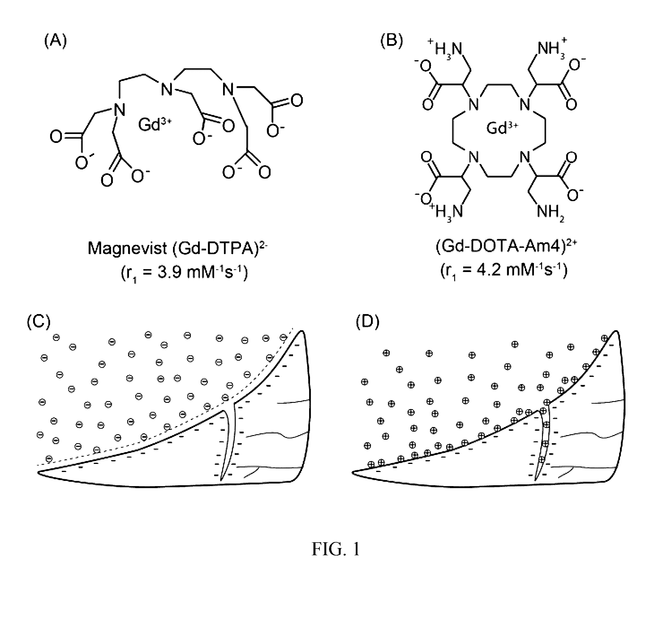
Indeed, this interaction at the tissue-fluid interface may predispose the agents to poor penetration into narrow crevices, making it difficult to identify fine features, tears, and thinning cartilage (FIG. 1C).

Method used


Image

  • Cationic contrast agents and methods of using the same
  • Cationic contrast agents and methods of using the same
  • Cationic contrast agents and methods of using the same

Examples


example 1

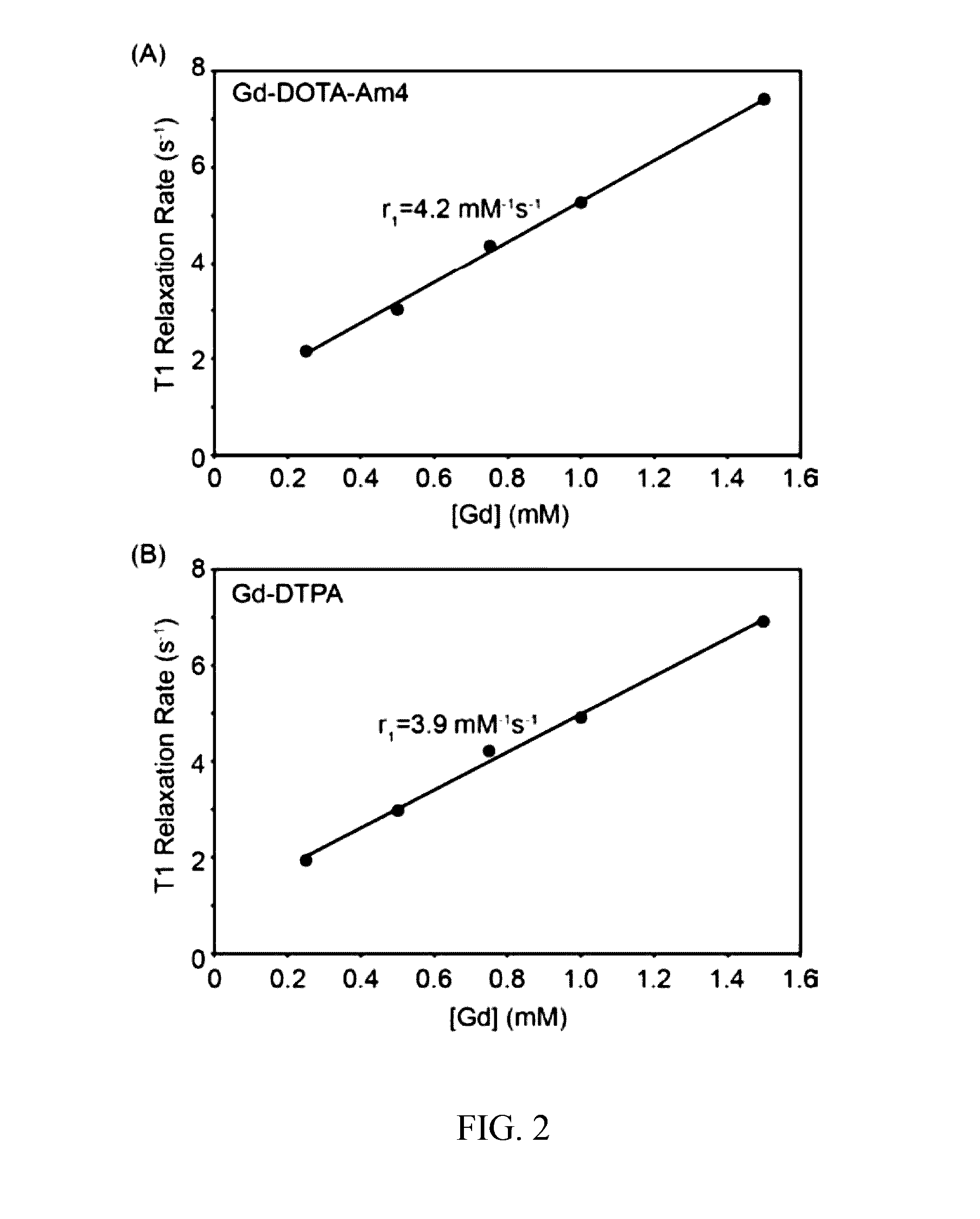
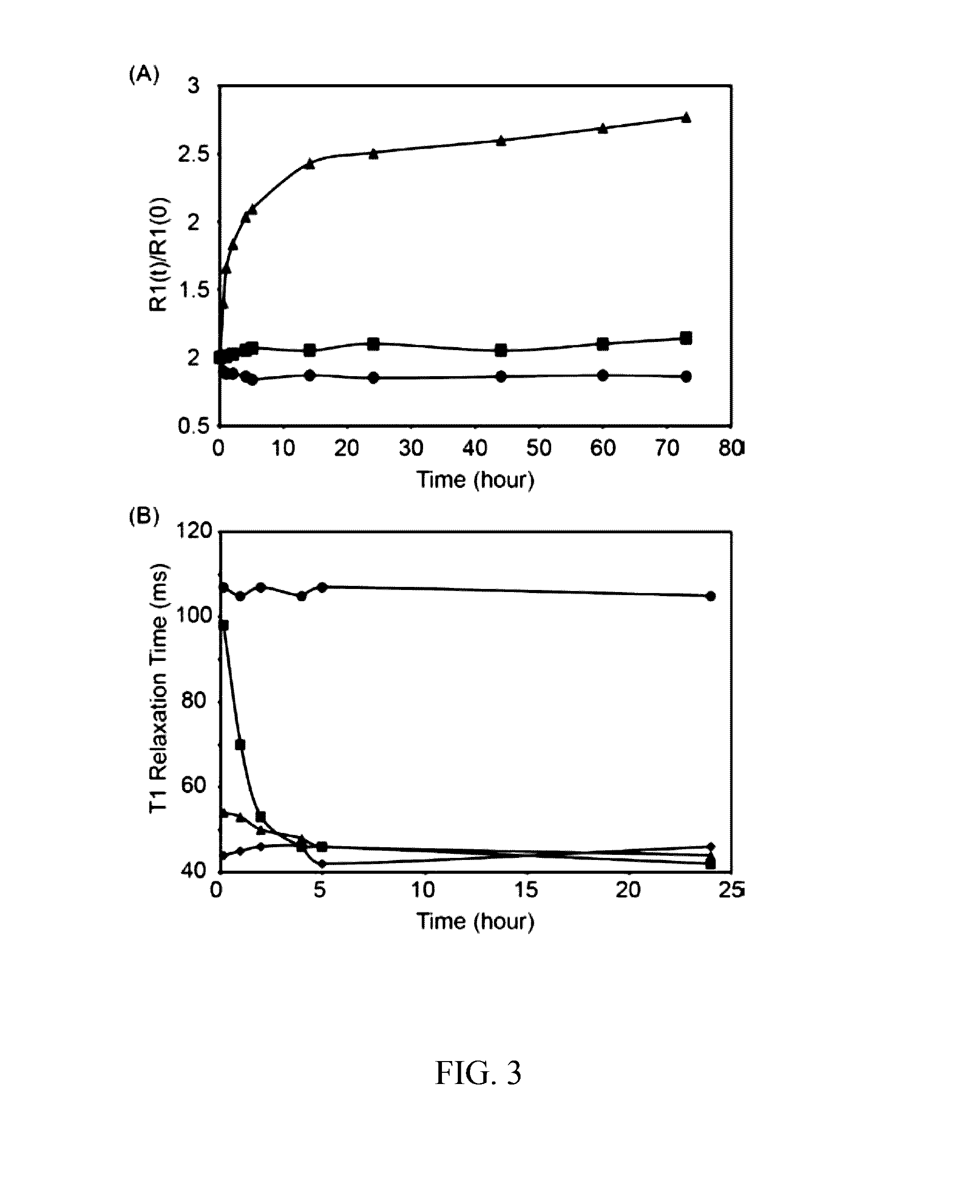
[0045]As described below, the efficacy of Complex A in highlighting soft tissue tears was evaluated in comparison to a clinically employed contrast agent (Magnevist®) using explants obtained from adult bovine menisci, a fibrocartilaginous tissue in the knee joint. Complex A appeared to improve the ability to detect soft tissue defects by providing increased signal intensity along the length of the tear. Magnevist® showed a strong signal near the liquid-meniscus interface, but less contrast was observed within the defect at greater depths compared to what was observed with Complex A.

[0046]Compound Ia was synthesized as shown below:

[0047]Compound2 was prepared by bromination of methyl acrylate using bromine in chloroform at room temperature, while compound 3 was prepared by slow addition of diethylamine into compound 2 in ether at 0° C. The methyl and ethyl protecting groups were efficiently removed with a methanol:NaOH (0.5 M) solvent cocktail (40:60) for 48 h. The solution was neutr...

example 2

[0079]Compound Ib (“DOTA-AmS4”) was synthesized as shown below:

[0080]Compound Ib was prepared as follows: Chlorosulfonyl isocyanate was reacted with tert-butanol and tert-butylamine in toluene to yield tert-butyl N-tert-butylsulfamoylcarbamate followed by removal of Cert-butylcarbamate using trifluoroacetic acid. The resulting N-tert-bytulsulfamide was reacted with brominated methylacrylate followed by a subsequent reaction with 1,4,7,10-tetraazacyclododecane (cyclen). The tert-butyl groups were removed via treatment with 2M hydrochloric acid in dioxane (2M-HCl-Dioxane) while methyl groups were removed by treatment with 1M NaOH to yield compound Ib.

[0081]Compound Ic (“DOTA-Ac4”) was synthesized as shown below:

[0082]Compound Ic was prepared as follows: L-serine was brominated using bromine and sodium nitrite, followed by protection of carboxylates using methanol with a catalytic amount of sulfuric acid. The alcohol was protected with tert-butyl group using magnesium sulfate and tert-...


PUM


Abstract

Gadolinium complexes for use as contrast agents, and methods for making and using the gadolinium complexes, are described. The contrast agent complexes preferably have a net positive charge, and can electrostatically interact with glycosaminoglycans to improve the delineation of fine tears within cartilage, detection of cartilage degeneration, or assessment of cartilage thickness, morphology, or glycosaminoglycan content via magnetic resonance imaging.

Description

CROSS REFERENCE TO RELATED APPLICATIONS[0001]This application claims priority to U.S. Provisional Application No. 62 / 143401, entitled CATIONIC CONTRAST AGENTS AND METHODS OF USING THE SAME, filed Apr. 6, 2015, the contents of which are incorporated by reference herein in their entirety.STATEMENT REGARDING FEDERALLY SPONSORED RESEARCH[0002]This invention was made with government support by the National Institutes of Health NIBIB R01EB012065 (AT) and NCI R01CA157766 (AT). The government has certain rights in the invention.FIELD OF THE INVENTION[0003]Embodiments of the present invention relate to contrast agents and methods of using the contrast agents.BACKGROUND OF THE INVENTION[0004]Diagnostic imaging of joints usually begins with a radiographic evaluation, which can help detect obvious sources of disease such as advanced arthritis, tumor, dislocation, or impingement. However, X-ray and computed tomography (CT) scans cannot detect tears in cartilage and other soft tissues. The early ...

Claims


Application Information

Patent Timeline
Patent Type & Authority Applications(United States)
IPC IPC(8): A61K49/10
CPCA61K49/106A61K49/108
Inventor TSOURKAS, ANDREWNWE, KIDO
Owner THE TRUSTEES OF THE UNIV OF PENNSYLVANIA
Learn More