Device and method for standardizing myoblast differentiation into myotubes

a technology of myoblasts and devices, applied in biochemistry apparatus, skeletal/connective tissue cells, biochemistry apparatus and processes, etc., can solve the problems of high variability, poor quantification of morphological parameters, and inability to develop robust cell based assays at high variability levels

Active Publication Date: 2016-10-27
CYTOO
View PDF1 Cites 2 Cited by
  • Summary
  • Abstract
  • Description
  • Claims
  • Application Information

AI Technical Summary

Benefits of technology

The invention aims to provide a device that can standardize the parameters of myotubes, such as their width and length, to better conduct cell-based assays. This standardization can help detect subtle effects of compounds on the physiology of myotubes, making it a useful tool for studying myotoxicity and drug discovery.

Problems solved by technology

However, although myotube orientation is extensively studied in the literature, their morphological parameters are poorly quantified and highly variable, due to difficulties in individualizing full mature structures and standardizing their morphology.
This high variability level does not permit robust cell based assay development.

Method used


Image

  • Device and method for standardizing myoblast differentiation into myotubes
  • Device and method for standardizing myoblast differentiation into myotubes
  • Device and method for standardizing myoblast differentiation into myotubes

Examples


example

[0176]The invention was used in a proof of principle screen with selected compounds (10 μM concentration) known for their myotoxic activity.

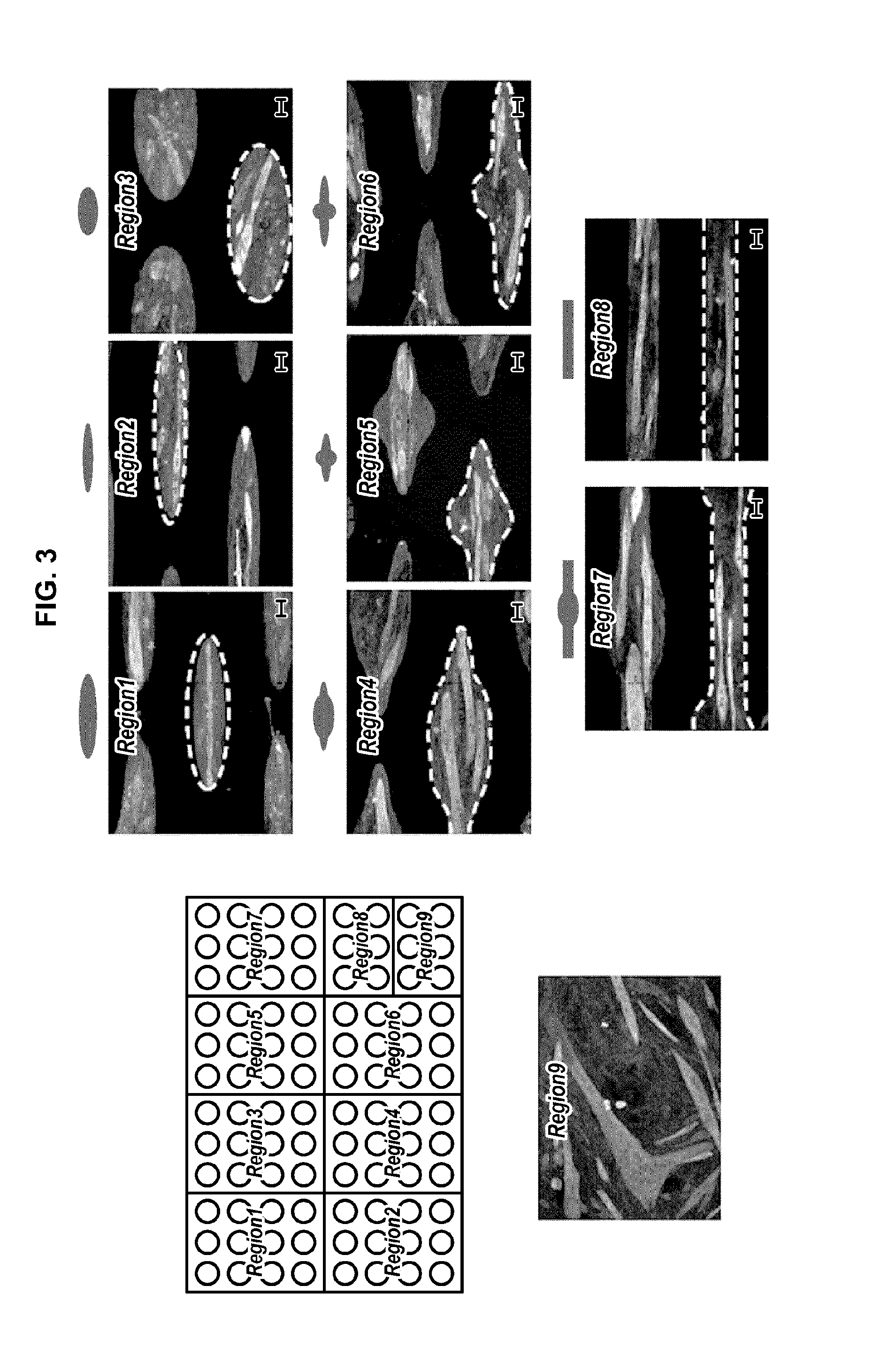
[0177]In this screening method, human primary myoblasts (HSMM, Lonza) are cultured on fibronectin coated surfaces (Region 5 patterns) within growth medium (Lonza SkGM™-2 cell culture Kit) during 24 hours. Then cells are cultured within differentiation medium (DMEM / F12, 2% Horse Serum, 0.5% P / S) for 24 hours. 60 different compounds, known as inducers of myotoxicity (including rhabdomyolysis syndromes, lysosomal myopathies, myofibrillar myopathies, inflammatory myopathies, hypokalemia myopathies, corticosteroid myopathies, myositis, myosin deficiency) and control, are added to the forming myotubes during 72 hours. Cells are fixed and an immunostaining is realized against myosin heavy chain, myogenin and nuclei. Images are acquired using an Operetta high content imaging system (Perkin Elmer).

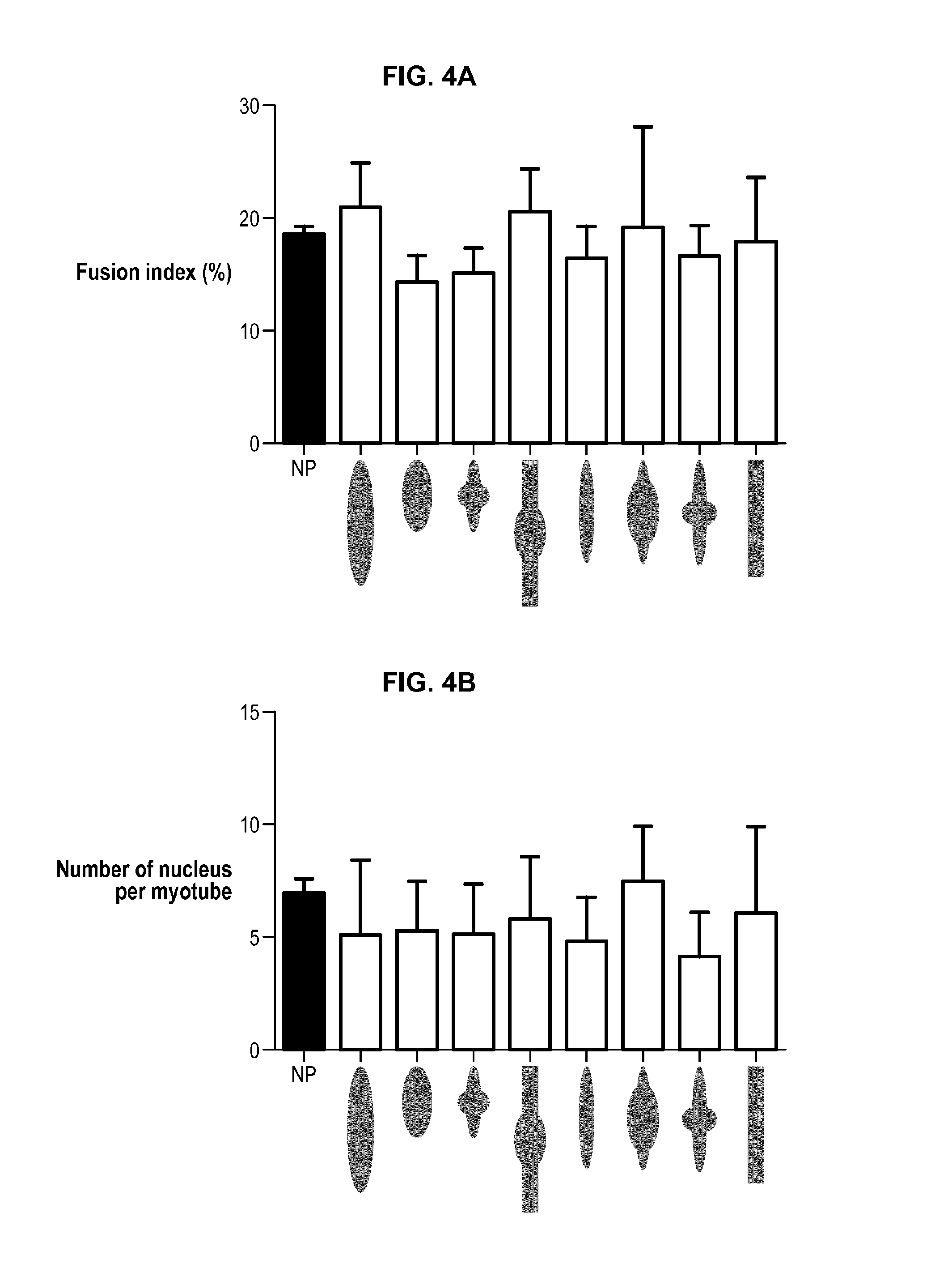
[0178]High responders, i.e. myotubes having a maximal wid...


PUM


Abstract

The invention relates to a device for standardizing myoblast differentiation into myotubes, comprising a substrate (1) and at least one cell-adhesive pattern (2) for culturing myoblasts on said substrate, wherein:said pattern (2) has an elongated surface comprising a central region (2C) and two lateral regions (2L) extending from said central region in both directions along a longitudinal axis of the pattern with a contour discontinuity between the central region (2C) and each lateral region (2L),the ratio between the maximum width (WC) of the central region (2C) and the maximum width (WL) of the lateral regions (2L) is greater than or equal to 2,the ratio between the length (L) and the maximum width (WC) of the pattern (2) is less than or equal to 4.The invention is also a method for standardizing myoblast differentiation into myotubes, comprising:(i) providing a device as described above,(ii) depositing myoblasts on at least one cell-adhesive pattern of said device,(iii) culturing said myoblasts in a differentiation medium during a determined incubation time so as to promote cell differentiation into myotubes and constrain elongation of the myotubes on the cell-adhesive pattern.Another aspect of the invention is a method for compounds screening in cell-based assays related to myotoxicity and drug discovery.

Description

FIELD OF THE INVENTION[0001]The invention relates to a device and a method for standardizing myoblast differentiation into myotubes, and a method for compound screening using such a device.BACKGROUND OF THE INVENTION[0002]Myogenesis is an in vivo process occurring during embryogenesis and tissue repair. Satellite cells begin to proliferate and form myoblasts. A myoblast is a mononucleate cell type that, by fusion with other myoblasts (this is a part of a process being also called “differentiation”), gives rise to myotubes that maturate and later eventually develop into muscle fibers. Maturation of myotubes can easily be characterized through the striated organization of myosin heavy chain molecules within sarcomeres, and the location of nuclei at the center of the structure [Abmayr 2012]. When myotubes are grown in a cell culture device, the growth method is intended to influence the growth of myotubes that are as close as possible as physiological myotubes.[0003]Instead of substrat...

Claims


Application Information

Patent Timeline
IPC IPC(8): C12N5/077C12M3/00G01N15/14C12M1/00C12M1/34C12M1/12G01N33/50
CPCC12N5/0658C12M25/00C12M21/08G01N33/5014G01N33/5061G01N2015/0065C12M41/46G01N15/1463C12N2535/10G01N2015/1497G01N2015/1006C12M23/20C12M23/04C12M25/06C12N2503/02G01N15/1433G01N15/01
Inventor FERNANDES, MATHIEUDEGOT, SEBASTIENMARGARON, YORANLIU, JIEBORNENS, MICHELCALVO MUNOZ, MARIA LUISABERTHELOT, AURELIEFUCHS, ALEXANDRAYOUNG, JOANNEMORALES, DELPHINE
Owner CYTOO
Why Eureka
Try Eureka