
Methods for extracellular vesicle isolation and selective removal

a technology of extracellular vesicles and selective removal, which is applied in the field of isolation of extracellular vesicles, can solve the problems of time-consuming and laborious methods, requiring the use of expensive and specialized ultracentrifuges, and using ultracentrifuges

Active Publication Date: 2016-11-17
NORGEN BIOTEK CORP
View PDF3 Cites 14 Cited by
  • Summary
  • Abstract
  • Description
  • Claims
  • Application Information

AI Technical Summary

Benefits of technology

The patent describes a method for isolating extracellular vesicles from liquid samples, such as cell culture media or biological fluids. The method involves adjusting the pH of the sample to a preselected binding pH and then contacting it with silicon carbide. The silicon carbide binds to the extracellular vesicles, which can then be separated from the sample. The isolated extracellular vesicles can include exosomes and are useful for various applications such as research and diagnostics. The patent also describes a method for producing a liquid sample depleted of extracellular vesicles.

Problems solved by technology

There are numerous drawbacks associated with the use of ultracentrifugation for exosome isolation.
First, this method is not specific for exosomes and will co-purify larger vesicles, protein aggregates and even ribosomes.
In addition, this method is time consuming and labour intensive, as it can involve as many as 5 centrifugation steps, with some of the steps requiring speeds of 100,000× g for several hours.
Furthermore, this method requires the use of expensive and specialized ultracentrifuges.
While these antibody based methods eliminate the need for ultracentrifugation, these methods are not suitable for the purification of large amounts of exosomes and are still quite costly.
While the use of precipitation agents eliminate the need for ultracentrifugation and are less expensive that antibodies and beads, there is still the problem of contamination of the exosomes with protein aggregates and macromolecular complexes.

Method used


Image

  • Methods for extracellular vesicle isolation and selective removal
  • Methods for extracellular vesicle isolation and selective removal
  • Methods for extracellular vesicle isolation and selective removal

Examples


example 1

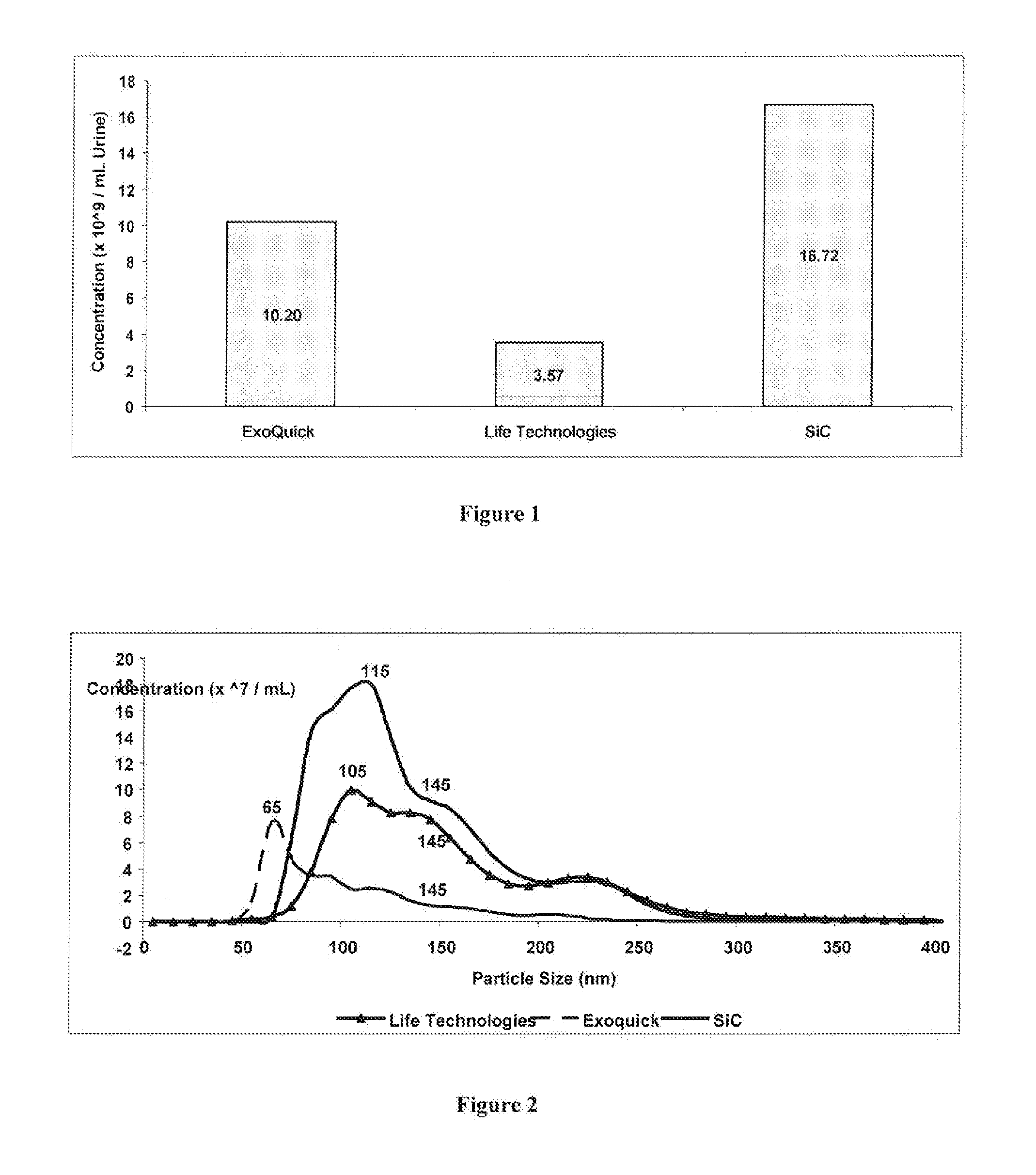
Comparing the Efficiency of SiC versus Commercially Available Kits in Isolating Extracellular Vesicles, including Exosomes, from Urine

[0063]SiC in a slurry format was tested for its ability to isolate exosomes from 5 mL of urine, and the performance of the SiC was compared to two different commercially available exosome precipitation reagents. The SiC slurry was prepared by adding 6.25 grams of SiC (G / S 2500) to 10 mL of 1× PBS (pH 7). Fifteen mL of urine was collected into a conical tube and centrifuged at 200× g (˜1,000 RPM) for 10 minutes to remove urine exfoliated cells and debris. The cell-free urine was decanted into a new tube and centrifuged at 1,800× g (˜3,000 RPM) for 10 minutes to remove any residual debris or bacterial cells. Five mL of the cell-free urine was transferred to a new tube and 400 μL of the SiC slurry was added to the urine samples. The pH was then adjusted to pH 8.5 using 1 M Tris Base (pH 11) to allow for the exosomes to bind to the SiC resin. The sample w...

example 2

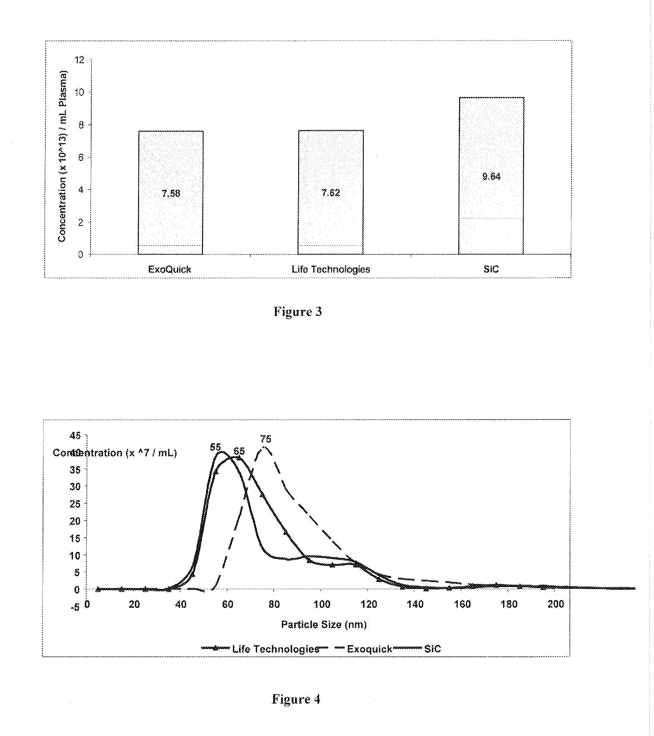
Comparing the Efficiency of SiC versus Commercially Available Kits in Isolating Extracellular Vesicles, including Exosomes, from Plasma

[0066]SiC in a slurry format was tested for its ability to isolate extracellular vesicles, including exosomes, from 1 mL of plasma, and the performance of the SiC was compared to two different commercially available exosome precipitation reagents. The SiC slurry was prepared by adding 6.25 grams of SiC (G / S 2500) to 10 mL of 1× PBS (pH 7). Blood was collected on EDTA or citrate using a standard blood collection tube. The collection tube was centrifuged at 2000 RPM for 15 minutes. The upper plasma fraction was collected and transferred to a fresh tube and centrifuged at 2000 RPM for 10 minutes. The clear supernatant containing the purified plasma was collected. One mL of the prepared plasma was transferred to a tube and 200 μL of the SiC slurry was added to the plasma samples. The pH was then adjusted to pH 8.5 using 1M Tris Base (pH 11) to allow for ...

example 3

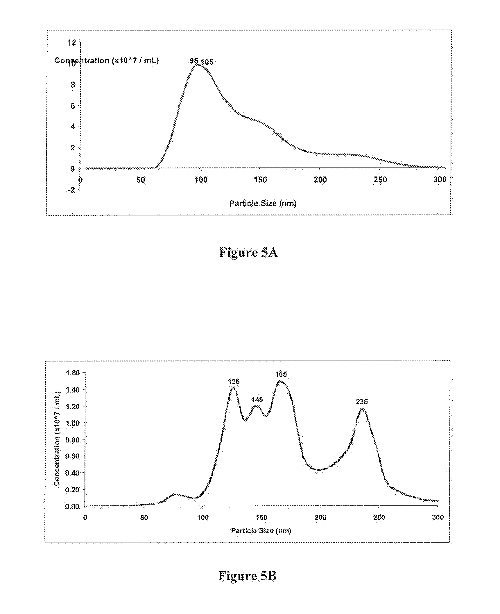
Comparing the Efficiency of SiC versus Ultracentrifugation in Isolating Extracellular Vesicles, including Exosomes, from Urine

[0069]SiC in a slurry format was tested for its ability to isolate exosomes from 10 mL of urine, and the performance of the SiC was compared to the traditional method of ultracentrifugation. The SiC slurry was prepared by adding 6.25 grams of SiC (G / S 2500) to 10 mL of 1× PBS (pH 7). Ten mL of urine was collected into a conical tube and centrifuged at 200× g (˜1,000 RPM) for 10 minutes to remove urine exfoliated cells and debris. The cell-free urine was decanted into a new tube and centrifuged at 1,800× g (˜3,000 RPM) for 10 minutes to remove any residual debris or bacterial cells. The remaining cell-free urine sample was transferred to a new tube and 400 μL of the SiC slurry was added to the urine samples. The pH was then adjusted to pH 8.5 using 1M Tris Base (pH 11) to allow for the exosomes to bind to the SiC resin. The sample was mixed by vortexing for 10...


PUM

PropertyMeasurementUnit
Diameteraaaaaaaaaa
Diameteraaaaaaaaaa

Abstract

Disclosed is a method for the isolation of extracellular vesicles, including exosomes, from a liquid sample, the method comprising the steps of: adjusting the pH of a liquid sample comprising extracellular vesicles to a preselected, binding pH; contacting the liquid sample with silicon carbide, wherein at the preselected, binding pH, the extracellular vesicles bind to the silicon carbide; and eluting the bound extracellular vesicles from the silicon carbide. The liquid samples can comprise bodily fluids. Further disclosed is a method for producing a liquid sample, substantially depleted of extracellular vesicles, including exosomes.

Description

CROSS-REFERENCE TO RELATED APPLICATION[0001]This application claims priority from U.S. provisional application No. 62 / 160,647, the entirety of which is hereby incorporated by reference.FIELD OF INVENTION[0002]The present invention relates to methods for the isolation of extracellular vesicles, including exosomes, from liquid samples, including bodily fluids. The present invention further relates to methods for the production of extracellular vesicle-depleted liquid samples, including serum.BACKGROUND[0003]There are a number of different types of vesicles released by cells, which are known as extracellular vesicles (EV). It is generally accepted that there are 3 main types of EVs which include apoptotic bodies (50-5,000 nm), microvesicles (100-1000 nm), and exosomes (40-150 nm). Apoptotic bodies are shed from dying cells whereas microvesicles are shed from the plasma membrane of viable cells. Exosomes are of endocrytic origin and are formed intracellularly by inward budding of the me...

Claims


Application Information

Patent Timeline
IPC IPC(8): C12N15/10
CPCC12N15/1006C12N15/10
Inventor HAJ-AHMAD, YOUSEF
Owner NORGEN BIOTEK CORP
Learn More