
Method of improved multiple-phase dynamic contrast-enhanced magnetic resonance imaging
a magnetic resonance imaging and contrast-enhanced technology, applied in the field of magnetic resonance imaging system, can solve the problems of degrading image quality, spatial displacement of images, and insufficient breath-holding capability of the subject of interes
- Summary
- Abstract
- Description
- Claims
- Application Information
AI Technical Summary
Benefits of technology
Problems solved by technology
Method used
Image

Examples
Embodiment Construction
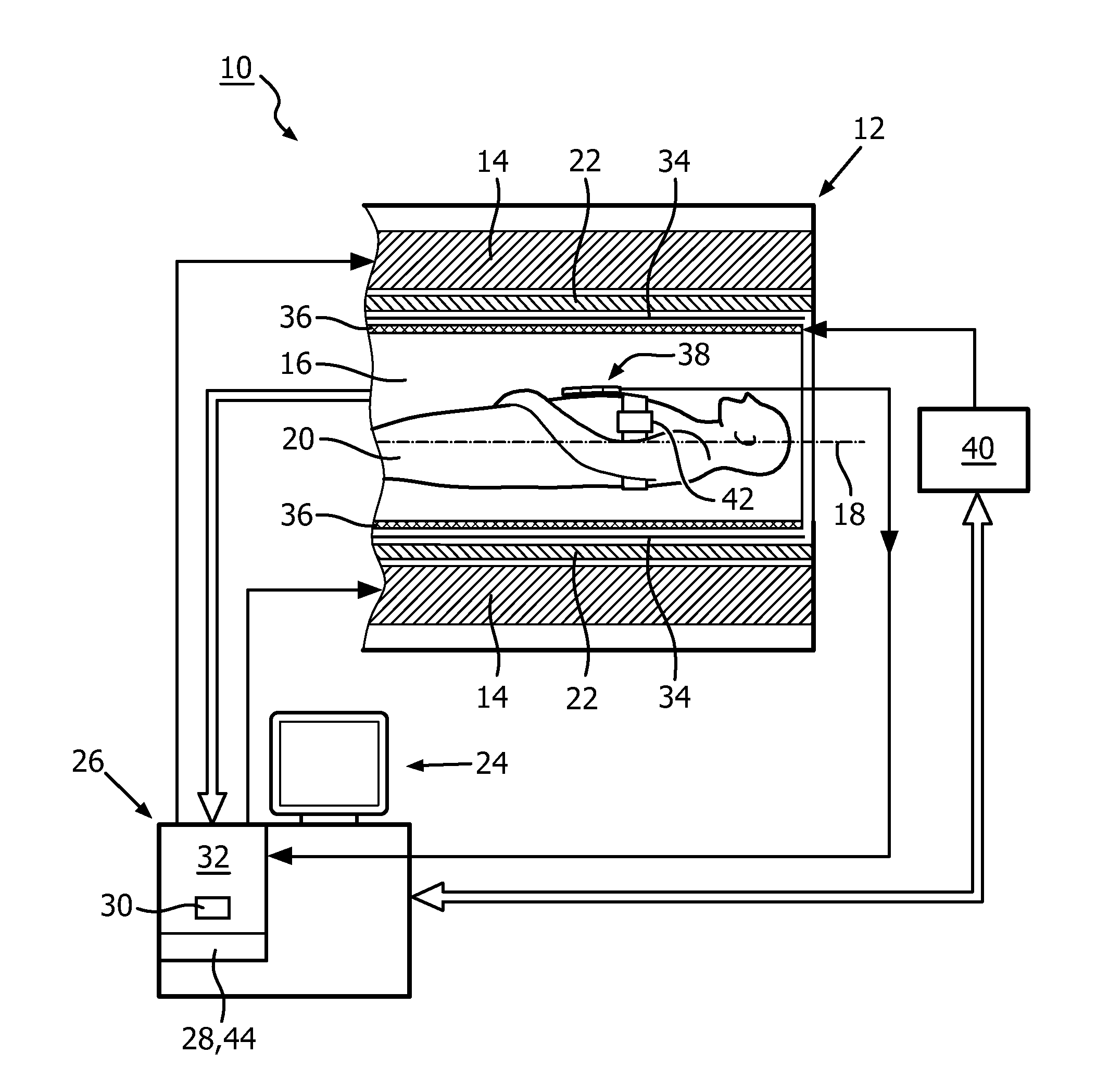
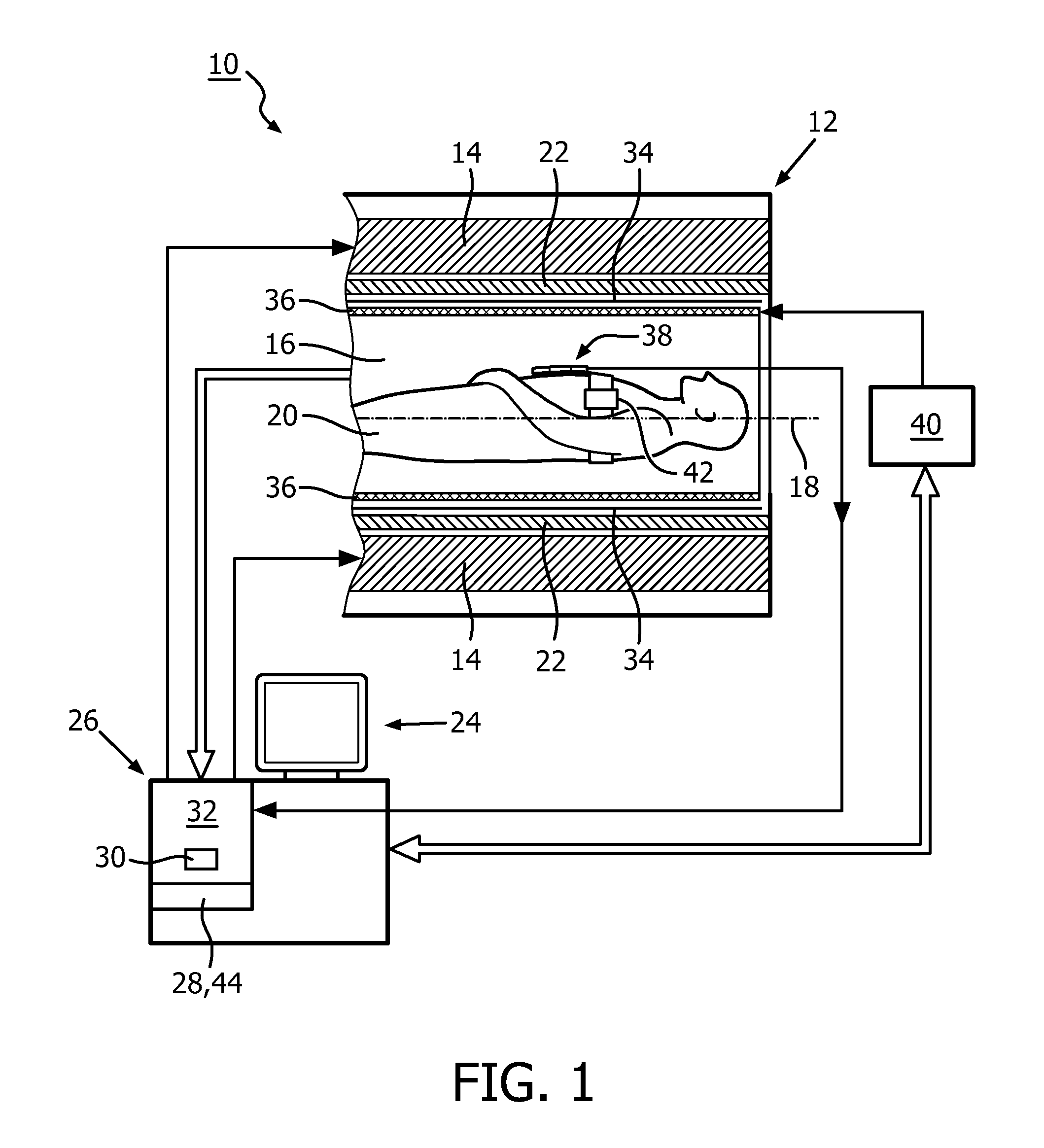
[0051]FIG. 1 shows a schematic illustration of a part of an embodiment of a magnetic resonance imaging system 10 configured for acquiring magnetic resonance images of at least a portion of a subject of interest 20, usually a patient. The magnetic resonance imaging system 10 comprises a scanning unit 12 having a main magnet 14. The main magnet 14 has a central bore that provides an examination space 16 around a center axis 18 for the subject of interest 20 to be positioned within, and is further provided for generating a static magnetic field B0 at least in the examination space 16. For clarity reasons, a customary table for supporting the subject of interest 20 has been omitted in FIG. 1. The static magnetic field B0 defines an axial direction of the examination space 16, aligned in parallel to the center axis 18. It is appreciated that the invention is also applicable to any other type of magnetic resonance imaging systems providing an examination region within a static magnetic fi...
PUM
 Abstract
Description
Claims
Application Information