Materials and methods for diagnosis of peri-implant bone and joint infections using prophenoloxidase pathway

a technology of prophenoloxidase and peri-implant bone, applied in the direction of material analysis, biochemistry apparatus and processes, instruments, etc., can solve the problems of inability to detect infection, difficult diagnosis of infection following surgery, and high risk of infection, and achieve the effect of high sensitiveness, convenient and accessibl

Inactive Publication Date: 2017-02-16
PARVIZI JAVAD +1
View PDF0 Cites 1 Cited by
  • Summary
  • Abstract
  • Description
  • Claims
  • Application Information

AI Technical Summary

Benefits of technology

[0006]This invention is based on the idea that a combination of at least two biomarkers, (one specific to the pathogenic microbe and the other specific to destruction of peri-implant tissues) that are detectable in the peripheral blood can be used for diagnosis of peri-implant infections. Any fluid or tissue samples drawn from a peri-implant environment (such as joint aspirate, tissue aspirate, tissue biopsy, etcetera) can be tested for the dual biomarker concept. Moreover, a blood or urine test that is highly sensitive for detecting specific structural microbial molecules and peri-implant tissue components can be convenient and accessible sources for diagnosing peri-implant infection of the bone and joints.

Problems solved by technology

There is a risk of infection following implantation of any prosthetic material or device in a physiologically sterile site or organ such as bone and joints.
Diagnosis of infection following surgery can be difficult and in many cases the infection cannot be detected at an early stage.
On the other hand, a sufficiently competent immune system will elicit an inflammatory reaction in response to the presence of the microbes which can also cause damage to host tissues as a secondary adverse effect.

Method used


Image

  • Materials and methods for diagnosis of peri-implant bone and joint infections using  prophenoloxidase pathway
  • Materials and methods for diagnosis of peri-implant bone and joint infections using  prophenoloxidase pathway
  • Materials and methods for diagnosis of peri-implant bone and joint infections using  prophenoloxidase pathway

Examples


example 1

Feasibility of PPO Test Using an In Vitro Model of Infected Synovial Fluid

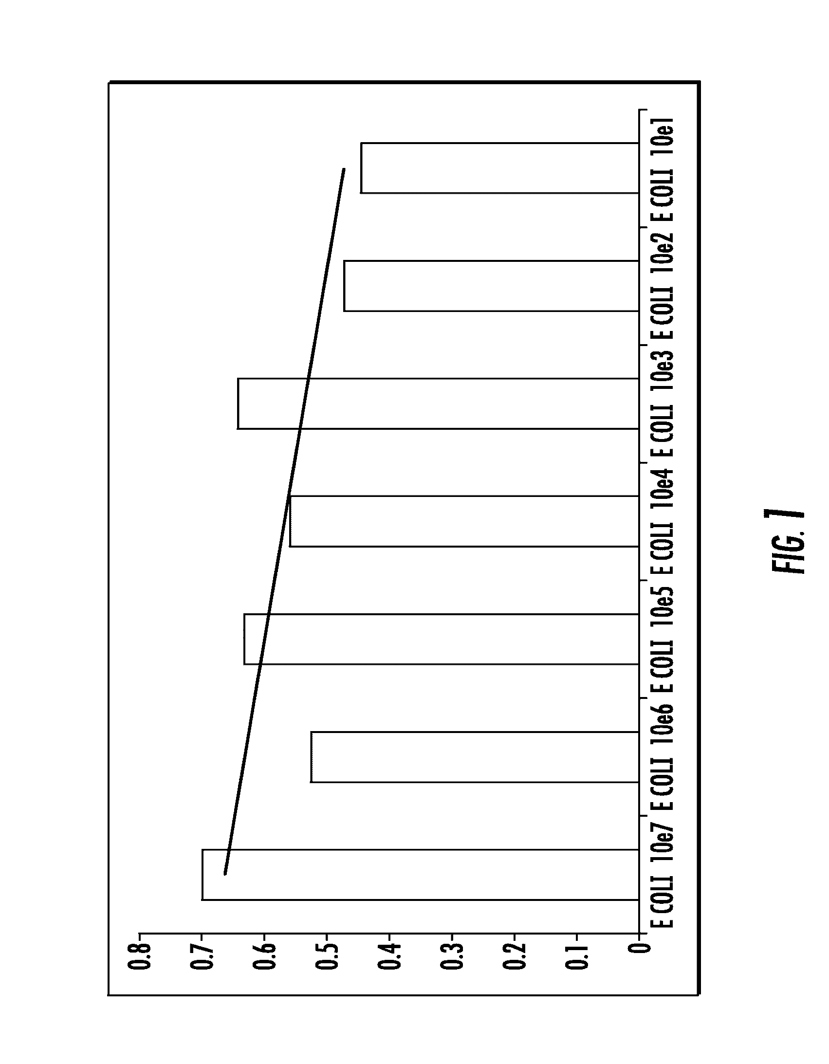
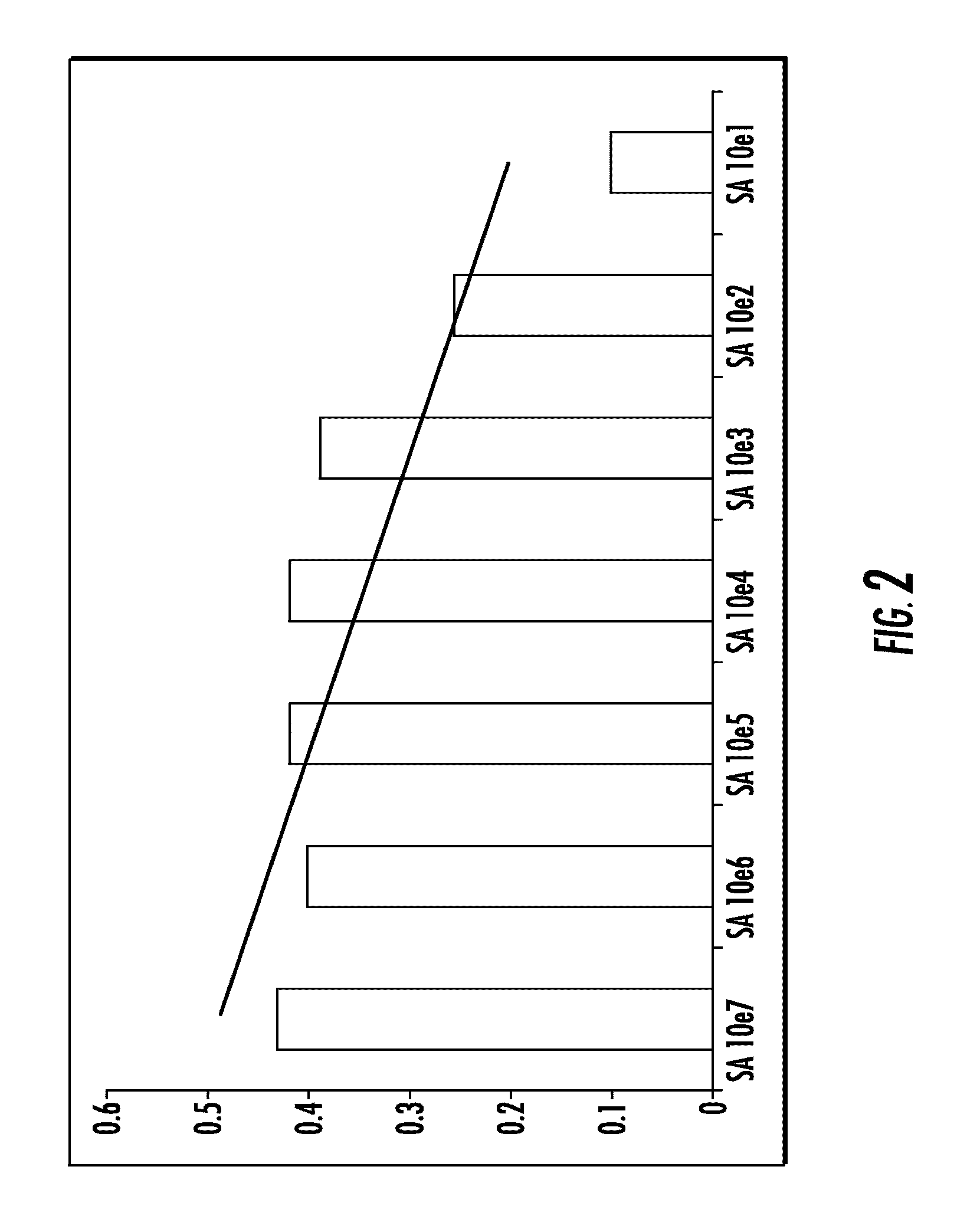
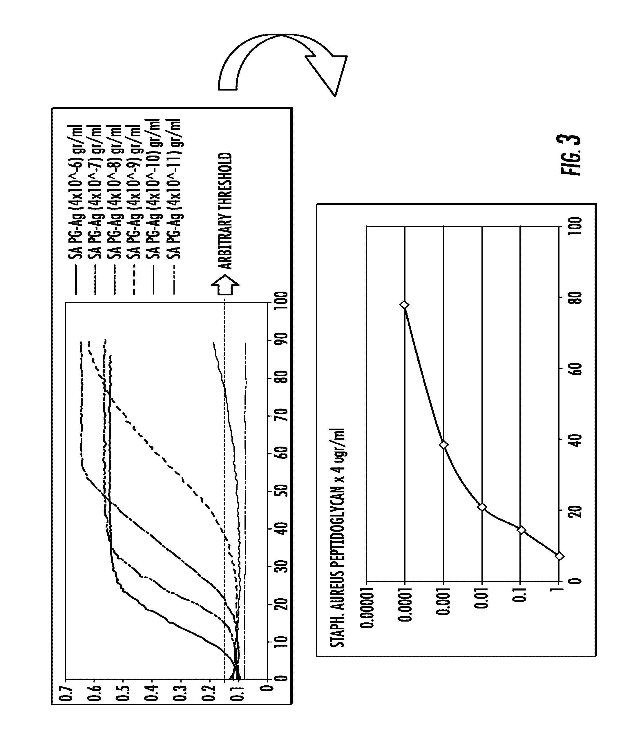
[0112]To assess feasibility and sensibility of the assay, in vitro models of infected synovial fluid samples were developed using Staphylococcus aureus (S. aureus) type ATCC 25923 and Escherchia coli (E. coli) type ATCC 25922. Non-infected synovial fluid samples were obtained from patients undergoing primary total hip or knee replacement in whom the procedure was performed to treat advanced joint osteoarthritis without any past history of infection in the joint. Bacteria were cultured in 10 ml of Trypticase Soy Broth (Becton Dickinson) in a shaker incubator (New Brunswick Scientific Inc, Edison, N.J.) overnight at 37° C. The following morning, bacteria were washed using sterile phosphate buffer saline (PBS) solution and centrifugation (14000 rpm for 5 min). Bacteria were added to sterile PBS solution. Serial 100 ul amounts of whirled bacterial solution were added to 900 ul of sterile PBS until turbidity meter ...

example 2

Experiments for Evaluating PPO Test on Clinical Samples from Non-Infected Synovial Fluid Samples

[0115]In another series of experiments synovial fluid samples from three different patients with non-infected knee joints including one with primary degenerative osteoarthritis, one with rheumatoid arthritis and one with inflamed Baker's cyst were tested with SLP reagent in undiluted state and dilution of 1:50 using water for injection. The water used for dilution was also tested. All negative controls were assayed using SLP reagent in the same microplate wells and using similar microplate reader setting as described earlier (FIG. 5).

example 3

Feasibility of the PPO Test for Real Clinical Samples

[0116]Following the preliminary experiments with in vitro models and negative controls, several experiments were conducted using samples from preoperative aspiration or intraoperative sampling of synovial fluid from prosthetic knee or hip joints that underwent revision surgery because of already confirmed or suspected prosthetic infection. SLP assay was performed for these clinical synovial fluid samples in several dilutions including 1:1 (undiluted), 1:10, 1:50, 1:100 and 1:200 using water for injection for dilution. In one clinical scenario, a 66-year old male patient (hereby named as patient #1) presented with progressive right knee pain. As past surgical history, patient #1 had undergone primary total knee replacement due to osteoarthritis two years ago. The patient had complications regarding his surgical wound healing that took several weeks to heal. However, he did not have any sign of deep infection. Shortly after recovery...


PUM

PropertyMeasurementUnit
colorimetric assayaaaaaaaaaa
light transmittanceaaaaaaaaaa
light absorbanceaaaaaaaaaa

Abstract

Diagnostic assays and related systems, test samples and methods are described for detection of infection following implantation of a prosthetic material or device on and / or into bones and joints. A combination of at least two biomarkers is used, one specific to the pathogenic microbe and the other specific to the destruction of peri-implant tissues. The biomarkers are detectable in the peripheral blood, urine, fluid or tissue samples drawn from a peri-implant environment. They can also be detected with a blood or urine test that is highly sensitive for detecting specific structural microbial molecules and peri-implant tissue components.

Description

BACKGROUND OF THE INVENTION[0001]Field of the Invention[0002]The present invention has to do with the detection of infection following implantation of a prosthetic material or device on and / or into bones and joints. The invention has both human and veterinary applications.[0003]The Related Art[0004]There is a risk of infection following implantation of any prosthetic material or device in a physiologically sterile site or organ such as bone and joints. Any kind of internal or external orthopedic implants, including but not limited to breast implants, prosthetic heart values, neurosurgical implants (such as vascular clips, shunt tubes and vascular stents) are just a few examples of prosthetic materials and devices that can be the subject of this invention. Diagnosis of infection following surgery can be difficult and in many cases the infection cannot be detected at an early stage. Early stage detection is important because it greatly enhances successful surgical outcomes.[0005]Each ...

Claims


Application Information

Patent Timeline
Patent Type & Authority Applications(United States)
IPC IPC(8): G01N33/68C12Q1/04C12Q1/26
CPCG01N33/6887G01N2800/245C12Q1/04C12Q1/26G01N33/569
Inventor PARVIZI, JAVADALIJANIPOUR, POUYA
Owner PARVIZI JAVAD
Why Eureka
Try Eureka