Method for detecting l-tyrosine by using graphene-modified graphite pencil electrode system

a graphene pencil and tyrosine technology, applied in the field of graphene (gr)modified graphite pencil electrode system and a method for detecting ltyrosine in a solution, can solve the problems of inability to make tyrosine, inability to process phenylalanine properly, and inability to detect ltyrosine,

Inactive Publication Date: 2017-03-23
KING FAHD UNIVERSITY OF PETROLEUM AND MINERALS
View PDF1 Cites 14 Cited by
  • Summary
  • Abstract
  • Description
  • Claims
  • Application Information

AI Technical Summary

Benefits of technology

[0022]In one or more embodiments, the oxidation peak potential of L-tyrosine in the solution ranges from about 0.5 V to about 1.0 V. In one or more embodiments, the sweeping the potential of the graphene-modified graphite pencil working electrode from the adsorption potential is to adsorb the L-tyrosine in the solution to the surface of the graphene-modified graphite pencil working electrode. In some embodiments, the adsorption time is about 60-120 seconds.
[0023]In one or more embodiments, the lowest detectable L-tyrosine concentration in the solution is about 0.08 μM.
[0024]In one or more embodiments, the solution further comprises at least one selected from the group consisting of phenylalanine, alanine, glucose, fructose, L-methionine, uric acid, ascorbic acid, Na+, K+, Li+, Ni2+, SO42−, and Cl−.
[0025]In one or more embodiments, the solution comprises at least one selected from the group consisting of whole blood, plasma, serum, saliva, sweat, urine, washes of tissues, extracts of tissues, amniotic fluid, placental fluid, a pharmaceutical composition, and a dietary composition.
[0026]In one or more embodiments, the method further comprises plotting the difference in current between the forward pulse current and the reverse pulse current during each square wave cycle, the difference in current represented by I, against the applied potential of the graphene-modified graphite pencil working electrode, the applied potential represented by E, to obtain a square wave voltammogram, and measuring the magnitudes of peak changes in I in the square wave voltammogram. In some embodiments, the magnitude of the peak change in I occurring at the L-tyrosine oxidation peak potential in the square wave voltammogram linearly correlates with the concentration of L-tyrosine ranging from about 1.3 μM to 80 μM in the solution. In some embodiments, the linear relationship between the magnitude of the peak change in I occurring at the L-tyrosine oxidation peak potential in the square wave voltammogram and the concentration of L-tyrosine in the solution is defined by a linear equation, and the slope of the linear equation is at least 1000 μA mM−1.
[0027]According to a third aspect, the present disclosure relates to a method of determining an L-tyrosine concentration in a solution. The method comprises contacting the solution with the graphene-modified graphite pencil electrode system of claim 1, and conducting square wave voltammetry to determine the L-tyrosine concentration in the solution. The conducting square wave voltammetry comprises (a) applying a pulsed potential to the graphene-modified graphite pencil working electrode while sweeping the potential of the graphene-modified graphite pencil working electrode from a potential that is less than an oxidation peak potential of L-tyrosine in the solution and defined as the adsorption potential positively to a potential that is at least the oxidation peak potential of L-tyrosine in the solution, and (b) recording the amount of a forward pulse current and a reverse pulse current during each square wave cycle. The square wave voltammetry includes conditions in which: the frequency is 20-30 Hz; the amplitude is 0.01-0.03 V; the voltage step is 2-10 mV; the adsorption potential is 0.0-0.4 V; the adsorption time is 80-100 seconds; and the pH value is 6-8.

Problems solved by technology

People who have this disorder can't process phenylalanine properly.
As a result, they can't make tyrosine.
Although all these methods display a good accuracy, most of them are tedious, require several preparatory steps prior to testing, are time-consuming, and require a skilled practitioner.
However, an electrochemical method for detecting L-tyrosine in a solution with an excellent detection limit, sensitivity, and linear range is urgently needed, since the existing electrochemical methods are not entirely satisfactory in all of the above three aspects.

Method used


Image

  • Method for detecting l-tyrosine by using graphene-modified graphite pencil electrode system
  • Method for detecting l-tyrosine by using graphene-modified graphite pencil electrode system
  • Method for detecting l-tyrosine by using graphene-modified graphite pencil electrode system

Examples


example 1

Preparation of the Graphene (GR)-Modified Graphite Pencil Electrode (GPE)

[0087]Graphene oxide (1 mg / mL, Sigma-Aldrich, USA) was dispersed in a 0.1 M acetate buffer at pH 4.8, and a uniform dispersion was obtained by sonicating the solution for 30 minutes. The graphene oxide solution was then transferred into a 3 mL glass cell. The three-electrode system comprising the graphite pencil working electrode (GPE), the platinum wire counter electrode (CHI 115, CH instrument Inc.), and the Ag / AgCl reference electrode (in 3 M KCl, CHI 111, CH instruments Inc.) was inserted through a Teflon cap into the 3 mL glass cell containing the graphene oxide solution. The graphene oxide was electrochemically reduced on the surface of the GPE under a cyclic sweeping potential from −1.4 V to 0.3 V applied at a scan rate of 10 mV / s for 4 cycles. All experiments were conducted at room temperature.

example 2

Morphological and Electrochemical Characterization of the Bare GPE and GR-Modified GPE

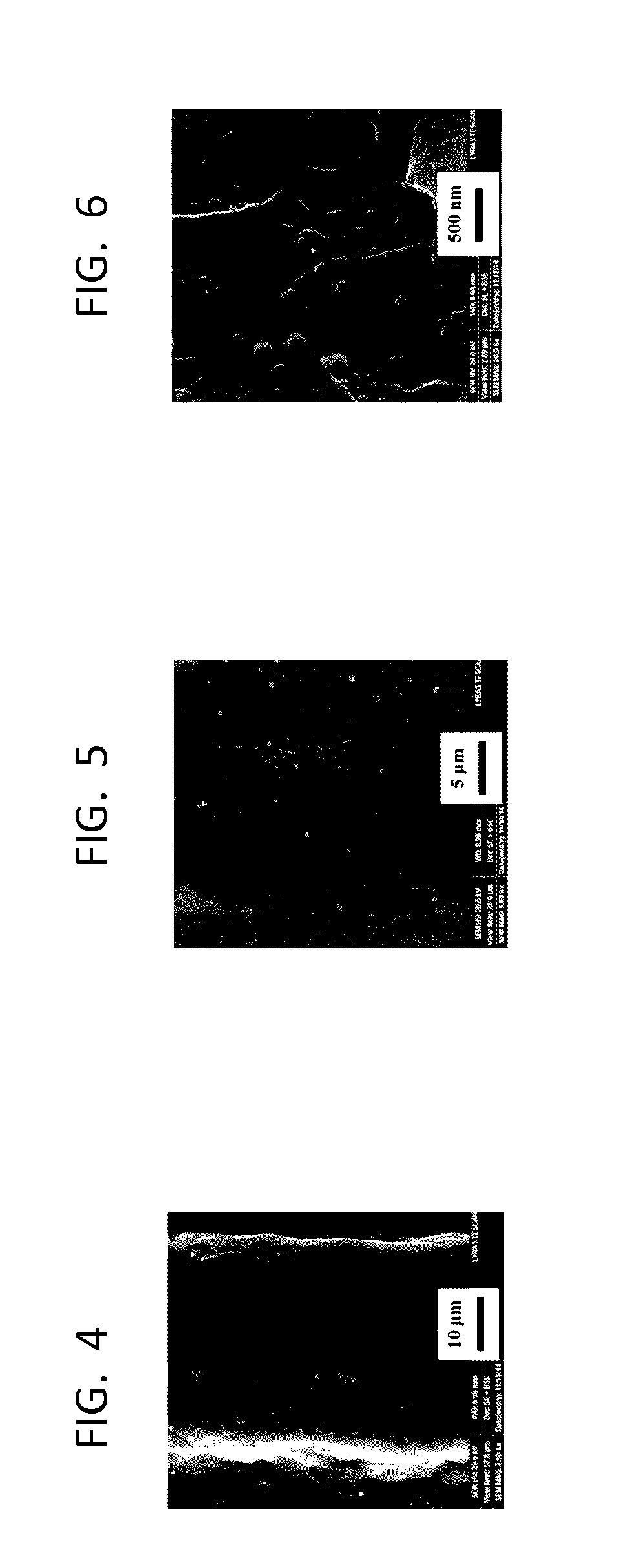
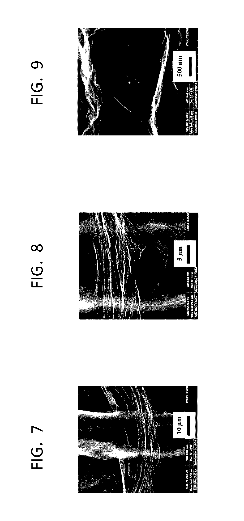
[0088]The surface morphologies of the bare GPE, i.e. the graphite pencil base electrode, and GR-modified GPEs were analyzed using field emission scanning electron microscopy (FE-SEM, TESCAN LYRA 3, Brno, Czech Republic). The FE-SEM images were collected from the bare and the GR-modified GPEs at three different magnifications, i.e. 10 μm in FIGS. 4 and 7, 5 μm in FIGS. 5 and 8, and 500 nm in FIGS. 6 and 9, to optically image the electrode surface. FIGS. 4-6 and FIGS. 7-9 show the bare GPE and GR-modified GPE, respectively. A comparison of FIG. 4 showing the bare GPE surface at 10 μm magnification with FIG. 7 showing the GR-modified GPE surface also at 10 μm magnification indicated the formation of the graphene layer on the GPE surface. FIG. 7 reveals that a small region uncovered by graphene was present on the GR-modified GPE surface, whereas the rest of the GR-modified GPE surface was covered by gr...

example 3

The Effect of pH on the Detection of L-Tyrosine in a Solution Using the Graphene-Modified Graphite Pencil Electrode System and Square Wave Voltammetry (SWV)

[0094]The effect of pH on the detection of L-tyrosine in a solution using the graphene-modified graphite pencil electrode system and square wave voltammetry was examined in a PBS solution (0.1 M) containing 50 μM of L-tyrosine at a pH ranging from 6.5 to 8.0 determined by a pH meter (Accumet® XL50), with the results presented in FIG. 18. pH significantly affected the oxidation peak current of L-tyrosine as well as the peak height corresponding to the magnitude of the peak change in I occurring at the oxidation peak potential of L-tyrosine in the square wave voltammograms shown in FIG. 18, where I is the difference in current between the two current measurements during each square wave cycle, one at the end of the forward pulse, and the other at the end of the reverse pulse. In addition to the oxidation peak current presented in F...


PUM

PropertyMeasurementUnit
thicknessaaaaaaaaaa
voltageaaaaaaaaaa
frequencyaaaaaaaaaa

Abstract

A graphene-modified graphite pencil electrode (GPE) system and a method for detecting L-tyrosine in a solution. The electrode system includes a graphene-modified graphite pencil working electrode comprising a graphite pencil base electrode and a layer of graphene comprising wrinkled graphene sheets on the surface of the graphite pencil base electrode, a counter electrode, and a reference electrode. The method comprises contacting the solution with the graphene-modified GPE system and conducting voltammetry, preferably square wave voltammetry, to detect the L-tyrosine concentration in the solution.

Description

BACKGROUND OF THE INVENTION[0001]Technical Field[0002]The present disclosure relates to a graphene (GR)-modified graphite pencil electrode (GPE) system and a method for detecting L-tyrosine in a solution. The graphene-modified graphite pencil electrode system includes a graphene-modified graphite pencil working electrode comprising a graphite pencil base electrode and a layer of graphene comprising wrinkled graphene sheets on the surface of the graphite pencil base electrode, a counter electrode, and a reference electrode. The method for detecting L-tyrosine in a solution comprises contacting the solution with the graphene-modified graphite pencil electrode system and conducting voltammetry to detect the L-tyrosine concentration in the solution.[0003]Description of the Related Art[0004]The “background” description provided herein is for the purpose of generally presenting the context of the disclosure. Work of the presently named inventors, to the extent it is described in this back...

Claims


Application Information

Patent Timeline
Patent Type & Authority Applications(United States)
IPC IPC(8): G01N27/327G01N27/48G01N27/30
CPCG01N27/3275G01N27/48G01N27/308G01N27/3278G01N33/68
Inventor KAWDE, ABDEL-NASSER METWALLY ALYBAIG, NADEEM
Owner KING FAHD UNIVERSITY OF PETROLEUM AND MINERALS
Why Eureka
Try Eureka