Genetic correction of myotonic dystrophy type 1

a myotonic dystrophy and gene therapy technology, applied in the field of genetic correction of myotonic dystrophy type 1, can solve the problems of affecting the use of dm1 primary cells, affecting the proliferation capacity of cells, and eventually dying out, and achieves limited proliferative capacity of primary dm1-derived cells, limited possible application for transplantation, and limited life-span.

Inactive Publication Date: 2017-03-30
THE GENERAL HOSPITAL CORP +1
View PDF6 Cites 24 Cited by
  • Summary
  • Abstract
  • Description
  • Claims
  • Application Information

AI Technical Summary

Benefits of technology

[0006]The present inventors have found that it is virtually impossible to expand DM1 patient derived precursor cells, such as myogenic cells (e.g. myoblasts or mesoangioblasts) or neuronal cells. While such cells may be isolated and to a certain extent propagated in vitro, after a few passages, the cells lose their proliferative capacity, and eventually die out. Without wishing to be bound by theory, it seems plausible that the toxic accumulation of defective DMPK mRNA may contribute to this effect. This undermines for instance the use of DM1 primary cells (such as muscle derived cells, be it myoblast or mesoangioblasts, or neuronal cells) for drug screening, disease investigation, and regenerative medicine. The present inventors have however, found that it is possible to derive induced pluripotent stem cells (iPS) from cells originating from subjects afflicted with myotonic dystrophy type 1 (DM1). More surprising, these DM1 patient derived iPS cells are capable of being differentiated into myogenic precursor cells, such as cardiomyogenic cells, myoblast- or mesoangioblast-like cells, or alternatively or neuronal or neurogenic cells. Importantly, and unexpectedly, both the DM1 derived iPS as well as the myogenic precursor cells derived therefrom display a DM1 specific phenotype, i.e. nuclear foci, which are characteristic for nuclear RNA accumulation associated with DM1. The iPS-derived precursors, such as the myogenic or neurogenic precursors, provide for an unprecedented opportunity to replicate both normal and pathologic human tissue, such as muscle or nerve tissue, formation in vitro, that impacts on disease investigation, drug development and regenerative medicine. The availability of such platform overcomes some of the bottlenecks intrinsic to the use of patient-derived primary cells, such as myoblasts or mesoangioblasts, but also neuronal or neurogenic cells, which have a much more restricted life-span. An additional important advantage of the DM1 derived iPS, in particular the nuclear foci phenotype, is that differentiation of cellular commitment, such as myogenic or neurogenic commitment, is not necessary if drug screening purposes can be done from the DM1 undifferentiated iPS cells. In addition, the present inventors have found that DM1-derived iPS, as well as the precursors derived therefrom, such as the myogenic or neurogenic precursors derived therefrom, are less fragile than primary DM1-derived cells, such as primary myogenic or neurogenic cells, and can advantageously be subjected to gene transfer, with minimal loss of cell death and proliferative capacity, in contrast to primary DM1-derived cells, which experience a vast amount of cell death and the surviving cells often even fail to grow anymore after transfection. In view of the limited proliferative capacity of primary DM1-derived cells, their possible application for transplantation is severely limited. In contrast, the DM1-derived iPS and their progeny, such as myogenic or neurogenic progeny, provide for a more robust cell system platform, which, in view of their continued proliferative capacity, may be readily used not only for a variety of in vitro assays, but also for transplantation, for instance after in vitro and in vivo gene correction.

Problems solved by technology

While such cells may be isolated and to a certain extent propagated in vitro, after a few passages, the cells lose their proliferative capacity, and eventually die out.
This undermines for instance the use of DM1 primary cells (such as muscle derived cells, be it myoblast or mesoangioblasts, or neuronal cells) for drug screening, disease investigation, and regenerative medicine.
In view of the limited proliferative capacity of primary DM1-derived cells, their possible application for transplantation is severely limited.

Method used


Image

  • Genetic correction of myotonic dystrophy type 1
  • Genetic correction of myotonic dystrophy type 1
  • Genetic correction of myotonic dystrophy type 1

Examples


example 1

n of iPS Cells from DM1 Patient Cells

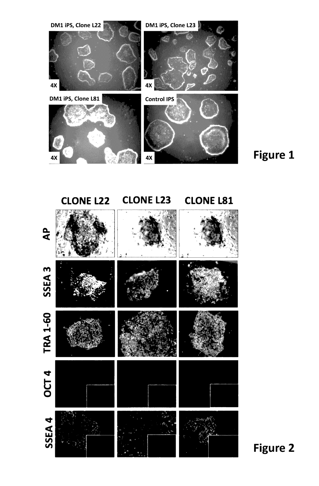
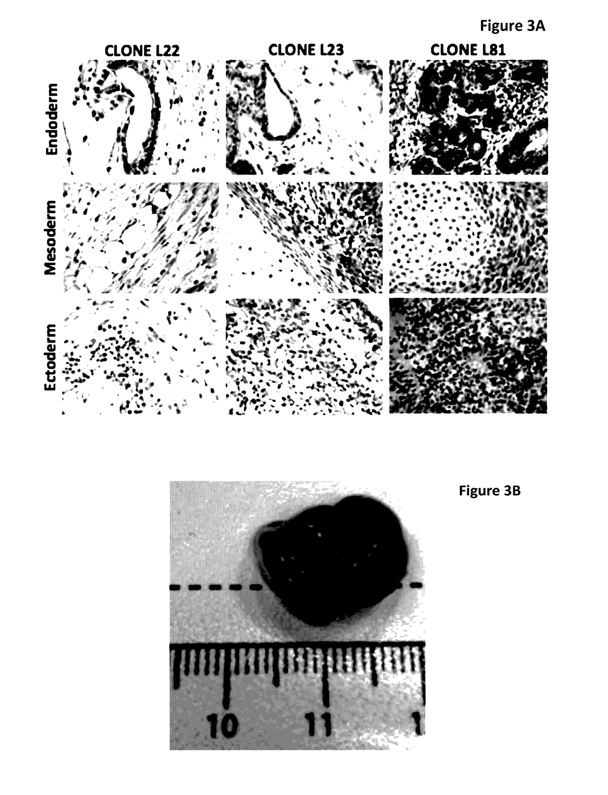
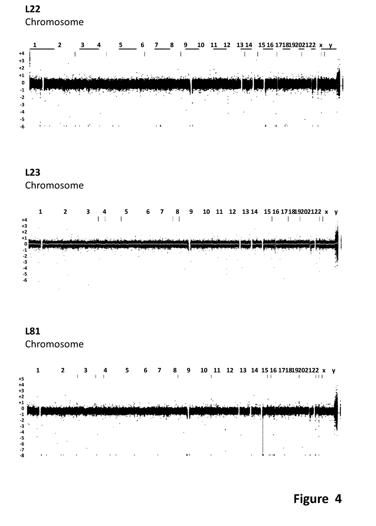
[0180]Human iPS cells were derived from myoblast cells from DM1 patients with extensive CTG repeats and from fibroblast of normal donors. For patient details see Table 2. The DM1 iPS cells were generated from skeletal myoblasts obtained from a 46 year old female suffering from DM1 with clinical manifestation of ptosis, slight atrophy, weakness of distal muscle, neckflexors and facial muscles; myotonia; cataract; ECG conduction abnormalities and daytime somnolence. This patient was selected because of the presence of expanded 1250 CTG repeats and because she manifested a severe DM1 phenotype. From a population of DM1 iPS colonies generated from this patient, three distinct iPS clones were selected and isolated for further expansion for subsequent characterizations experiments and for extensive characterization of the DM1 phenotype in differentiated and non-differentiated iPS cells. These expanded clones were designated as L22, L23 & L81, as shown ...

example 2

rdiomyogenic and Myogenic Differentiation of iPS from DM1 Patient Cells

[0198]Coaxed myogenic differentiation was induced in human iPS cells derived from cells of normal (healthy) subjects or DM1 patients, making it possible to study the effects of the mutated DMPK gene on myocardial differentiation and functionality.

[0199]The DM1 iPS clones were expanded and subsequently subjected to myogenic differentiation. For the myogenic differentiation, we follow a 5-step feeder-free differentiation procedure (Tedesco et al. (2012) Sci Transl Med 4, 140ra89); see also FIG. 7. The differentiation protocol was carried out using iPS cells cultured on inactivated feeder cells (inactivated MEF) as per the protocol published by Tedesco et al. (2012). We also used iPS cells cultured on feeder free condition to differentiate by the same protocol. For the clone DM1 L81, DM1 L23 and Control iPS we generated HIDEMs derived from iPS cells cultured both under feeder free and feeder (inactivated MEF) condit...

example 3

oci Staining Experiment on DM1 L81 iPS Corrected with dTALEN

[0203]In this experiment, iPS cells derived from DM1 patient were used and iPS cells derived from normal donor were used as control. In order to obtain genetic correction of the expanded CTG repeats in the patient cells, the dTALEN genome-editing tool was used. The dTALEN approach as a ‘molecular scissors’ in combination with a donor molecule was used to specifically target the DMPK gene. Two dTALENs were designed to bind at the appropriately spaced positions of the complementary DMPK strands in order for the FokI to generate a double-strand break in the DMPK gene. A donor molecule (or homology molecule) containing a puromycin expression cassette flanked by left and right homology arms was used for homologous recombination (FIG. 14). The donor molecule incorporated a polyA tail, which prevents transcription of downstream sequences (i.e. the CTG repeats). The donor molecule is as set forth in SEQ ID NO: 7. The left homology ...


PUM

PropertyMeasurementUnit
humidityaaaaaaaaaa
humidityaaaaaaaaaa
timeaaaaaaaaaa

Abstract

The invention relates to polynucleotides suitable for reducing or eliminating the expression of expanded repeat RNA (CUGexp) of the dystrophy myotonic-protein kinase (DMPK) gene in a cell of a DM-1 patient. The polynucleotides are a combination of a polynucleotide for a site specific nuclease targeting the dystrophy myotonic-protein kinase (DMPK) gene locus, and a donor polynucleotide having 5′ and 3′ regions which are homologous with the sequence of DMPK gene which flank the target site of the nuclease. The invention further relate to in vivo and in vitro methods to reduce or eliminate CTG repeats in the DMPK gene. The invention further relates to the medical use of polynucleotides and cells for treating DM-1 patient.

Description

FIELD OF THE INVENTION[0001]Provided herein are compositions and methods for the treatment of myotonic dystrophy type 1 (DM1). The present invention in particular relates to compositions and methods involving genetic correction of DM1-derived induced pluripotent stem cells (iPS) or its differentiated progeny, in particular muscle-like or myogenic cells, as well as in vitro and in vivo use of DM1-derived iPS or its differentiated progeny, in particular muscle-like or myogenic cells.BACKGROUND OF THE INVENTION[0002]Myotonic dystrophy type 1 (DM1) is a dominantly inherited neurodegenerative disorder that afflicts 1 in 8000 individuals. There is currently no cure or effective treatment available. DM1 is not caused by expression of a mutant protein, but instead is due to expression of a pathogenic RNA. Indeed, expression of the mutated DMPK gene gives rise to an expanded repeat RNA (CUGexp) that is directly toxic to cells by interfering with splicing, expression and function of multiple ...

Claims


Application Information

Patent Timeline
Patent Type & Authority Applications(United States)
IPC IPC(8): C12N5/074C12N9/12A61K35/34C12N15/90
CPCC12N5/0696C12N15/907A61K48/00A61K35/34C12N2800/80C12N9/1205C12N5/0658A61K2039/515A61K2039/53C12N2810/10C12N15/113C12N2310/10C12N2310/20
Inventor VANDENDRIESSCHE, THIERRYCHUAH, MARINEEJOUNG, J. KEITHFU, YANFANGREYON, DEEPAK
Owner THE GENERAL HOSPITAL CORP
Why Eureka
Try Eureka