Methods of linearly amplifying whole genome of a single cell

a single cell, whole genome technology, applied in the field of nucleic acid amplification, nucleic acid manipulation, genetics, medicine, etc., can solve the problems of inconvenient reporting of whole genome whole-genome sequencing, unsatisfactory whole-genome representation, and low coverage of methods used to date, so as to improve the accuracy of whole-genome sequencing and improve the accuracy of the method , the effect of high uniformity

Active Publication Date: 2017-06-29
BAYLOR COLLEGE OF MEDICINE
View PDF0 Cites 4 Cited by
  • Summary
  • Abstract
  • Description
  • Claims
  • Application Information

AI Technical Summary

Benefits of technology

[0007]Embodiments of the disclosure include methods for performing whole genome amplification for single cells (or optionally multiple cells) with high uniformity and high fidelity across the genome. Such methods allow accurate detection of copy number variations (CNVs) and single nucleotide variations (SNVs), for example, and optionally the presence of the CNVs or SNVs, for example, are detected by standard high throughput sequencing platforms or microarray or PCR-based genotyping following the methods of the invention.
[0008]Embodiments of methods of the disclosure provide a significant improvement of presently used methods in the art. For example, the SNV detection accuracy by linear amplification from original single cell DNA fragments covering the whole genome, i.e. creating independent copies of amplicons from single cell DNA templates, is greatly improved over methods in the art. Linear amplification allows the efficient filtering of amplification errors, therefore achieving the accuracy comparable to bulk sequencing for SNP detection, for example. Specific embodiments of methods provided herein introduce a “barcode” for each independently copied amplicon. This barcode allows the determination of the false positive rate at each loci across the genome.
[0009]Methods are provided herein that can be used to perform the amplification of DNA fragments extracted from one single cell, although in some cases the DNA may be extracted from more than one cell. Therefore, at least certain methods allow uniform whole genome amplification for one single cell, which allow accurate detection of copy number variations (CNVs) and single nucleotide variations (SNVs) by standard high throughput sequencing platforms.
[0010]Embodiments of the disclosure allow one to utilize an exact linear amplification method, which will provide accurate SNV detection with one and only one cell. No kindred cells are required to filter out the false positives, in particular cases. Embodiments of the disclosure are an improvement over the art by allowing the use of histological clinical samples as a source of cells for the nucleic acid to be amplified.
[0011]Methods are provided herein for removing the bias in amplification by separating the different DNA fragments into millions of small volume reaction compartments and conducting the amplification without the interference and competition between different amplicons. Thus, in particular embodiments a single amplicon is present in a reaction well for amplification, such as by PCR. In certain aspects, the volume of the reaction in the reaction compartment is femtoliter to nanoliter volumes.
[0012]Embodiments of methods provided herein significantly improve the uniformity by performing the amplification of each DNA fragment in separated reaction compartments in microfluidic device or separated reaction droplets created by emulsions, for example. The individual amplification is done in the tens of millions or more reaction compartments with as small as femtoliter volume, in some cases. In specific aspects, the amplification is saturated in each of the individual reactions and the nonlinear amplification (sequencing dependent PCR bias or the amplification bias of MDA) is minimized.

Problems solved by technology

However, the methods used to date are generally hampered by relatively low coverage.
Relying on exponential amplification with random primers, PCR-based whole-genome amplification methods introduce strong sequence dependent bias, and hence are not ideal for uniform representation of the whole genome.
For these reasons, whole-genome sequencing of single human cells, which allows the accurate detection of single nucleotide variants (SNVs), has not been convincingly reported.
To achieve whole-genome SNV calling for a single cell with the accuracy that is comparable to the bulk sequencing, the main technological barrier is the amplification errors produced and propagated in nonlinear amplification in the current state of the art.
Eventually in the sequencing data, these errors cannot be discriminated from true heterozygous variants in the single cell.
More importantly, this false positive rate cannot be reduced simply by increasing sequencing depth.
As a result, the amplification errors are independently generated and can be diluted among the linearly amplified products.

Method used


Image

  • Methods of linearly amplifying whole genome of a single cell
  • Methods of linearly amplifying whole genome of a single cell
  • Methods of linearly amplifying whole genome of a single cell

Examples


example 1

Isolation of Complete Single Cells from Tissue Sample

[0072]Histological tissue slides are prepared using commercial cryostat. Sections were prepared of 100-200 μm thickness of paraformaldehyde-fixed solid tissue. The cells of interest are cut from the section slide by laser microdissection microscopes (Leica, MMI etc.). The individual cells are dissociated from the microdissected tissue section using standard cell dissociation protocols. The complete single cells are collected into individual tubes. The individual cells are lysed in 3 to 5 μl Lysis buffer (30 mM Tris-Cl PH 7.8, 2 mM EDTA, 20 mM KCl, 0.2% Triton X-100, 12 μg / ml Qiagen Protease) is added to the side of the PCR tube and span down. The captured cell is then thermally lysed using the using following temperature schedule on PCR machine: 50° C. 2 hours, 75° C. 20 minutes, 80° C. 5 minutes.

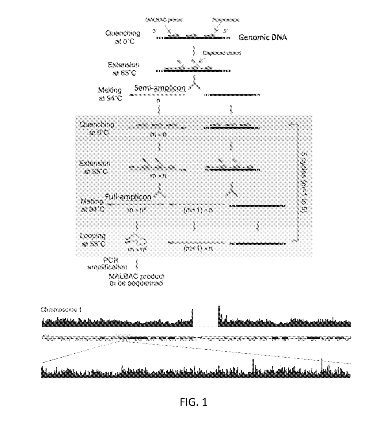
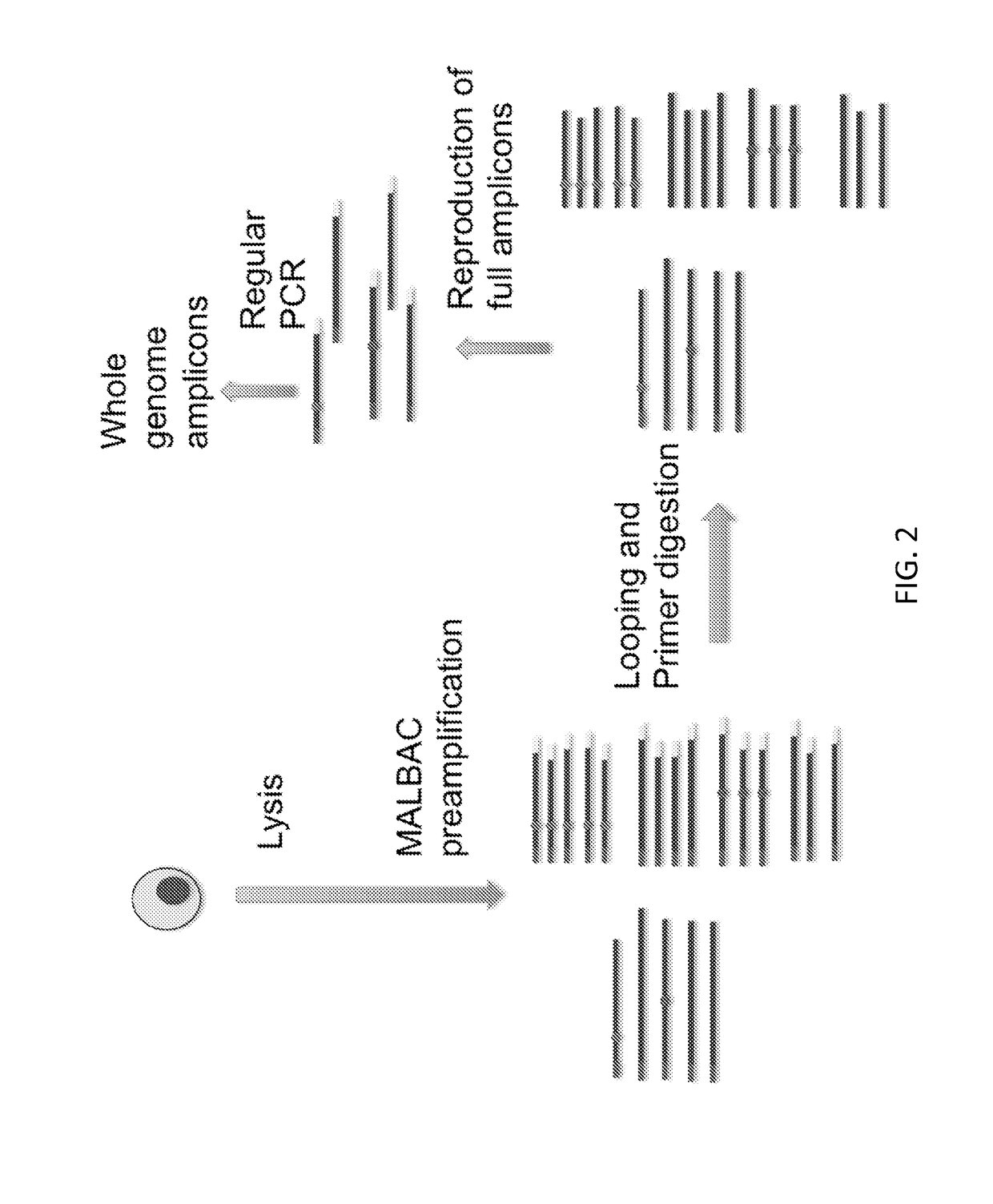
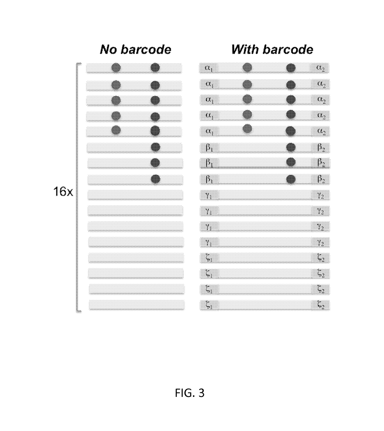
[0073]Multiple Cycles for Linear Production of Semi-amplicons (FIG. 4)

[0074]In the first round of amplification, a pair of quasi-degener...

example 2

Single Tube or Multiple Tube Amplification of Amplicons Using Standard Methods

[0109]The following reaction buffer is prepared and added to the PCR tube which is being maintained on ice.

[0110]3.0 μl ThermoPol Buffer (NEB)

[0111]1.0 μl dNTP (10 mM)

[0112]26 μl H2O (UV treated)

0.1 μl primer (100 μM)(5′-GTGAGTGATGGTTGAGGATGAGTG-3′; SEQ ID NO: 5)

[0113]The amplification buffer can be split into multiple tubes. The linearly amplified products will be split into each tube (FIG. 10). Amplification is performed with standard PCR procedures as follows to generate 1-2 μg of DNA material.

[0114]94° C.-20 seconds

[0115]58° C.-20 seconds

[0116]65° C.-1 minutes

[0117]72° C.-1 minutes

[0118]Repeat the above cycle 20×

[0119]72° C.-5 minutes

[0120]4° C.-∞

[0121]After the second round of amplification, DNA can be purified using a Qiagen column and stored for a next procedure to remove the primer end of the DNA amplicons.

[0122]The amplicon products can be used for whole genome or targeted (e.g., exome and any lis...

example 3

Whole Genome Single Fragment Amplification in Micro-Droplets / Wells

[0123]Linearly amplified amplicons from EXAMPLE 1 cover the whole genome of the single cell DNA and can be used as the starting materials for the following procedure (see FIG. 6).

[0124]Additional methods of amplification known to those of skill in the art can be used as follows.

[0125]The product is split into tens of millions of picoliter micro-droplets / wells or femtoliter micro-droplets / wells. Commercial available microfluidic based droplet emulsifiers or microfluidic devices with picoliter or femtoliter microwells can be used. By reaching to saturation of the amplification (limited either by available primers or dNTPs) in each reaction micro-droplet / well, the individual DNA fragments are amplified to similar level.

[0126]Additional methods of creating large scale of individual reactions known to those of skill in the art can be used as follows.

[0127]PCR reactions can be performed for amplifying single amplicons in ea...


PUM

PropertyMeasurementUnit
temperatureaaaaaaaaaa

Abstract

Embodiments of the disclosure encompass methods of amplifying nucleic acid from one or more cells using MALBAC (multiple annealing and looping-based amplification cycles) primers. In particular embodiments, the nucleic acid is amplified as amplicons in a linear manner. Specific embodiments include the removal or effective destruction of nonlinearly produced amplicons.

Description

[0001]This application claims priority to U.S. Provisional Patent Application Ser. No. 61 / 989,002, filed May 6, 2014, which is incorporated by reference herein in its entirety.TECHNICAL FIELD[0002]Embodiments of the disclosure include the fields of nucleic acid amplification, nucleic acid manipulation, genetics, medicine, and so forth. The field of embodiments of the disclosure concerns genome or transcriptome amplification, including from one or more cells, for example.BACKGROUND OF THE INVENTION[0003]Great interest in single cell heterogeneity has led to recent endeavors toward single cell genome sequencing with whole-genome amplification and robustness (Navin et al., 2011; Fan et al., 2011; Lao et al., 2008; Hou et al., 2012; Cheng et al., 2011; Telenius et al., 1992; Zhang et al., 2006; Zhang et al., 1992). However, the methods used to date are generally hampered by relatively low coverage. Polymerase chain reaction (PCR) has been a gold standard for DNA amplification of specifi...

Claims


Application Information

Patent Timeline
Patent Type & Authority Applications(United States)
IPC IPC(8): C12Q1/68
CPCC12Q1/6844C12Q1/6804C12Q1/6846C12Q1/6806C12Q2521/301C12Q2525/131C12Q2525/161C12Q2531/119C12Q2537/143
Inventor ZONG, CHENGHANGGUNDRY, MICHAELSHENG, KUANWEI
Owner BAYLOR COLLEGE OF MEDICINE
Why Eureka
Try Eureka