
Method for Treating Aortic Arch Diseases

a technology for aortic arch and disease, applied in the field of aortic arch disease treatment, can solve the problems of reducing the time of operation and difficulty of operation, serious threats to human health, and difficulty in performing an operation at the aortic arch

Active Publication Date: 2017-07-06
LIFETECH SCIENTIFIC (SHENZHEN) CO LTD
View PDF2 Cites 7 Cited by
  • Summary
  • Abstract
  • Description
  • Claims
  • Application Information

AI Technical Summary

Benefits of technology

The present invention provides a method for treating aortic arch disease by reducing the flow of cerebral perfusion while forming an incision. After the incision, a stent is released and expanded in the aorta and arch branch vessel. Before anastomosis, a blocking device is used to prevent blood flow through the intraoperative stent and incision, reducing the circulatory arrest time and ischemic time of visceral organs and lower limbs of the patient.

Problems solved by technology

Aortic diseases, mainly including aortic dissection and aortic aneurysm, are very dangerous diseases, with a mortality higher than 50% after the diseases come on within 48 hours, and a mortality higher than 85% after the diseases come on within two weeks, so the diseases pose serious threats to the human health.
At the aortic arch, a human body generally has three arch branch vessels for supplying blood to the brain, and it is required to establish a blood extracorporeal circulation during a surgery to maintain the blood supply to the brain, so it is very difficult to perform an operation at the aortic arch.
Although the hydrid method reduces both the time of operation and the difficulty of the operation in comparison to a pure thoracotomy, it is still required to recover the low body perfusion (e.g., femoral artery perfusion) after the vessel is completely anastomosed and to recover the flow of a cerebral perfusion through the right axillary artery to a normal flow.
Therefore, the time of circulatory arrest and the time of the cerebral perfusion time which is less than the normal perfusion amount are still long, so the surgical risk is relatively high.

Method used


Image

  • Method for Treating Aortic Arch Diseases
  • Method for Treating Aortic Arch Diseases
  • Method for Treating Aortic Arch Diseases

Examples


Embodiment Construction

[0036]To understand the technical features, objectives and effects of the present invention more clearly, specific implementations of the present invention will be described in detail with reference to the accompanying drawings. For convenience, in the following description, the distal direction and the proximal direction are defined by a common method in this art. That is, the blood flows from a proximal end to a distal end.

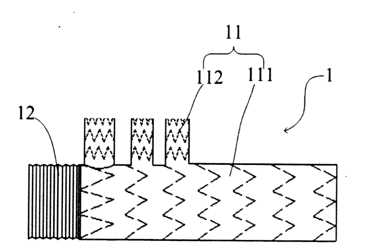
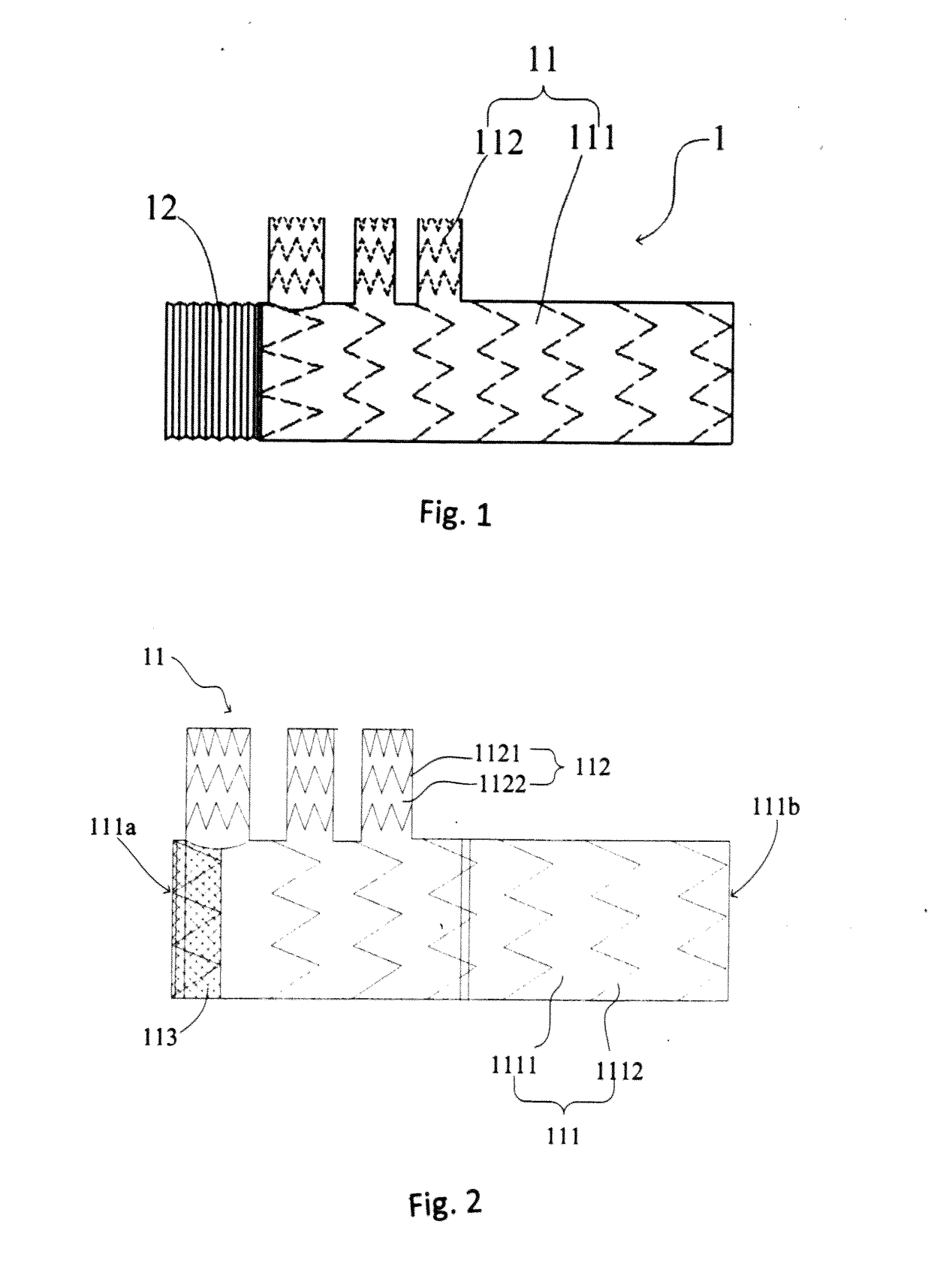
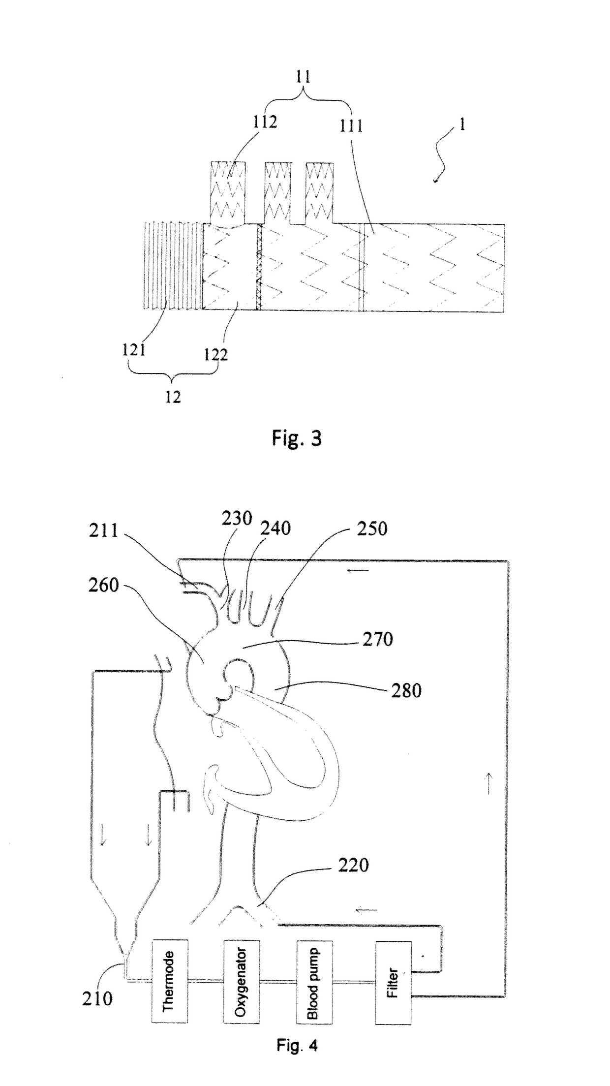
[0037]FIGS. 1-3 show an exemplary intraoperative stent 1 available for a lesion region of an aortic arch. It should be understood that the intraoperative stent 1 is merely illustrative and is not intended to limit the scope of the present invention, as those skilled in the art may utilize the illustrated intraoperative stent 1 to implement the method according to the embodiments of the present invention to treat diseases (e.g., Stanford A aortic dissection) at an aortic arch or utilize any other suitable stent to implement the method according to the embodiments...


PUM


Abstract

In a method for treating an aortic arch disease, after an intraoperative stent is expanded in an aorta and at least one arch branch vessel, and before the vessel is exactly anastomosed with an incision, by blocking a thoracic aorta between the incision and an arch branch vessel closest to the incision, the intraoperative stent in the thoracic aorta is also blocked, so that blood is prevented from flowing from the incision, and a proximal orifice of the intraoperative stent, and the low body perfusion may be recovered in advance, thereby reducing the circulatory arrest time of the low body perfusion and reducing the time of ischemia of visceral organs and lower limbs of a patient. Meanwhile, the flow of a cerebral perfusion through the right axillary artery may be recovered to a normal flow in advance, and the time of ischemia of the brain of the patient is reduced.

Description

TECHNICAL FIELD[0001]The present invention relates to a method for treating aortic arch diseases, and in particular to a method for treating aortic arch diseases by using stents in conjunction with surgical methods and endovascular techniques.BACKGROUND OF THE PRESENT INVENTION[0002]Aortic diseases, mainly including aortic dissection and aortic aneurysm, are very dangerous diseases, with a mortality higher than 50% after the diseases come on within 48 hours, and a mortality higher than 85% after the diseases come on within two weeks, so the diseases pose serious threats to the human health. With the arrival of the aging trend in China, the mortality rates will constantly rise.[0003]Stanford A aortic dissection is one common disease among the aortic arch diseases and occupies about 70% of the whole dissection diseases. At the aortic arch, a human body generally has three arch branch vessels for supplying blood to the brain, and it is required to establish a blood extracorporeal circu...

Claims


Application Information

Patent Timeline
Patent Type & Authority Applications(United States)
IPC IPC(8): A61B17/11A61F2/856
CPCA61B17/11A61B2017/1107A61F2/856A61B2017/1132A61B2017/12004A61F2/07A61F2002/061A61F2002/075
Inventor XU, ZHIYUNZHANG, XIANGMANHAN, LINZHANG, DEYUANXIAO, BENHAO
Owner LIFETECH SCIENTIFIC (SHENZHEN) CO LTD
Learn More