Methods for Mass Spectrometric Based Characterization of Biological Molecules

a mass spectrometry and biological molecule technology, applied in the field of mass spectrometry, can solve the problems of many proteins that are difficult to isolate and purify, the amount of information that can be obtained when reducing intact proteins to their constituent peptides, and the top-down analysis, etc., to achieve enhanced spectral information and simplify spectral analysis

Active Publication Date: 2017-07-20
THERMO FINNIGAN
View PDF8 Cites 4 Cited by
  • Summary
  • Abstract
  • Description
  • Claims
  • Application Information

AI Technical Summary

Benefits of technology

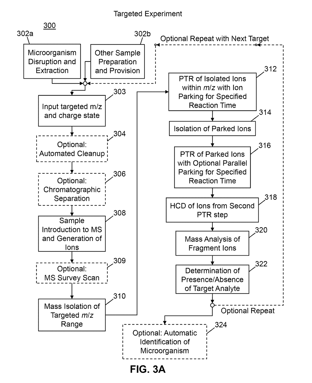
[0016]The present disclosure teaches mass spectrometric methods that employ proton transfer reactions (PTR) in combination with ion parking techniques followed by higher-collisional energy dissociation (HCD) so at to yield broad m / z-band activation and fragmentation. The intended results provide simplified spectral analysis with enhanced spectral information. The present disclosure teaches the methods of targeted, data-dependent and data-independent mass spectral analysis using the PTR and HCD techniques in combination.

Problems solved by technology

Although this technique is routine in many laboratories, there are limitations as to the amount of information that can be obtained when reducing intact proteins to their constituent peptides.
However, top-down analysis has a disadvantage when compared to a bottom-up analysis in that many proteins can be difficult to isolate and purify.
The process of analyzing intact proteins in cell lysates by mass spectrometry (MS) is associated with a number of difficulties.
Firstly, electrospray ionization (ESI) of protein mixtures from cell lysates can generate extremely complex mass spectra due to the presence of multiple proteins, each comprising its own charge state envelope, where each charge state envelope is the collection of mass spectral lines corresponding to plural charge states, and where each charge state correlates directly with the number of positively charged protons that are adducted to an otherwise charge-free molecule.
Commonly used techniques in MS are often insufficient for simplifying these spectra because of the inherent peak overlapping as well as the inherent wide range of magnitudes of MS lines of ionized constituents, where such constituents may range from uninteresting small molecules to interfering biomolecules to the proteins of interest, themselves.
Isolation of a specified charge state of a protein within such complex spectra does not typically alleviate the burden of multiple protein peaks overlapping, since the isolation of ions of a particular protein charge state will generally result in co-isolation of one or more additional ions.
This co-isolation makes it a challenge not only to dissociate the protein in an attempt to identify it based on the fragments produced, but also to accurately determine the intact mass and sequence coverage of that protein.
Unlike mixtures of proteolytic peptides typically analyzed in bottom-up experiments, intact proteins mixtures contain a wide range of molecular weights, isoelectric points, hydrophobicities, and other physiochemical properties that make it challenging to analyze these mixtures via any single separation technique in a comprehensive manner.
Liquid chromatography tends to require significant amounts of time per sample to separate individual proteins, although it is still common to have two or more proteins co-elute.
The IMS method also suffers from ionization competition, requires extensive optimization and typically involves dynamic conditions to observe a full mass spectrum containing all charge states.
Another difficulty associated with the mass spectrometric analysis of proteins in cell lysates by (MS) is that the fragmentation behavior for each charge state of a protein is generally unknown prior to the dissociation event.
In particular, ions comprising some charge states can dissociate well while ions comprising other charge states may dissociate poorly.
Isolation and dissociation of ions of a particular charge state therefore does not guarantee efficient dissociation or dissociation into a set of diagnostic fragments.
A third challenge associated with intact protein analysis is the wide distribution of charge states produced for high molecular weight proteins typically in excess of 50 kDa.
The ability to produce ample sequence coverage for protein identification can therefore be difficult with a single tandem mass spectrum.

Method used


Image

  • Methods for Mass Spectrometric Based Characterization of Biological Molecules
  • Methods for Mass Spectrometric Based Characterization of Biological Molecules
  • Methods for Mass Spectrometric Based Characterization of Biological Molecules

Examples


example 1

tral Analysis of Ubiquitin Mixture

[0104]FIGS. 4A, 4B, 4C and 4D illustrate application of the combined techniques of PTR and HCD to analysis of a solution of ubiquitin containing abundant polymeric contaminants. FIG. 4A is a graphic depiction of a survey scan (MS1 scan) of the sample. Peaks which may be recognized as relating to the ubiquitin compound are labeled with their charge states and m / z positions. Most or all of the remaining peaks, a few select ones of which are labeled with their m / z positions, correspond to polymeric contaminants. In order to simplify the spectrum, a sub-population of ions within an isolation window of width 2 Th around the expected position of the ubiquitin +13-charge-state peak were isolated (mass spectrum of isolate shown inFIG. 4B) and the isolated original ions were exposed to PTR reagent, without ion parking, for 50 ms. A mass spectrum of the first-generation product ions produced by this PTR step is illustrated in FIG. 4C. After the PTR step, seve...

example 2

tral Analysis of Two-Protein Mixture

[0106]FIGS. 5A, 5B, 5C, 5D, 5E and 5F illustrate application of the combined techniques of PTR and HCD to analysis of a mixture of carbonic anhydrase and myoglobin. FIG. 5A is an MS1 survey spectrum of the mixture of carbonic anhydrase and myoglobin in solution. The tall peak at 616 Th is the signature of the heme moiety that is otherwise non-covalently attached to myoglobin. The sample preparation results in separation of the heme (holo-myoglobin becoming apo-myoglobin). The heme peak is commonly observed. In order to simplify the spectrum, a sub-population of ions within an isolation window of width 2 Th around the expected position of the myoglobin +21-charge-state peak were isolated (mass spectrum of isolate shown in FIG. 5B) and the isolated original ions were exposed to PTR reagent, without ion parking, for 30 ms. A mass spectrum of the first-generation product ions produced by this PTR step is illustrated in FIG. 5C. The general form of the...

example 3

tral Analysis of Myoglobin in Bacterial Lysate

[0109]FIGS. 6A, 6B, 6C, 6D, 6E, 6F and 6G illustrate application of the combined techniques of PTR and HCD to analysis of myoglobin within a bacterial lysate. The sample was prepared by adding a small amount of myoglobin into a lysate of the bacterium E. coli. FIG. 6A shows an MS1 survey scan of the sample. Peaks attributable to myoglobin (not labeled) in the mass spectrum of FIG. 6A are nearly invisible in the midst of many peaks attributable to bacterium-derived compounds. During mass spectral analysis of this sample, a sub-population of the original ions were isolated within an isolation window of width 2 Th centered at an m / z value of 808 Th, which is the expected position of the ion species comprising the multi-protonated myoglobin molecule of charge state +21. FIG. 6B shows a mass spectrum of the isolated ions.

[0110]FIG. 6C illustrates a test mass spectrum that was obtained after reacting a portion of the isolated ions depicted FIG...


PUM

PropertyMeasurementUnit
molecular weightaaaaaaaaaa
volumesaaaaaaaaaa
molecular weightaaaaaaaaaa

Abstract

Applications of ion-ion reaction chemistry are disclosed in which proton transfer reactions (PTR) combined with higher-collision-energy dissociation (HCD) are used to (1) simplify complex mixture analysis of samples introduced into a mass spectrometer, and (2) improve resolution and sensitivity for the analysis of large proteins in excess of 50 kDa by removing charge, reducing the collisional cross section, and, in several cases, enhancing the sequence coverage obtained.

Description

CROSS REFERENCE TO RELATED APPLICATIONS[0001]This application claims, under 35 U.S.C. §119(e), priority to and the benefit of the filing date of commonly-assigned, co-pending U.S. Provisional Application No. 62 / 278,942, filed on Jan. 14, 2016 and titled “Methods for Mass Spectrometric Based Characterization of Biological Molecules”, the disclosure of which is hereby incorporated by reference in its entirety. The subject matter of this application is also related to commonly-assigned and co-pending International Application No. PCT / US2015 / 040914, filed on Jul. 17, 2015 and titled “Methods for Mass Spectrometry of Mixtures of Proteins of Polypeptides Using Proton Transfer Reaction”, the disclosure of which is hereby incorporated by reference in its entirety.TECHNICAL FIELD[0002]The present invention relates to mass spectrometry and, more particularly, relates to methods for purifying and analyzing complex mixtures of proteins or polypeptides by mass spectrometry of proton transfer rea...

Claims


Application Information

Patent Timeline
Patent Type & Authority Applications(United States)
IPC IPC(8): G01N33/68H01J49/16H01J49/00
CPCG01N33/6848H01J49/0072H01J49/165H01J49/0031G01N27/622G01N27/68G01N30/06
Inventor MCGEE, WILLIAM M.CARDASIS, HELENE L.
Owner THERMO FINNIGAN
Why Eureka
Try Eureka