Subcellular Western Blotting of Single Cells
a technology of western blotting and single cells, applied in the field of single cell subcellular western blotting, can solve the problems of not being suitable for conventional methods and challenging proteomic analysis of rare cell populations
- Summary
- Abstract
- Description
- Claims
- Application Information
AI Technical Summary
Benefits of technology
Problems solved by technology
Method used
Image
Examples
example 1
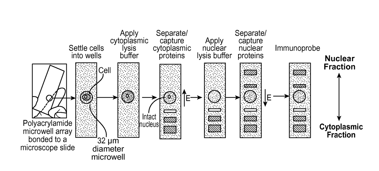
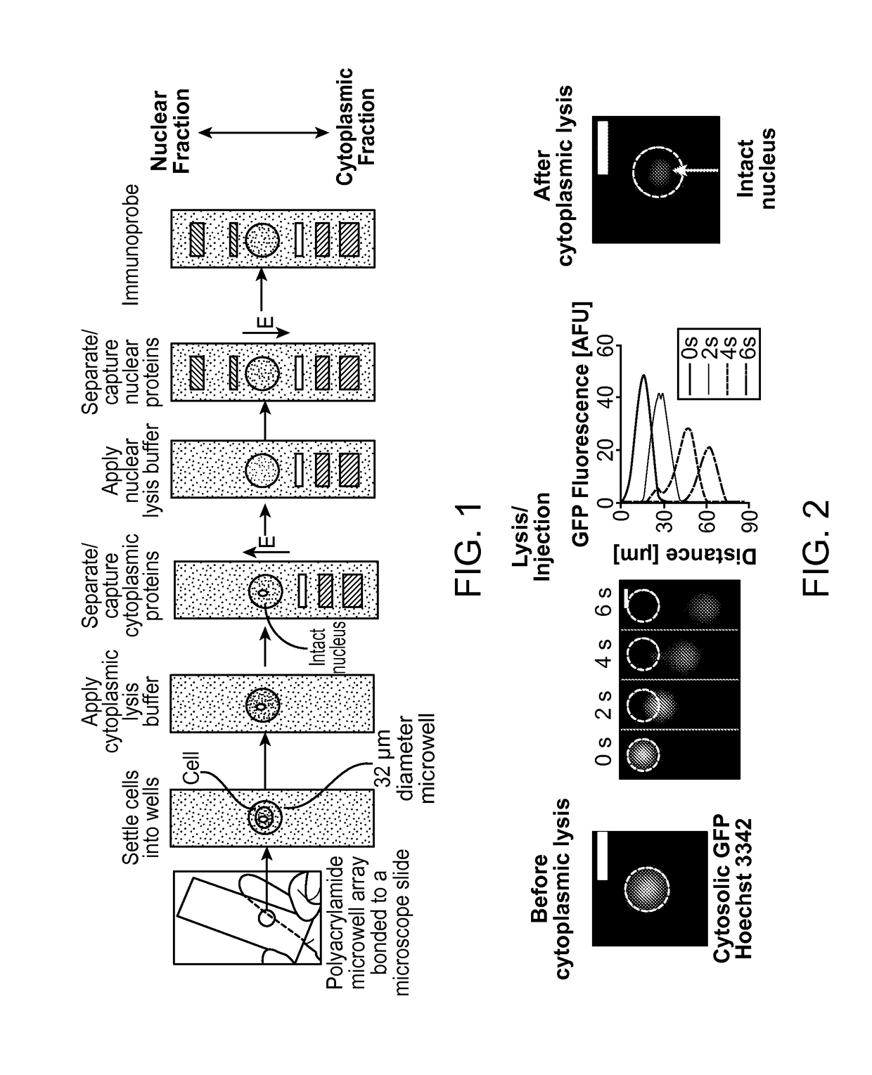
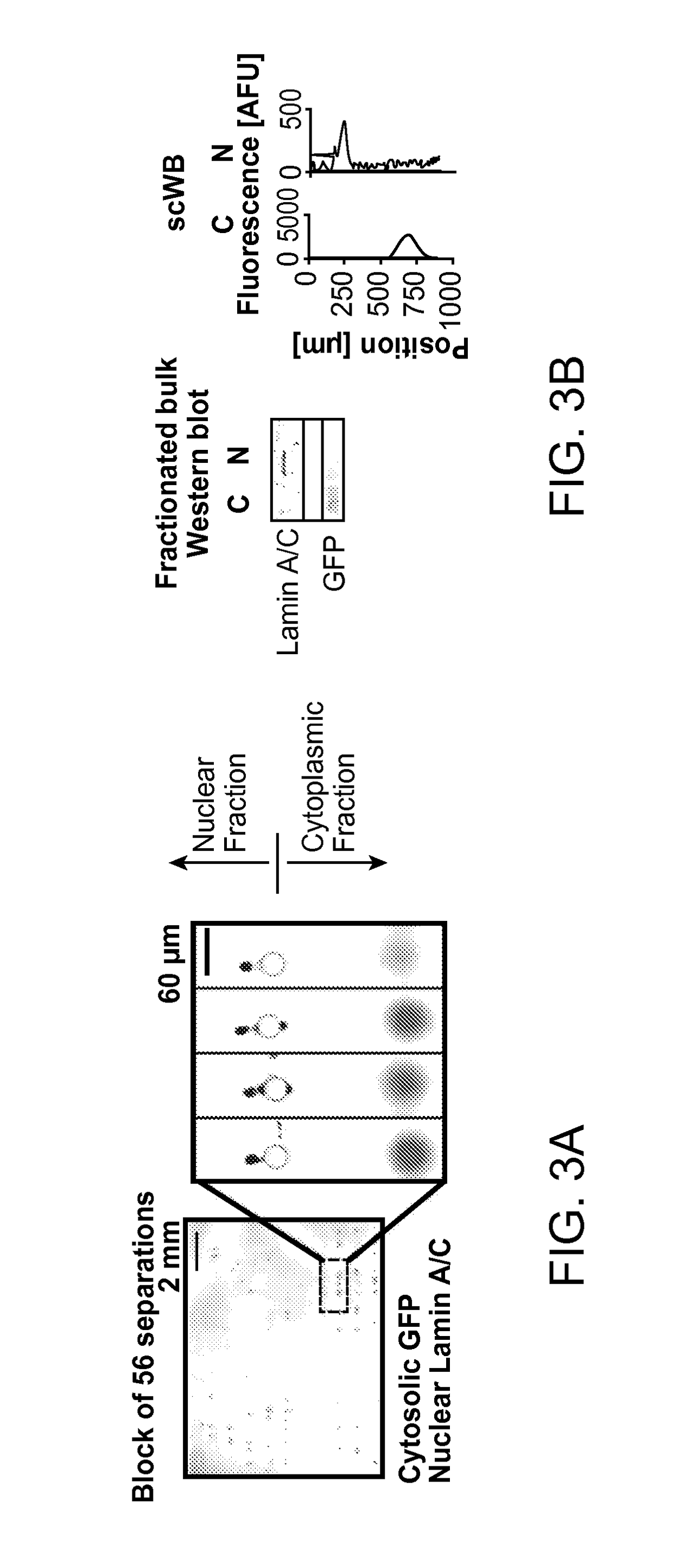
[0143]Embodiments included a single-cell Western blot for analyses of sub-cellular compartments (e.g., cytosol, nucleus). Protein sub-cellular localization is intrinsically linked to function, with aberrant localization linked to numerous diseases. Existing sub-cellular protein analyses are slow, offer low multiplexing (˜5 proteins), and are not quantitative. Single-cell Western blotting arrays (scWesterns) included a capacity to (1) differentially lyse specific sub-cellular compartments in single cells, (2) perform single-cell Western blotting solely on proteins in those targeted compartments (5+ proteins per compartment), and (3) serially assay multiple sub-cellular compartments in the same single-cells. The specificity of scWesterns with sub-cellular resolution was used to study of intracellular signaling pathways, which may facilitate the analysis of life processes that are difficult to measure.
[0144]The sub-cellular scWesterns provided selectivity by using a buffer system for s...
example 2
[0147]A polyacrylamide microwell array was fabricated on top of a functionalized glass microscope slide (30 μm thick, 30 μm diameter microwells). Next, GFP-expressing U373 cells in a PBS suspension were added into the microwell. The cell suspension was sufficiently diluted and the microwells were dimensioned such that most occupied wells contained only one cell. A cytosolic lysis buffer-soaked lid containing digitonin, Triton X-100, and tris-glycine was applied for 30 seconds. After cytosolic lysis, the electric field (40 V / cm) was applied for 15 seconds and the proteins were photocaptured to the polyacrylamide gel. Next, a nuclear lysis buffer-soaked lid (SDS, sodium deoxycholate, Triton X-100, tris-glycine) was applied for 30 seconds. After nuclear lysis, an electric field (40 V / cm) was applied in the opposite direction to the electric field applied for the cytosolic lysis separation for 25 seconds, and then the nuclear proteins were photocaptured to the polyacrylamide gel. Next, ...
example 3
[0150]The technical variance in the Subcellular Western blotting assay was assessed by comparing sequential measurements of the same cell type. The distribution of NF-KB localization using three nonparametric measures was compared: median, skew, and interquartile range. The technical variance to immunocytochemistry was compared (FIG. 4, panels A-C).
[0151]A polyacrylamide microwell array was fabricated on top of a functionalized glass microscope slide (40 μm thick, 32 μm diameter microwells). Next, U373 cells in a PBS suspension were settled into the microwell. The cell suspension was sufficiently diluted and the microwells were dimensioned such that most occupied wells contained only one cell. The cell suspension was divided across three different devices. A cytosolic lysis buffer-soaked lid containing digitonin, Triton X-100, and tris-glycine was applied for 25 seconds. After cytosolic lysis, the electric field (100 V / cm) was applied for 35 seconds and the protein was photocaptured...
PUM
| Property | Measurement | Unit |
|---|---|---|
| thickness | aaaaa | aaaaa |
| thickness | aaaaa | aaaaa |
| thickness | aaaaa | aaaaa |
Abstract
Description
Claims
Application Information
 - R&D
- Intellectual Property
- Life Sciences
- Materials
- Tech Scout
- Unparalleled Data Quality
- Higher Quality Content
- 60% Fewer Hallucinations
Browse by: Latest US Patents, China's latest patents, Technical Efficacy Thesaurus, Application Domain, Technology Topic, Popular Technical Reports.
© 2025 PatSnap. All rights reserved.Legal|Privacy policy|Modern Slavery Act Transparency Statement|Sitemap|About US| Contact US: help@patsnap.com


