Stealth nanocapsules for protein delivery
- Summary
- Abstract
- Description
- Claims
- Application Information
AI Technical Summary
Benefits of technology
Problems solved by technology
Method used
Image
Examples
example 1
Encapsulation with the pMPC Shell does not Compromise the Protein Structure
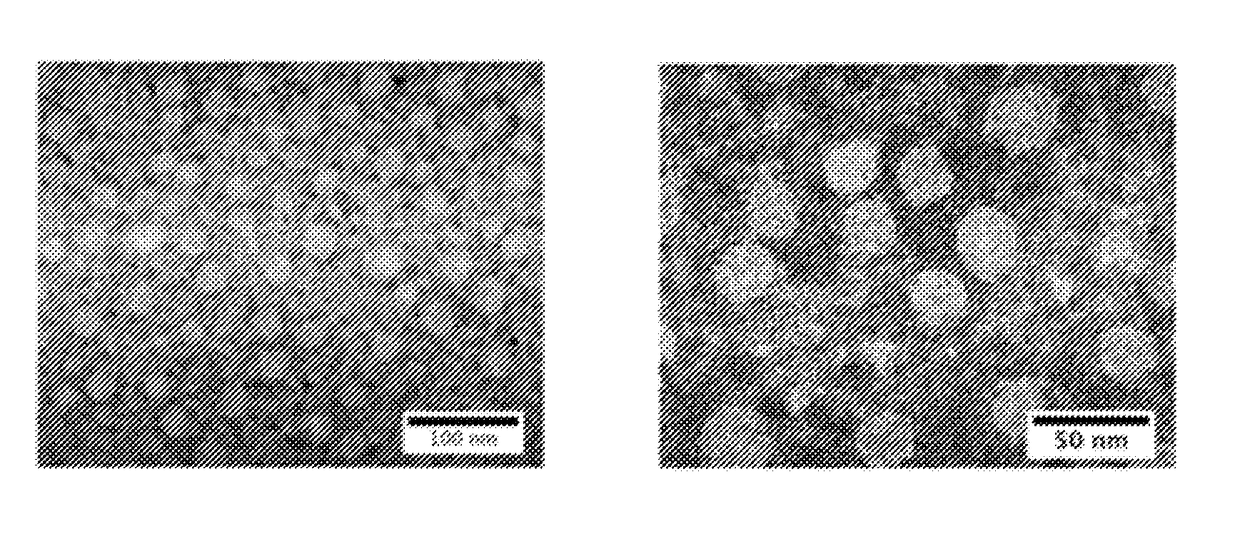
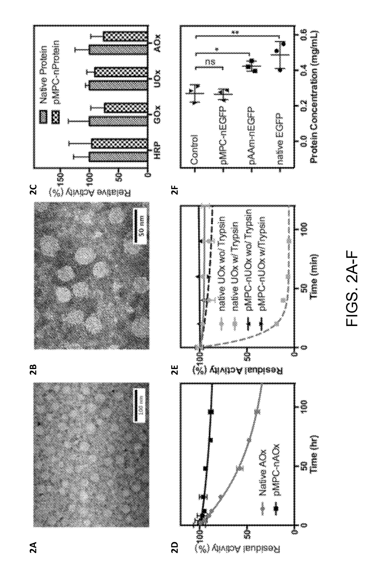
[0031]The successful preparation of pMPC protein nanocapsules was demonstrated using enhanced green fluorescence protein (EGFP) and ovalbumin (OVA). After encapsulation with pMPC, the protein nanocapsules showed a uniform, spherical morphology with an average diameter of 25±5 nm according to transmission electron microscope (TEM) images of pMPC-nEGFP (FIG. 2A) and pMPC-nOVA (FIG. 2B). Considering that the particle size of EGFP and OVA is around 8 nm, the average thickness of the pMPC shell is around 8-11 nm. Because the pMPC coating is formed using a very mild reaction in aqueous media, the proteins encapsulated inside are able to retain their structures and biological functions.
[0032]To verify this, four enzymes, including horseradish peroxidase (HRP), glucose oxidase (GOx), uricase (UOx), and alcohol oxidase (AOx), were encapsulated with the same method, and their enzymatic activities were compared with the...
example 2
Encapsulation with the pMPC Shell Enhances Protein Stability
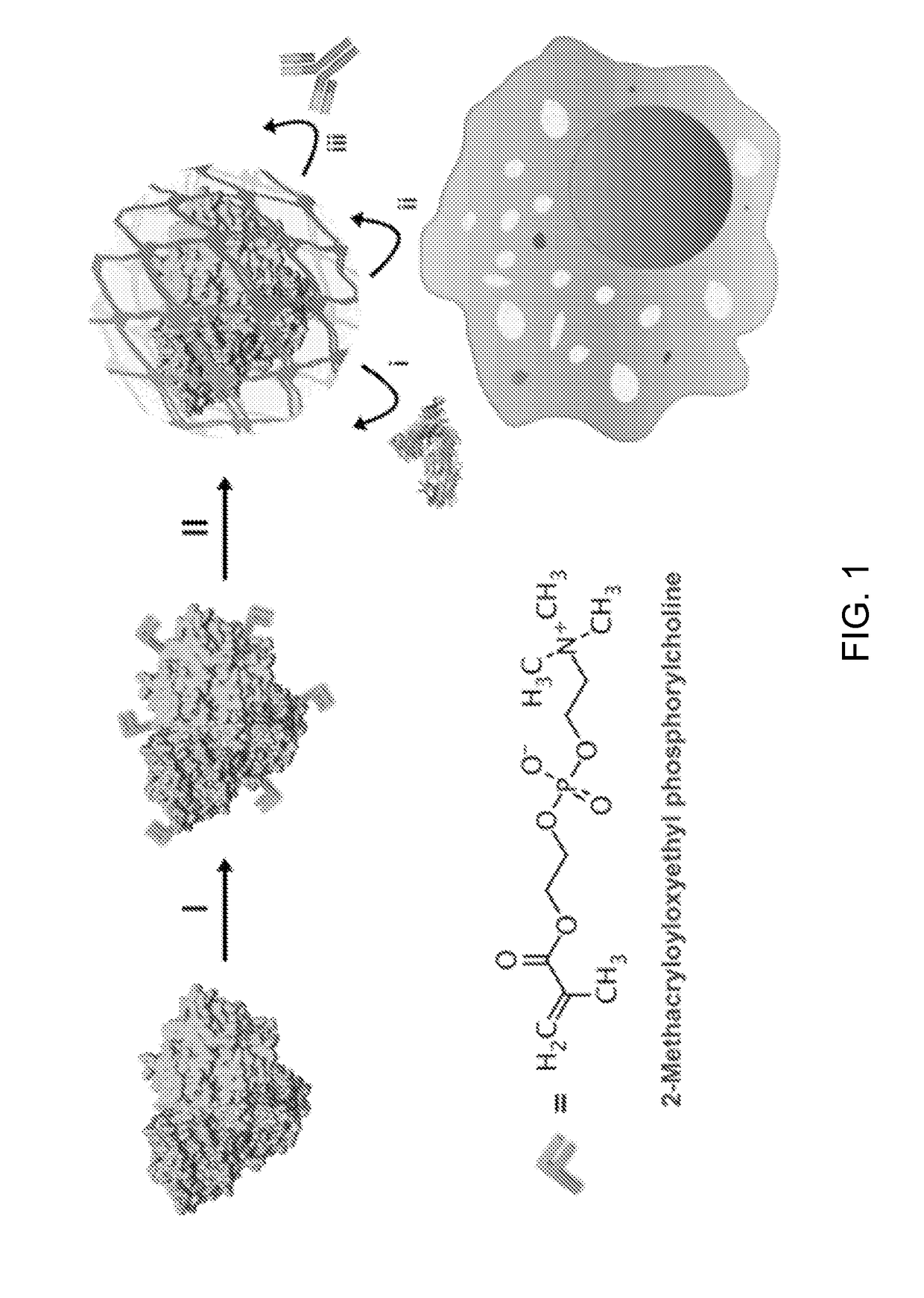
[0033]As illustrated in the scheme, the pMPC shell wrapped outside the protein is synthesized from the MPC monomer directly. Unlike traditional self-assembly and “graft-on” methods for coating the protein, this polymerization method prepares a cross-linked and dense polymeric network that ensures a full coverage of the inner protein during circulation in the blood, where the shearing force is high and the physiological condition is changing continuously. As a result, the nanocapsule disclosed herein provides a stable microenvironment for the protein inside, which effectively enhances its stability.
[0034]To verify this, the stability of protein nanocapsules was first challenged against thermal denaturation. Using AOx as a model protein, native AOx and pMPC-nAOx were incubated under 37° C. for 5 days and their enzymatic activities were monitored at different times. According to the activity comparison (FIG. 2D), native AOx lo...
example 3
Encapsulation with the pMPC Shell Prevents Proteolysis and Lowers Protein Adsorption
[0035]The pMPC shell further isolates the encapsulated protein from the outer environment. During circulation, proteins, cells, tissues and organs have to interact with the pMPC shell instead of the surface of the inner protein, which provides two major benefits for prolonging the circulating lifetime of the protein on a molecular level. First, the pMPC shell prevents the protein from proteolysis by inhibiting the binding of proteases. Exemplified with UOx (FIG. 2E), native UOx lost its activity completely within 40 min when incubating with trypsin, whereas the pMPC-nUOx retained more than 95% of its activity even after 90 min incubation. Second, the pMPC shell replaces the protein surface with a zwitterionic structure, resulting in low protein adsorption onto the pMPC protein nanocapsules. FIG. 2F compares the amount of protein adsorbed by different EGFP samples after 30 min incubation with mouse se...
PUM
| Property | Measurement | Unit |
|---|---|---|
| Temperature | aaaaa | aaaaa |
| Fraction | aaaaa | aaaaa |
| Fraction | aaaaa | aaaaa |
Abstract
Description
Claims
Application Information
 - R&D
- Intellectual Property
- Life Sciences
- Materials
- Tech Scout
- Unparalleled Data Quality
- Higher Quality Content
- 60% Fewer Hallucinations
Browse by: Latest US Patents, China's latest patents, Technical Efficacy Thesaurus, Application Domain, Technology Topic, Popular Technical Reports.
© 2025 PatSnap. All rights reserved.Legal|Privacy policy|Modern Slavery Act Transparency Statement|Sitemap|About US| Contact US: help@patsnap.com


