Methods and apparatus for x-genetics
a technology of x-genetics and methods, applied in the field of methods and apparatus for x-genetics, can solve the problem that light cannot penetrate deeply into biological tissue, and achieve the effect of less invasive stimulation and little attenuation
- Summary
- Abstract
- Description
- Claims
- Application Information
AI Technical Summary
Benefits of technology
Problems solved by technology
Method used
Image
Examples
example 1
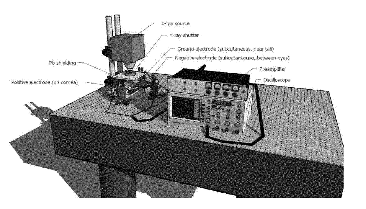
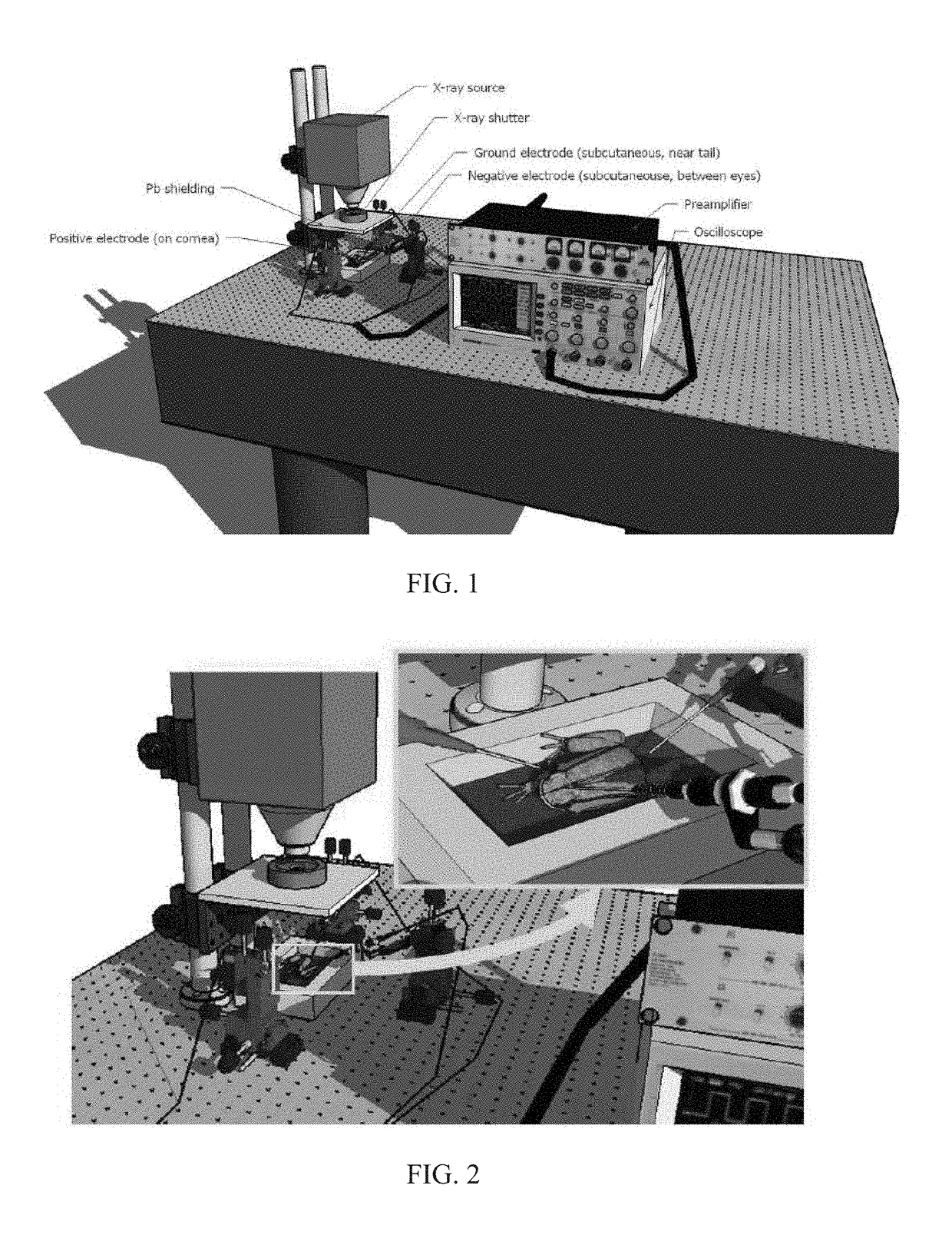
[0073]A visible / X-ray ERG prototype was applied to elicit and measure retinal responses of the Norther leopard frog (R. pipiens) from visible and X-ray stimuli. Eight Northern leopard frogs were used once, unless otherwise noted, in this round of signal acquisition with the ERG system. Each frog was dark-adapted overnight and handled in a room illuminated with a low intensity, 650 nm high pass filtered light (Roscolux #27, Rosco Laboratories). The frogs were anesthetized in the dark room by immersion in 1 g / L solution of MS-222 (pH buffered to −7 with NaHCO3) for 2-8 minutes. After pulsing, each animal was revived in dH2O until fully recovered. During the ERG acquisitions, the frogs did not receive supplemental oxygen. Table 1 shows each of the visible, X-ray, and solenoid-only ERG recordings of the frogs. The cumulative amount of radiation prior to the visible ERG recording is also listed.
[0074]GRASS subdermal, platinum needle electrodes (NATUS) were used for the ERG signal acquisi...
example 2
[0094]A hybrid visible / X-ray ERG system was used to measure ERGs in 30 Northern leopard frogs. Dark-adapted frogs were anesthetized, placed in an animal holder, and subject to a sequence of light and X-ray pulses. In the experiments, X-rays were pulsed using a lead shutter controlled by a solenoid. Platinum electrodes were placed on the corneas of the frogs and behind their eyes in order to measure the differential potential changes induced by the pulse stimulation. While the frog was stimulated with visible light or X-ray pulses, the ERG signals were recorded on an AD Instruments device.
[0095]In the best controlled experiments, 8 frogs were tested for X-ray sensitivity. Of those frogs, three registered X-ray responses that were distinguishable from the background noise and drift. After responding to X-ray stimuli, an increase in visible light ERG was generally observed in these frogs. The total absolute area under the a, b, and c waves of the visible light ERG signals was used as a...
example 3
[0098]An image reconstruction demonstration with deep learning was performed. A poor-quality initial image was reconstructed to a good-quality image. A 2D world of Shepp-Logan phantoms was defined. A field of view was a unit disk covered by a 128*128 image, 8 bits per pixel. Each member image was one background disk of radius 1 and intensity 100 as well as up to 9 ellipses completely inside the background disk. Each ellipse was specified by the following random parameters: center at (x, y), axes (a, b), rotation angle q, and intensity selected from [−10, 10]. A pixel in the image could be covered by multiple ellipses including the background disk. The pixel value is the sum of all the involved intensity values. From each image generated, 256 parallel-beam projections were synthesized, 180 rays per projection. From each dataset of projections, a simultaneous algebraic reconstruction technique (SART) reconstruction was performed for a small number of iterations. This provided blurry i...
PUM
 Abstract
Description
Claims
Application Information
 - R&D
- Intellectual Property
- Life Sciences
- Materials
- Tech Scout
- Unparalleled Data Quality
- Higher Quality Content
- 60% Fewer Hallucinations
Browse by: Latest US Patents, China's latest patents, Technical Efficacy Thesaurus, Application Domain, Technology Topic, Popular Technical Reports.
© 2025 PatSnap. All rights reserved.Legal|Privacy policy|Modern Slavery Act Transparency Statement|Sitemap|About US| Contact US: help@patsnap.com


