Novel light-activated compositions and methods using the same

a technology of light-activated compositions and compositions, applied in the direction of antibacterial agents, bacteria material medical ingredients, therapy, etc., can solve the problems of destroying biomolecules such as dna, rna and proteins, inducing cell death by heating the surrounding medium, etc., to reduce the growth of e, reduce the oxidation potential, and the effect of the same reduction potential

Active Publication Date: 2018-02-15
UNIV OF COLORADO THE REGENTS OF
View PDF1 Cites 3 Cited by
  • Summary
  • Abstract
  • Description
  • Claims
  • Application Information

AI Technical Summary

Benefits of technology

[0125]To evaluate the photoeffect of tuned QDs in a biological environment, CdTe and CdSe of the same bandgap (2.4 eV) with varying redox potentials were selected (FIG. 2c), hereafter referred to as CdTe-2.4 and CdSe-2.4, respectively. 1.9 eV bandgap CIS particles (FIG. 2C, hereafter referred to as CIS-1.9) were also evaluated, because they have the same reduction potential as the CdSe-2.4 particle and a lower oxidation potential than CdSe-2.4 and CdTe-2.4 along with a smaller bandgap. The comparison of CdSe-2.4 to CdTe-2.4 allowed for a direct observation of the effect of the redox potentials as opposed to the bandgap. The comparison of CIS-1.9 to CdSe-2.4 allows for direct observation of the effect of the oxidation potential as the reduction potential is the same for both particles.
[0126]QDs were added, in varying concentrations, to E. coli cultures and grown rich media while the optical density was monitored using a microplate reader. Cultures were grown in the absence and presence of QDs with and without visible light illumination (lamp spectra with UV and IR filters, FIG. 14).
[0127]Exposure in dark was used as a control, as cadmium based QDs can be intrinsically toxic to cells; however, the present studies were performed below reported toxic levels. Further, any intrinsic toxicity would be accounted for in the dark control analysis. Degradation studies of cadmium QDs upon light illumination showed a red shift in quantum dot emission as opposed to a blue shift that would be indicative of free, toxic cadmium release (FIGS. 19A, 19B, 20A, 20B, 21A, 21B).
[0128]The studies showed a significant reduction in growth of E. coli grown in the presence of CdTe-2.4 particles in light compared to dark (FIG. 3A). If the phototoxicity were an effect of generic ROS, as opposed to the tuned response, higher phototoxicity with illumination of CdSe-2.4 particles would be expected due to their higher oxidation potential. In contrast to this hypothesis, in the presence of CdSe-2.4 and illumination, no significant phototoxicity was observed in E. coli (FIG. 3A). Surprisingly, CIS-1.9 particles upon illumination displayed a photo-proliferative effect in E. coli.
[0129]Evaluating these particles over a concentration range allowed for the determination of the threshold where a photoeffect was observed. In order to assess the photoeffect of the QDs, parameters for phototoxicity (Equation 2) and photo-proliferation (Equation 3) were defined to observe the optical density of cultures in light relative to dark. There was a strong phototoxic effect above 35 nM CdTe-2.4 (FIG. 3B). Contrastingly, there was no significant photoeffect even up to a CdSe-2.4 concentration of 500 nM (FIG. 3B). Plating the cells post exposure confirmed that the CdTe-2.4 is bactericidal in light and not in dark, as evidenced by a large decrease in the number of viable cells whereas CdSe-2.4 did not exhibit a bactericidal photoeffect (FIGS. 3C and 17). Thus, the source of the photoeffect is the specifically tuned redox potentials of the QDs and not the bandgap, which remained constant at 2.4 eV.
[0130]Examining the concentration dependence of CIS-1.9 showed a statistically significantly region of photo-proliferation centered at 100 nM CIS-1.9 (FIG. 3B). There were thresholds below which the generated LARS were at too low of concentration to greatly impact the cell, and above which the LARS are in excess and overwhelm redox homeostasis. The measured photo-proliferation observed with CIS-1.9 differed greatly in the no photoeffect observed for CdSe-2.4. As the bandgap was not the source of the difference in photoeffect between CdTe-2.4 and CdSe-2.4, without wishing to be limited by any theory, a possible conclusion is that the CIS-1.9 photo-proliferation compared to CdSe-2.4 derives from the 0.5 V shift in oxidation potential between the two materials.

Problems solved by technology

Metal nanoparticles may act as infrared absorbers and induce cell death by heating the surrounding medium.
This toxic effect is generally attributed to the generation of generic reactive oxidative species (ROS) and free radicals, which can damage biomolecules such as DNA, RNA and proteins.

Method used


Image

  • Novel light-activated compositions and methods using the same
  • Novel light-activated compositions and methods using the same
  • Novel light-activated compositions and methods using the same

Examples


example 1

Characterization of Tunable Quantum Dot Properties

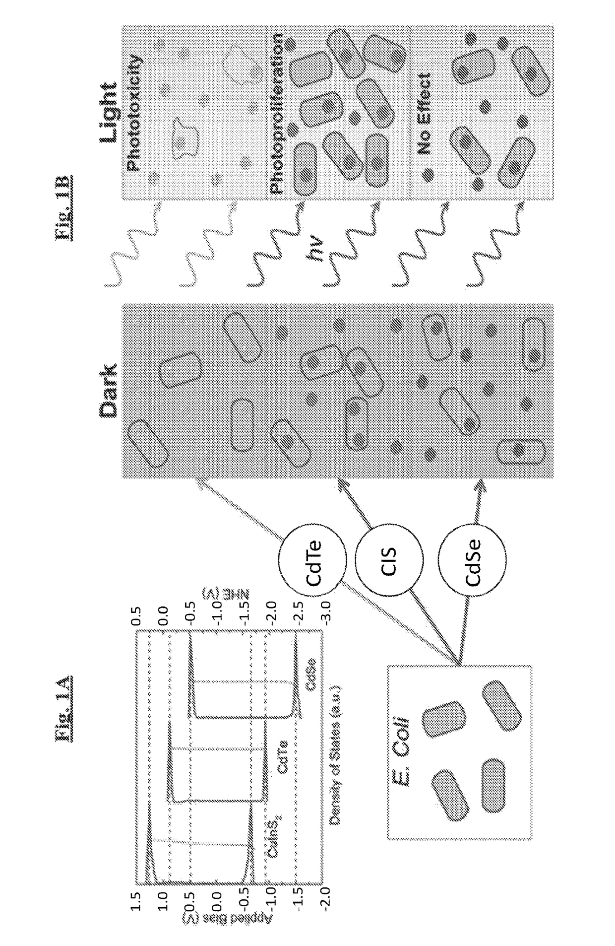
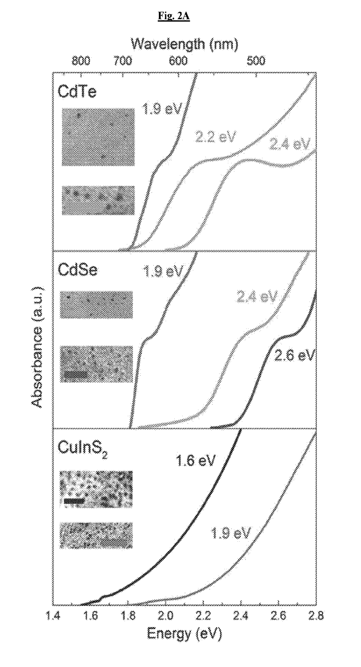
[0123]To examine the range of possible effects, cadmium telluride (CdTe), cadmium selenide (CdSe), and copper indium sulfide (CuInS2, CIS) QDs of different sizes, which absorb light across the visible and near-infrared spectra, were prepared (FIG. 2A). Infrared absorbing dots are highly attractive, as human tissue is generally transparent to light from 750-1000 nm allowing in vivo stimulation.

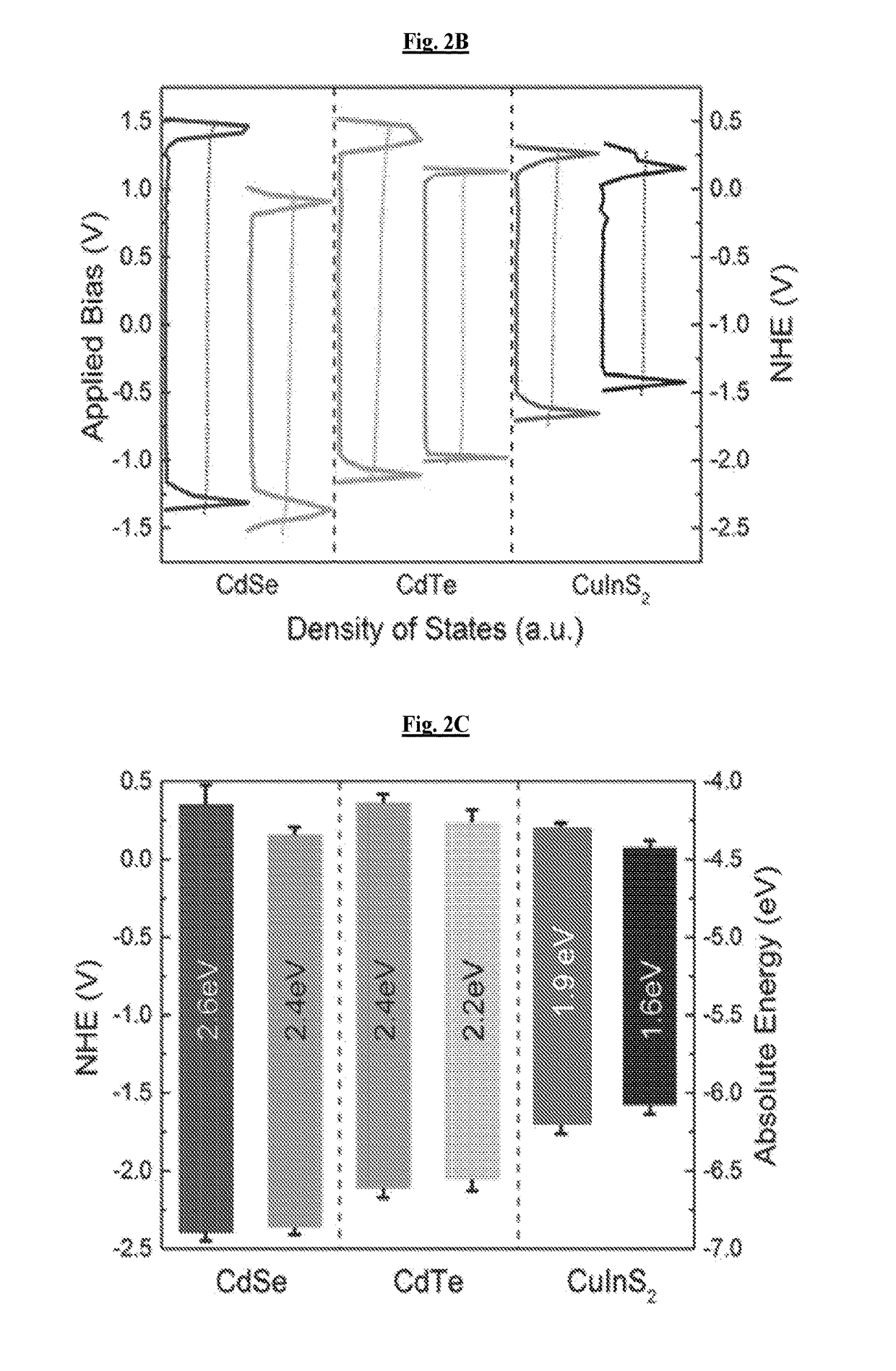
[0124]Electronic structures of these materials were quantified using scanning tunneling spectroscopy of individual nanocrystals (FIG. 2B). For clarity of comparison to biochemical systems, these measurements are compared relative to the normal hydrogen electrode potential. Based on these measurements, the band edge states of the various materials, which play the largest role in determining the redox potentials of each quantum dot, were identified. Because the samples were not perfectly mono-disperse, there was a distribution in band position for...

example 2

Photoeffect of Quantum Dots on Escherichia coli

[0125]To evaluate the photoeffect of tuned QDs in a biological environment, CdTe and CdSe of the same bandgap (2.4 eV) with varying redox potentials were selected (FIG. 2c), hereafter referred to as CdTe-2.4 and CdSe-2.4, respectively. 1.9 eV bandgap CIS particles (FIG. 2C, hereafter referred to as CIS-1.9) were also evaluated, because they have the same reduction potential as the CdSe-2.4 particle and a lower oxidation potential than CdSe-2.4 and CdTe-2.4 along with a smaller bandgap. The comparison of CdSe-2.4 to CdTe-2.4 allowed for a direct observation of the effect of the redox potentials as opposed to the bandgap. The comparison of CIS-1.9 to CdSe-2.4 allows for direct observation of the effect of the oxidation potential as the reduction potential is the same for both particles.

[0126]QDs were added, in varying concentrations, to E. coli cultures and grown rich media while the optical density was monitored using a microplate reade...

example 3

Confirming E. coli Response is Non-Material Dependent

[0131]To observe fine changes in redox potential, various sizes of the three materials were examined to confirm that these were not material dependent properties. The focus was on two CdTe particles, CdTe-2.3 with a bandgap of 2.3 eV and CdTe-2.2 with a bandgap of 2.2 eV. A higher energy CdSe particle with a bandgap of 2.6 eV and a lower energy CIS particle with a bandgap of 1.6 eV, hereafter referred to as CdSe-2.6 and CIS-1.6, respectively, were analyzed as well. CdTe materials showed decrease in the phototoxic effect with decreasing reduction potential (FIG. 4). As the bandgaps changed with increasing size, their reduction potentials moved closer to those of CdSe-2.4, implying that in certain embodiments the conduction band states relate to the phototoxic effect, given the consistency in valence band position (FIG. 2C). This data further precludes the suggestion that phototoxicity is arising from degradation of the QDs, as the ...


PUM

PropertyMeasurementUnit
reduction potentialaaaaaaaaaa

Abstract

The invention includes light-activated compositions and methods that are useful for promoting cell death or growth. In certain embodiments, the compositions comprise quantum dots (QD).

Description

CROSS-REFERENCE TO RELATED APPLICATIONS[0001]The present application claims priority under 35 U.S.C. § 119(e) to U.S. Provisional Application Ser. No. 62 / 136,128, filed Mar. 20, 2015, which application is incorporated herein by reference in its entirety.BACKGROUND OF THE INVENTION[0002]The small size and tunability of nanomaterials has engendered great interest in biological applications such as diagnostics and therapeutics. Silver and metal oxide nanoparticles show bactericidal effects in a range of microorganisms. Metal nanoparticles may act as infrared absorbers and induce cell death by heating the surrounding medium. This toxic effect is generally attributed to the generation of generic reactive oxidative species (ROS) and free radicals, which can damage biomolecules such as DNA, RNA and proteins.[0003]Cells that grow in aerobic environments have mechanisms to mitigate or use ROS through redox homeostasis processes. Within cell types, a specific redox homeostasis is maintained b...

Claims


Application Information

Patent Timeline
Patent Type & Authority Applications(United States)
IPC IPC(8): A61K41/00H01L29/26A61K35/74A61N5/06A61K31/496A61K31/7056A61K31/16A61K31/7036H01L29/22A61K31/546
CPCA61K41/0057H01L29/2203H01L29/26H01L29/22A61K35/74A61K31/546A61K31/496A61K31/7056A61K31/16A61K31/7036A61N5/062A61N2005/0661A61N2005/0662B82Y5/00A61K9/5115H01L29/0665H01L29/225A61P31/04Y02A50/30
Inventor NAGPAL, PRASHANTCHATTERJEE, ANUSHREECOURTNEY, COLLEENGOODMAN, SAMUEL MARTIN
Owner UNIV OF COLORADO THE REGENTS OF
Why Eureka
Try Eureka