Microvesicles

a technology of microvesicles and microvesicles, which is applied in the field of microvesicles, can solve the problems of difficult to obtain difficult to achieve significant numbers of autologous derived microvesicles, and difficult to store. achieve the effect of facilitating uptak

Inactive Publication Date: 2018-04-26
LYDAC NEUROSCIENCE LTD
View PDF0 Cites 0 Cited by
  • Summary
  • Abstract
  • Description
  • Claims
  • Application Information

AI Technical Summary

Benefits of technology

[0054]A “cosmetic” method or product is not directed to the therapy of a disease but is, rather, directed to the improvement of an individual's aesthetic appearance, particularly the appearance of the skin or hair of an individual. Examples of cosmetic effects include a reduction in skin wrinkles, an increase in skin firmness, an increase in hair growth or shine, a reduction in grey hairs, a regrowth of hair in cases of baldness (especially male pattern baldness), an aesthetic enhancement of breast size or shape, and a reduction in cellulite. Cosmetic effects also include a reduction in hair growth (especially facial hair growth).
[0056]“Derivative thereof” includes a fraction or extract (especially those containing RNA and / or DNA and / or protein) of the original microvesicle or population of cells which retains at least some biological activity (especially the ability to induce differentiation and / or the ability to provide therapeutic benefit) of the original. Also included by the term are complexed, encapsulated or formulated microvesicles or cells (for example, microvesicles that have been encapsulated, complexed or formulated to facilitate uptake into recipient cells). According to certain embodiments, intact cells and microvesicles or derivatives containing intact cells or microvesicles are used. According to other embodiments derivatives such as lysates, lyophilates and homogenates may be used. Such derivatives have the advantage that they may be easier to store (for example they may be stored at room temperatures). The literature on live cell therapy demonstrates that cell homogenates and lysates can retain biological activity.

Problems solved by technology

However, their origin (donor cell) is non-autologous with the intended recipient cell, thus their use in inducing phenotypic change may be problematic for various human and animal applications.
Microvesicles will carry donor cell markers, and this may limit their use in clinical applications.
Further, it would be difficult to obtain significant numbers of autologous derived microvesicles to alter the phenotype of a recipient stem cell, or stimulate endogenous stem cell differentiation, migration and integration.
Similarly, the use of microvesicles from a non-autologous source would carry significant risk of transferring pathogens from donor cell to recipient cells.

Method used


Image

  • Microvesicles
  • Microvesicles

Examples


example 1

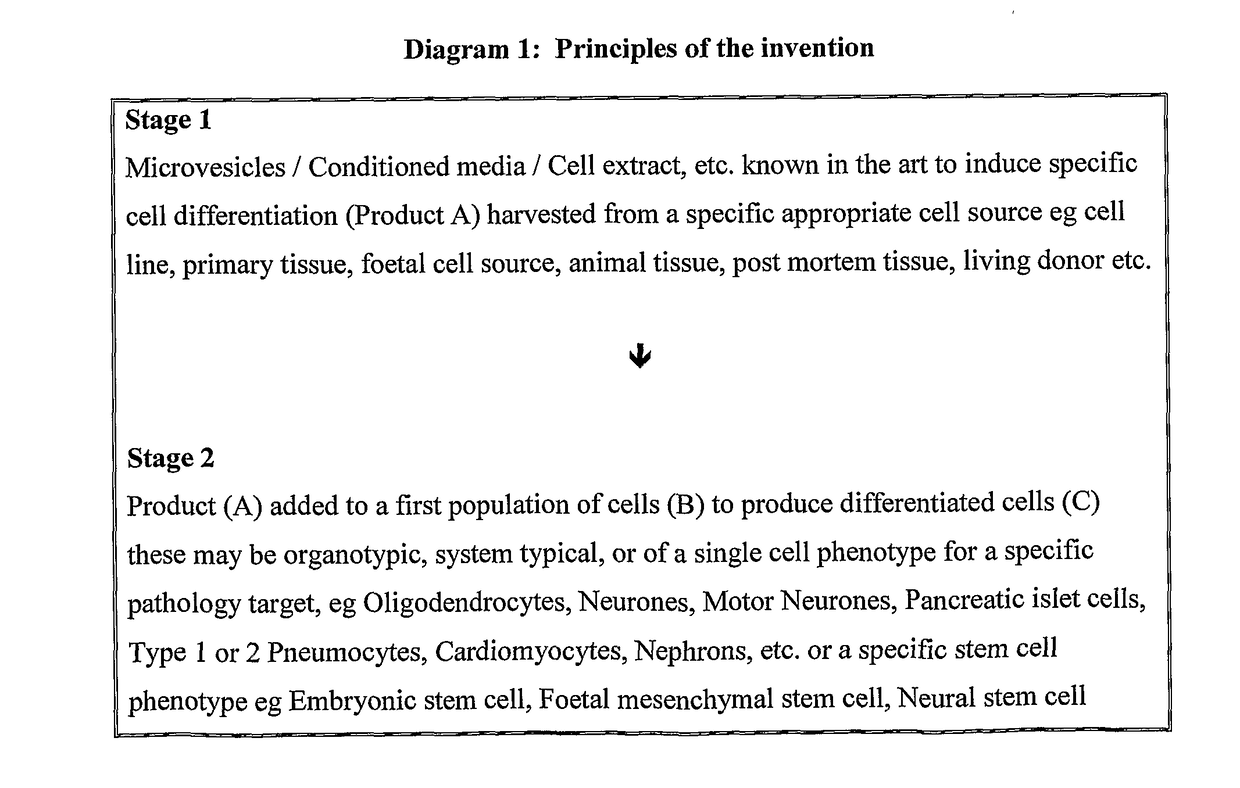
[0240]Improved Differentiation of Stem Cells Mediated by Autologous Microvesicles Manufactured by a Method of the Invention Compared to Non-Autologous Microvesicles Prepared by Methods Known in the Art.

[0241]Aim

[0242]To test the potential benefit of autologous microvesicles compared to non-autologous microvesicles in inducing stem cells to differentiate to a specific fate. It was hypothesised that the surface membrane properties of microvesicles may influence their efficiency in fusing with recipient cells. Further, that autologous microvesicles may show improved cell fusion and thus be a more powerful stimulant for specific stem cell differentiation.

[0243]Stem Cells

[0244]Stimulated bone marrow mesenchymal stem cells were harvested from peripheral blood of an adult male Hooded Lister rat following mobilisation of these cells using GCSF and plated into a 174 ml plastic culture flask (Falcon) in alpha-mem media with 1% penicillin and streptomycin and 10% foetal calf serum. Cells were ...

example 2

[0258]Autologous Microvesicle Primed Stem Cell Induced Repair in Aged Rat Compared to Existing Non-Autologous Equivalent Methods.

[0259]Having established an advantage to the methods claimed in the invention for autologous microvesicle induced differentiation, their efficacy in an in vivo model was explored. It was hypothesised that non-autologous microvesicle induced differentiation, known in the art may change the surface characteristics of a recipient cell by the nature of their contributed cell membrane. Thus, such stem cells may not be capable of reaching their full potential of repair, regeneration or other reparative functions as many cells may be destroyed by the host immune system.

[0260]The test employed was a cognitive test of spatial memory, the Morris water maze. Aged rats are known to be poor at both learning and recalling this task. In brief the water maze is a plastic tank 1.8 m in diameter filled with 21° C. water. The water is made opaque by the addition of powdered ...

example 3

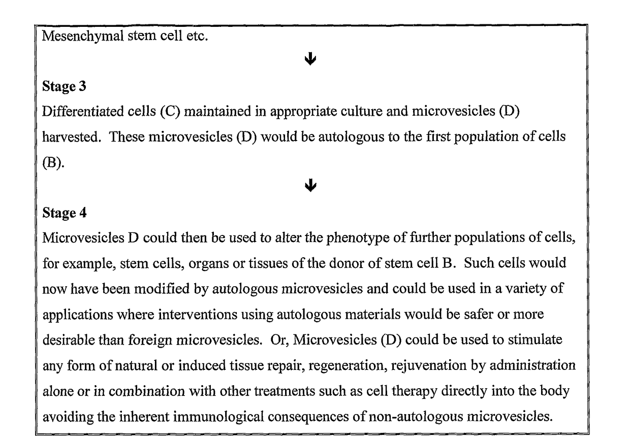
[0283]Recently, much interest has focused on microvesicles in a variety of areas of biological and medical science as a diagnostic, an inducer of stem cell differentiation and as mediators of various biological effects. All existent technology for cell modification relies on non-autologous microvesicles because autologous microvesicles would require some of the patients' damaged tissues to be removed and cultured in vitro to generate autologous microvesicles. This invention surmounts that issue with a novel two step approach. Further, microvesicle methods currently known in the art could not generate products which could be injected into a host organism as the microvesicles would mostly be destroyed by immune reaction. The invention disclosed in this patent application gets around that problem and provides methods to investigate microvesicle mediated direct repair in an organism. Thus a third example is provided investigating the potential of autologous microvesicles as direct media...


PUM

PropertyMeasurementUnit
buoyant densityaaaaaaaaaa
diameteraaaaaaaaaa
diameteraaaaaaaaaa

Abstract

A method of producing a population of differentiated cells comprising: a) inducing differentiation in a first population of cells by applying an inducer to said cells, b) harvesting microvesicles produced from first population of cells, and c) inducing differentiation in a second population of cells by applying said microvesicles or a derivative thereof to said second population of cells wherein, said first population of cells is autologous to said second population of cells and wherein the inducer applied to said first population of cells is not present in said second population of cells or is only present in trace amounts. Also related methods of producing microvesicles, methods of medical and / or cosmetic treatment and related products and uses.

Description

FIELD OF INVENTION[0001]The invention relates to microvesicles and methods for producing microvesicles, particularly microvesicles that are immunologically-matched autologous microvesicles for use in therapeutic and cosmetic and other applications.BACKGROUND OF THE INVENTION[0002]Microvesicles[0003]Microvesicles (MVs) are membrane-bound packets of cytoplasmic material shed by cells in various physiological states, either natural or induced. Long regarded as cellular debris, recently these structures have generated an ever expanding literature supporting the hypothesis that microvesicles are an important inter cellular signalling mechanism (eg Ratajczak et al. 2006).[0004]It has been reported that elevated levels of microvesicles are found in blood and other body fluids in various forms of pathology, most notably in cancers (reviewed Hoon & Taback, 2004). Consequently the value of microvesicles as a diagnostic tool is well established. More recently, it has been noted that microvesic...

Claims


Application Information

Patent Timeline
Patent Type & Authority Applications(United States)
IPC IPC(8): C12P1/00A61K35/28C12N5/071A61K8/98A61Q19/00
CPCC12N2506/1353A61K35/28C12N5/0602A61K8/981C12P1/00A61Q19/00A61K2800/10C12N2523/00A61P1/00A61P1/16A61P11/00A61P11/02A61P13/00A61P15/00A61P17/00A61P17/02A61P19/02A61P19/08A61P19/10A61P21/00A61P25/00A61P25/02A61P25/08A61P25/16A61P25/18A61P25/28A61P29/00A61P3/00A61P35/00A61P5/00A61P9/00A61P9/06A61P9/10
Inventor RAY, STEPHEN
Owner LYDAC NEUROSCIENCE LTD
Why Eureka
Try Eureka