Methods for monitoring polymorphonuclear myeloid derived suppressor cells

a technology of suppressor cells and polymorphonuclear myeloids, applied in the field of methods for monitoring polymorphonuclear myeloid derived suppressor cells, can solve the problems of time-consuming and inaccurate, shared phenotype, and introduction of errors into analysis

Active Publication Date: 2018-06-14
THE WISTAR INST OF ANATOMY & BIOLOGY
View PDF0 Cites 2 Cited by
  • Summary
  • Abstract
  • Description
  • Claims
  • Application Information

AI Technical Summary

Benefits of technology

[0005]In one aspect, a method for monitoring the population of polymorphonuclear myeloid derived suppressor cells (PMN-MDSCs) in a mammalian subject involves contacting a biological fluid sample from the subject containing polymorphonuclear neutrophils (PMNs) and PMN-MDSC with a ligand that specifically binds or forms a complex with LOX-1 on the cell surface. Detecting and distinguishing the complexes of ligand-bound LOX-1-cells from other cells not bound to the ligand in the sample enables the tracking of the number or changes in the number of PMN-MDSCs substantially free of PMN.

Problems solved by technology

One of the major obstacles in the identification of PMN-MDSC is that they share the same phenotype with normal polymorphonuclear cells (PMN).
Current methods for separating populations of PMN-MDSC from populations of PMN in biological fluids are complicated, time-consuming and inaccurate, requiring multiple gradient separation as well as multi-color flow cytometry analysis.
These conditions thus introduce errors into the analysis.
Additionally, these processes are inconvenient and difficult to standardize.
Thus, there are no useful methods currently exist that allow for discrimination of these two populations in blood and tissues.

Method used


Image

  • Methods for monitoring polymorphonuclear myeloid derived suppressor cells
  • Methods for monitoring polymorphonuclear myeloid derived suppressor cells
  • Methods for monitoring polymorphonuclear myeloid derived suppressor cells

Examples


example 1

ng Discriminatory Markers

[0076]In order to identify specific markers discriminating between these two populations, we performed genome-wide microarrays (Human HT-12 v4 expression Beadchip, Illumina) to compare the gene expression profiles between PMN-MDSC and PMN from the same cancer patients (7 patients) as well as age matching healthy donors (4 donors). All samples of peripheral blood (PB) were collected from patients at the Helen F. Graham Cancer Center and were analyzed within 3 hours of collection. PMN-MDSCs were evaluated in mononuclear fraction of PB after ficoll density gradient. PMN were evaluated from the cell fraction remaining after removal of mononuclear cells. Cells were resuspended in PBS and loaded on a step density gradient (Percoll 63% on top of Percoll 72%) to separate PMNs in a monolayer between the two Percoll phases. In an attempt to minimize the number of potential candidates and to identify true marker of PMN-MDSC, we analyzed the gene expression profiles of ...

example 2

g Validity of LOX-1 as a Biomarker

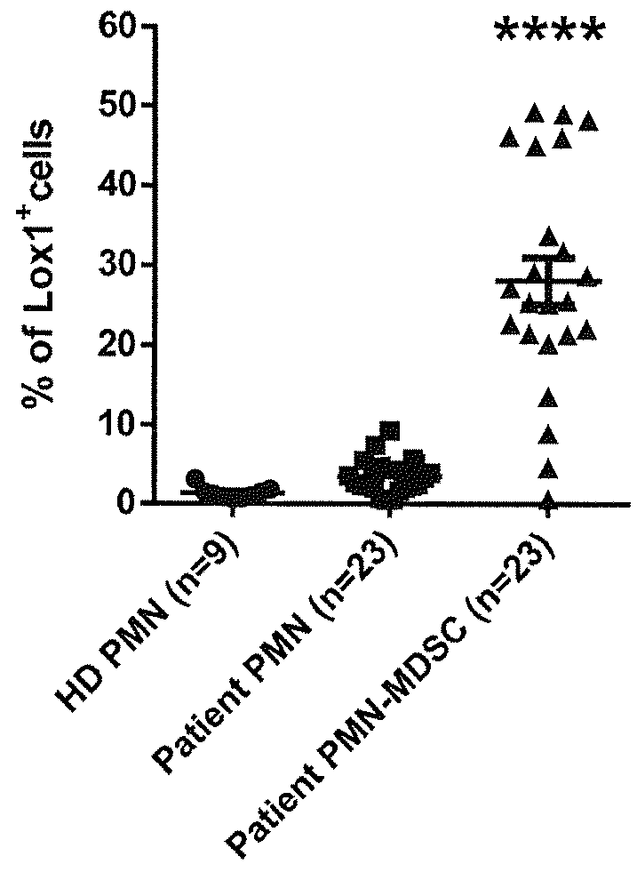
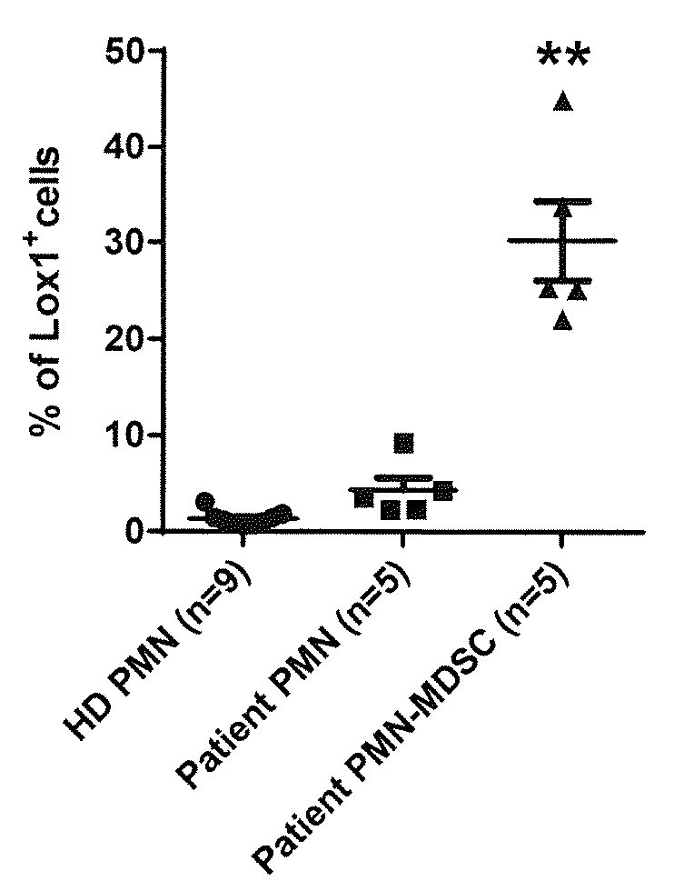
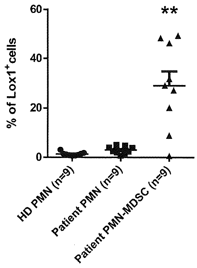
[0078]To confirm the validity of LOX-1 as a potential biomarker of PMN-MDSC, we analyzed the expression of this receptor by flow cytometry using an anti-LOX-1 monoclonal antibody (clone 15C4; Biolegend Inc., San Diego, Calif.) in blood samples from patients with 4 different types of cancer: head and neck, breast, non-small lung, or colon cancer.

[0079]We first analyzed the expression of LOX-1 using the classical definitions of PMN-MDSC (CD11b+ CD14− CD15+ and CD33+ from the low density mononuclear cells fraction) and PMN (cells with the same phenotype from high density fraction). The results of this experiment are reported graphically when healthy donors (HD) were compared with all cancer patients in FIG. 1. About 30% of the PMN-MDSC from all cancer patients (n=23) was found to express LOX-1 on their surface compared to less than 3% of the PMN from matching patients or about 1% from PMN from healthy donor (n=9) (p<0.001).

[0080]The results of this exp...

example 3

of Whole Blood Samples

[0082]We also performed an analysis of unseparated whole blood samples. As shown in FIGS. 4A and 4C, as expected, about 1% or less of the CD11b+ CD14− CD15+ and CD33+ PMN from healthy donors expressed LOX-1 on their surface. However, in samples from cancer patients (both head and neck and lung cancer patients), almost 5% of the PMN types of cells exhibit a positive staining for LOX-1 (≈2.3% of the total leukocytes) strongly supporting the designation of LOX-1 as a specific marker of PMN-MDSC. These results were confirmed by analyzing the disease separately, as reported in FIGS. 4B and 4D.


PUM

PropertyMeasurementUnit
size exclusionaaaaaaaaaa
concentrationaaaaaaaaaa
densityaaaaaaaaaa

Abstract

A method of obtaining a population of cells enriched in human polymorphonuclear myeloid derived suppressor cells (PMN-MDSCs) comprises isolating from a cell suspension those cells which express LOX-1 to provide a population of cells enriched with PMN-MDSCs. A method of monitoring the population of LOX-1+ cells in a cell-containing biological sample is useful for determining the efficacy of treatment or the metastasis or increasing progression of cancer. Other cell isolation and diagnostic methods are also described.

Description

CROSS-REFERENCE TO RELATED APPLICATIONS[0001]This application claims the benefit of the priority U.S. Provisional Patent Applications No. 62 / 187,991 filed Jul. 2, 2015 and No. 62 / 169,368, filed Jun. 1, 2015, which applications are incorporated by reference herein.BACKGROUND OF THE INVENTION[0002]Myeloid-derived suppressor cells (MDSC) represent a heterogeneous population of immature myeloid cells. These cells accumulate to a great extent in cancer patients and play a major role in regulating immune responses in cancer1. MDSC suppress T cells activation and proliferation as well as function of natural killer (NK) cells2,3. Ample evidence links these cells with tumor progression and outcome of the disease in cancer patients4,5. MDSC have been divided in two different sub-populations6, monocytic myeloid-derived suppressor cells (M-MDSC) and polymorphonuclear myeloid-derived suppressor cells (PMN-MDSC). About 20-30% of MDSC consists of monocytic cells, i.e., M-MDSC, and are generally as...

Claims


Application Information

Patent Timeline
Patent Type & Authority Applications(United States)
IPC IPC(8): G01N33/569G01N33/92G01N33/574C12N5/0787
CPCG01N33/56972G01N33/92G01N33/57492C12N5/0642G01N2333/705G01N2333/70596G01N33/56977G01N33/574C12N2503/00
Inventor GABRILOVICH, DMITRY I.CONDAMINE, THOMAS C.
Owner THE WISTAR INST OF ANATOMY & BIOLOGY
Why Eureka
Try Eureka