

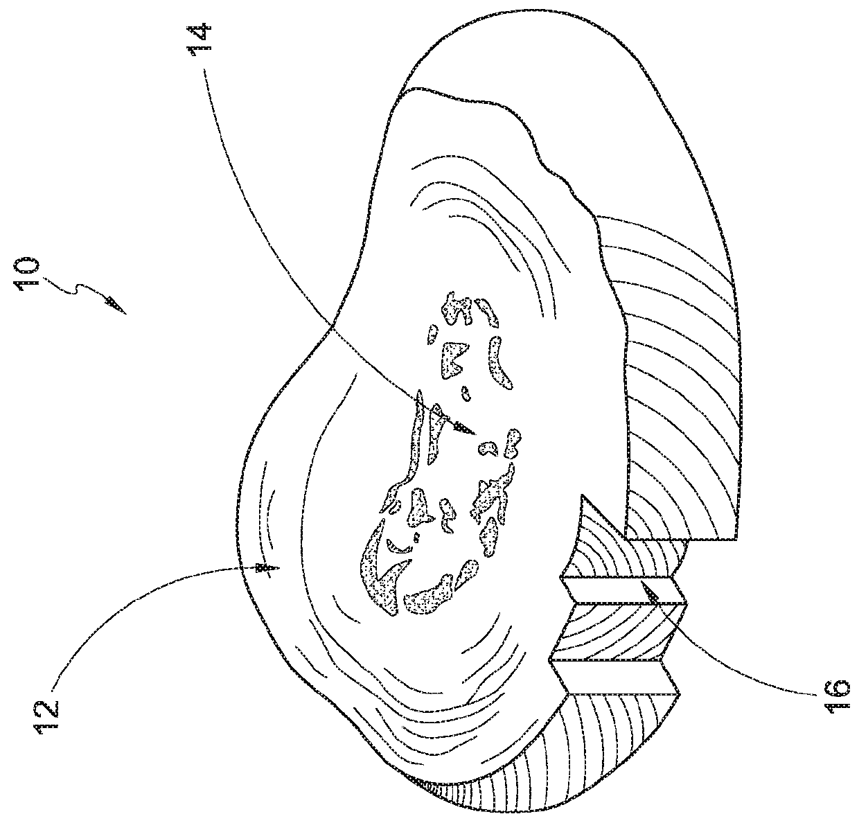
Tissue visualization and modification devices and methods
a technology applied in the field of tissue visualization and modification devices and methods, can solve the problems of reducing the blood flow through a blood vessel, adversely affecting the functioning and/or health of that organ, and compromising the function of one or more nerves
- Summary
- Abstract
- Description
- Claims
- Application Information
AI Technical Summary
Benefits of technology
Problems solved by technology
Method used
Image

Examples
experimental examples
[0264]The following examples are offered by way of illustration and not by way of limitation.
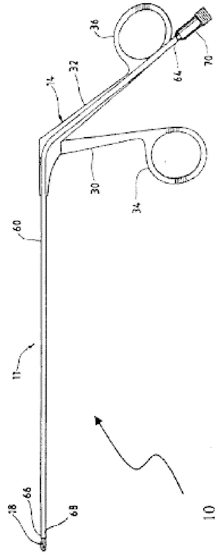
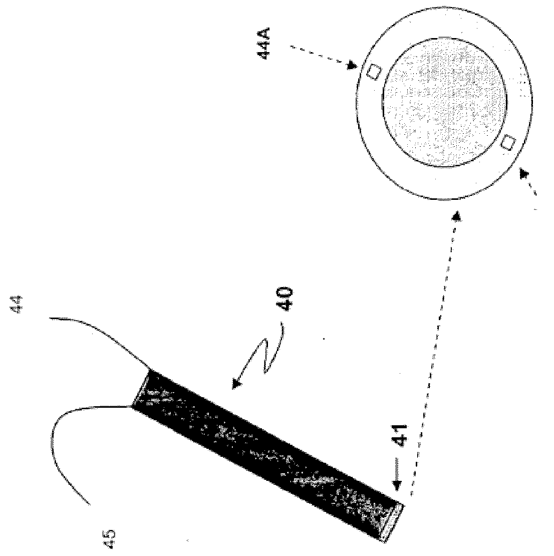
[0265]A hand-held minimally dimensioned diagnostic device having integrated distal end visualization was constructed as follows. The device consisted of an outer SLA shell in the form of a hand-held unit housing batteries, a 3.5″ monitor, a control board, and wires that connect to 2 LEOS and a visualization module at the distal tip of a steel 4 mm hypodermic tube that was connected to the handle. The tubing was bent about an inch back from the distal tip to about 30 degrees. A manual wheel was provided on the hand-piece connected to the tube, and when actuated, rotated the tube 180 degrees in each direction. Considering a field of view for the camera of roughly 120 degrees (diagonal), the rotation of the tube allowed the camera to view at least a full hemisphere of space. The visualization module at the 4 mm outer diameter distal tip of the hypodermic tube included an Omnivision 6920 QVGA im...
PUM
 Abstract
Description
Claims
Application Information