
Protein nanostructure based drug delivery system for the delivery of therapeutic agents to the anterior segment of the eye

a nanostructure and drug technology, applied in the direction of drug compositions, sense disorders, organic active ingredients, etc., can solve the problems of inability to meet the patient's requirements for intensive medication, lack of patient compliance of intensive medication courses, and difficulty in designing a successful system for keratitis

Inactive Publication Date: 2018-11-15
COUNCIL OF SCI & IND RES
View PDF1 Cites 0 Cited by
  • Summary
  • Abstract
  • Description
  • Claims
  • Application Information

AI Technical Summary

Benefits of technology

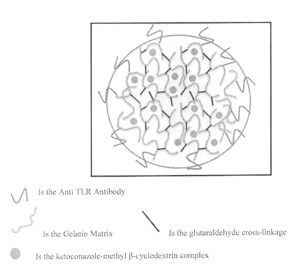
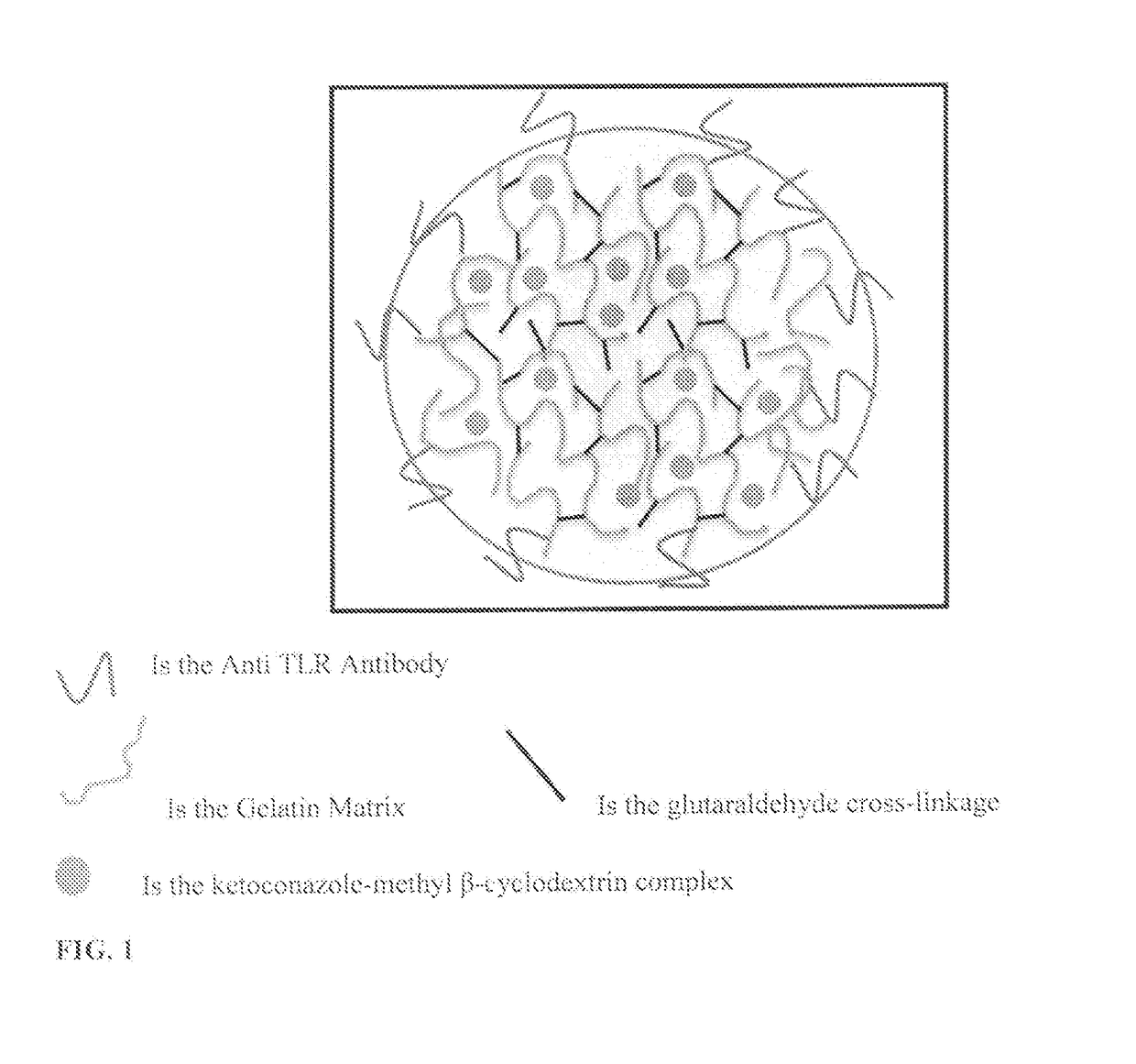
The present invention provides a nanostructure based drug delivery system that can deliver therapeutic agents to treat infections and inflammations. The system consists of a gelatin matrix, an anti-TLR4 ligand, and a therapeutic agent. The gelatin matrix is held together by covalent cross-linking through glutaraldehyde, and the anti-TLR4 ligand is chemically conjugated to the surface of the nanostructure. The therapeutic agent can be hydrophobic or hydrophilic, and can be complexed with cyclodextrins or used with double desolvation methods. The nanostructure is about 10-1000 nm in diameter, and can be used for the treatment of infectious and sterile keratitis. The pharmaceutical composition and kit for treatment of infectious or sterile keratitis are also provided.

Problems solved by technology

Although well protected (by the eyelids, the eye socket, tears, and the sclera), the cornea may be damaged by foreign objects which may lead to infection and inflammation, a condition commonly referred to as keratitis.
Topically applied antibiotics require high dosage frequency due to pre-corneal clearance, whereas application of anti-inflammatory drugs requires precise control over dosage, as a high dose may exacerbate the infection while a low dose may exaggerate the host response and lead to inflammation mediated corneal damage.
Such an intensive medication course lacks patient compliance and often needs hospitalization.
Designing a successful system for keratitis requires is challenging.
However, despite the advances in ocular drug delivery, an efficient system for corneal infections and inflammation (keratitis) is yet to be developed.
In some cases, there may be an exaggerated immune response, leading to irreversible tissue destruction.
This may result in degradation and disorganization of the extracellular matrix and scleral layer of the cornea, and eventually compromised or permanent visual loss.
But, the use of steroids is a point of concern as they can exacerbate the infection by suppressing host immunity and may promote recrudescence.
Although these adverse effects are taken care off while developing NSAIDs based formulations, the use of these drugs without a concomitant use of steroids has been associated with the development of corneal infiltrates.

Method used


Image

  • Protein nanostructure based drug delivery system for the delivery of therapeutic agents to the anterior segment of the eye
  • Protein nanostructure based drug delivery system for the delivery of therapeutic agents to the anterior segment of the eye
  • Protein nanostructure based drug delivery system for the delivery of therapeutic agents to the anterior segment of the eye

Examples


example 1

on of Ketoconazole with methyl-β-cyclodextrin

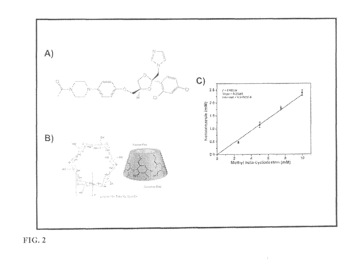
[0155]2.5 mM, 5 mM, 7.5 mM, 10 mM, 15 mM & 20 mM methyl-β-cyclodextrin was used to dissolve an excess amount of ketoconazole in 10 ml milliQ water. The contents of each tube were stirred for 3 days until equilibrium. After equilibration undissolved ketoconazole was separated by filtration with a 0.45 μm PVDF membrane syringe filter. The resulting solution was then analyzed for ketoconazole content by a Lambda Model UV-Visible spectrophotometer (Perkin Elmer, USA) at 260 nm after dilution of the samples.

example 2

Synthesis and Anti-TLR4 Antibody Conjugation

[0156]Step 1: The double-desolvation method was used for nanoparticle synthesis, partially modified by the method described by Coester et al. 2000. 2.5 g of gelatin type B (Bloom225) was dissolved in 50 ml water (5% w / w) under gentle heating (50° C.). In the first desolvation step 50 ml of acetone was added rapidly to the gelatin solution. After sedimentation of the precipitated gelatin fraction, the supernatant containing soluble low molecular weight gelatin was discarded. The sediment was redissolved again by the addition of 50 ml water under gentle heating (50° C.). The redissolved gelatin containing the high molecular weight fraction was snap freezed in liquid nitrogen and lyophilized. The lyophilized gelatin was stored at 4° C. till further use. 0.1 g of the freeze dried high molecular weight gelatin was dissolved in 10 ml water under gentle heating (50° C.) and the pH was adjusted to 3.5. Nanoparticle formation was initiated by the d...

example 3

Size Measurements

[0157]Gelatin nanoparticles were resuspended in PBS pH 7.4 and analyzed for their size and polydispersity in a nanopartica nanoparticle analyzer system (Horiba Scientific).


PUM

PropertyMeasurementUnit
diameteraaaaaaaaaa
sizeaaaaaaaaaa
diameteraaaaaaaaaa

Abstract

A multifunctional system in which a nanostructure (size range of about 10-1000 nm) degrades on exposure to an infection and its associated inflammatory milieu. The degraded nanostructures release the encapsulated drug during the process of degradation, where the kinetics of drug release is determined by the severity of the infection and inflammation. This degradation is triggered by proteases secreted by the pathogen, host polymorphonuclear leucocytes and other host cells. The nanostructures are conjugated to anti-TLR (Toll-like receptor) ligands for targeting the corneal epithelium and blocking the inflammatory pathway.

Description

FIELD OF INVENTION[0001]The present invention provides nanostructure based drug delivery system for therapeutic applications. The present invention provides nanostructure based drug delivery system wherein nanostructure loaded with an anti-microbial drug or an anti-inflammatory therapeutic agent, the release of which is regulated by the degradation of the nanostructure.BACKGROUND OF THE INVENTION[0002]The corneal tissue comprises mainly of a multilayered epithelium followed by a layer of densely packed collagen bundles called the stroma and an innermost single cell endothelial layer. The stromal layer consists of relatively few cells, mainly the collagen secreting keratocytes and unevenly distributed immune cells (dendritic cells, langerhans cells, B- and T-lymphocytes) mostly in the peripheral cornea and relatively less in the centre. Apart from the immune cells in the corneal stroma, the conjunctiva associated lymphoid tissue (CALT) and the lacrimal drainage associated lymphoid ti...

Claims


Application Information

Patent Timeline
Patent Type & Authority Applications(United States)
IPC IPC(8): A61K47/69A61K47/68A61K47/61A61K31/496A61P27/02
CPCA61K47/6931A61K47/6849A61K47/61A61K31/496A61P27/02
Inventor SAAD, MOHAMMAD AHSANCHINTALAGIRI, MOHAN RAO
Owner COUNCIL OF SCI & IND RES
Learn More