Partially degradable stents for controlled reduction of intraocular pressure

a stent and stent technology, applied in the field of biodegradable stents, can solve the problems of devices in which the lumen diameter increases, and achieve the effects of reducing protein adsorption and in vivo inflammatory response, improving glideability, and improving the effect of elasticity

Pending Publication Date: 2019-02-14
THE JOHN HOPKINS UNIV SCHOOL OF MEDICINE
View PDF6 Cites 2 Cited by
  • Summary
  • Abstract
  • Description
  • Claims
  • Application Information

AI Technical Summary

Benefits of technology

[0011]A tubular device having a biodegradable inner core and a non-degradable or slowly degradable outer layer, and optionally delivering therapeutic, prophylactic, and / or diagnostic agents, has been developed. The degradable core can result in a device in which the lumen diameter increases over time as a function of degradation. Optionally, the device contains an exterior coating, which in some embodiments is fluid-impermeable, releases a therapeutic and / or prophylactic agent, is degradable, and / or improves the glideability (e.g., by inclusion of glyconate). The device may include micro- or nano-fibers which are woven around a rotating cylindrical template. Removal of the template results in a tubular device with a lumen. Devices made with nano-structured fibers reduce protein adsorption and in vivo inflammatory response.

Problems solved by technology

The degradable core can result in a device in which the lumen diameter increases over time as a function of degradation.

Method used


Image

  • Partially degradable stents for controlled reduction of intraocular pressure
  • Partially degradable stents for controlled reduction of intraocular pressure
  • Partially degradable stents for controlled reduction of intraocular pressure

Examples


example 1

inning to Generate Partially Bio-Degradable Stents (“Pressure Control Shunt”) and In Vitro Characterization of Degradation in an Aqueous Environment

[0136]Materials and Methods

[0137]Materials used to generate the tubular shaped stents were generally regarded as safe (GRAS) materials approved by the United States Food and Drug Administration (FDA).

[0138]Sequential Electrospinning

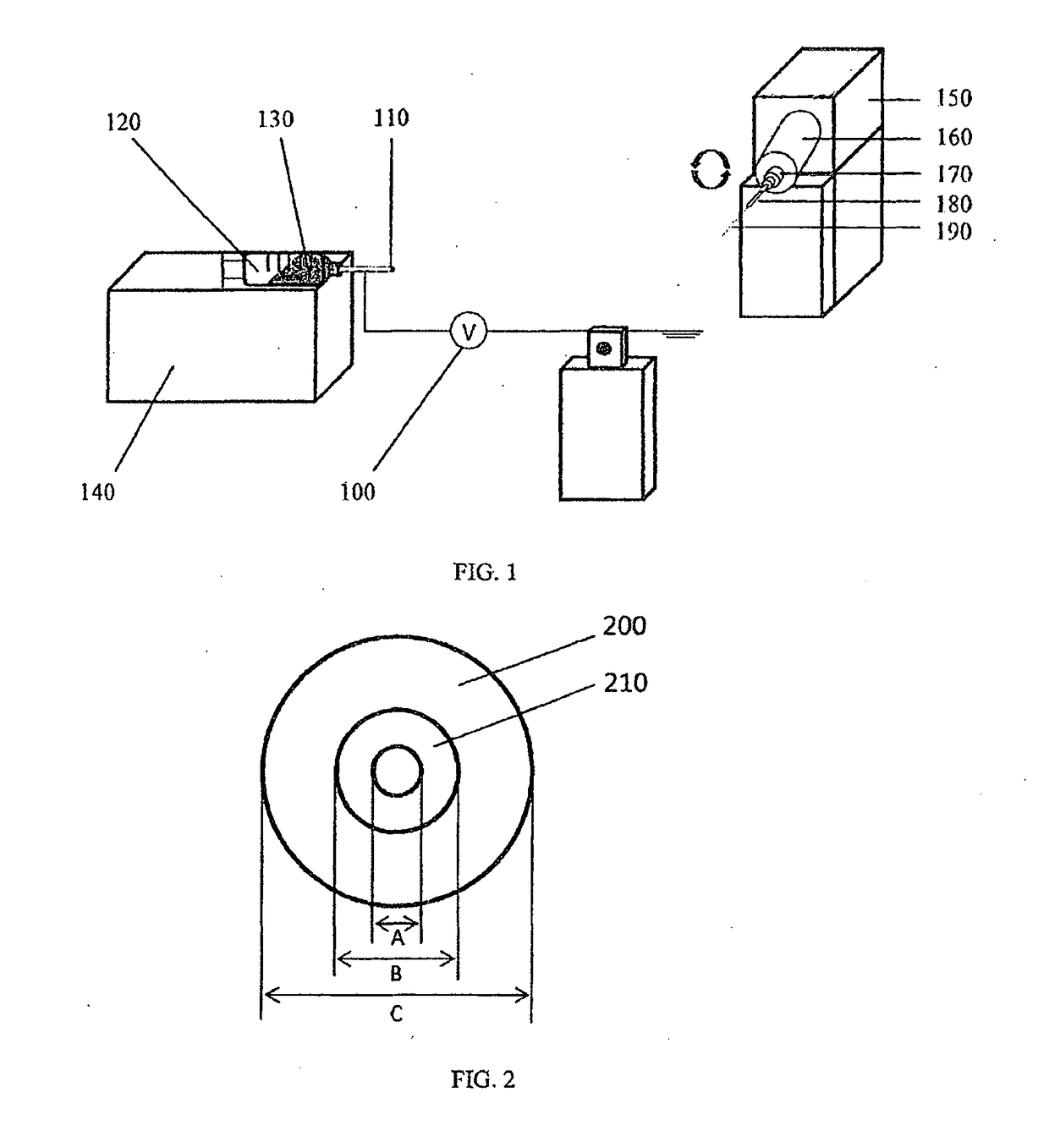
[0139]Biodegradable polyglycolide (PGA) was dissolved at 10 wt % in hexafluoroisopropanol (HFIP). Dissolved PGA was electrospun onto the surface of a template steel wire of 50 μm in diameter, followed by heating above the glass transition temperature of the polymer to improve its mechanical properties.

[0140]Next, non-degradable polyethylene terephthalate (PET) dissolved at 10 wt % in HFIP was sprayed on top of the PGA layer, forming the outer wall, which was also treated with heat afterwards.

[0141]Finally, the template wire was removed, leaving a hollow, tubular shaped stent made from PGA and PET.

[0142]Degrada...

example 2

tal Measurements and Mathematical Modeling of the Pressure Change of Flows Through Stents

[0151]Materials and Methods

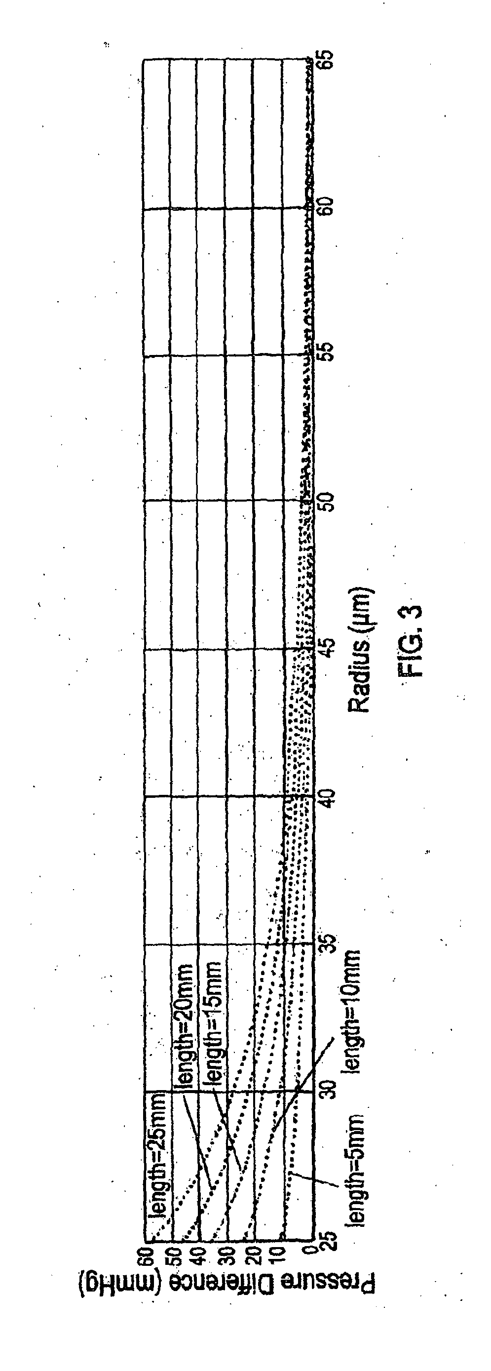
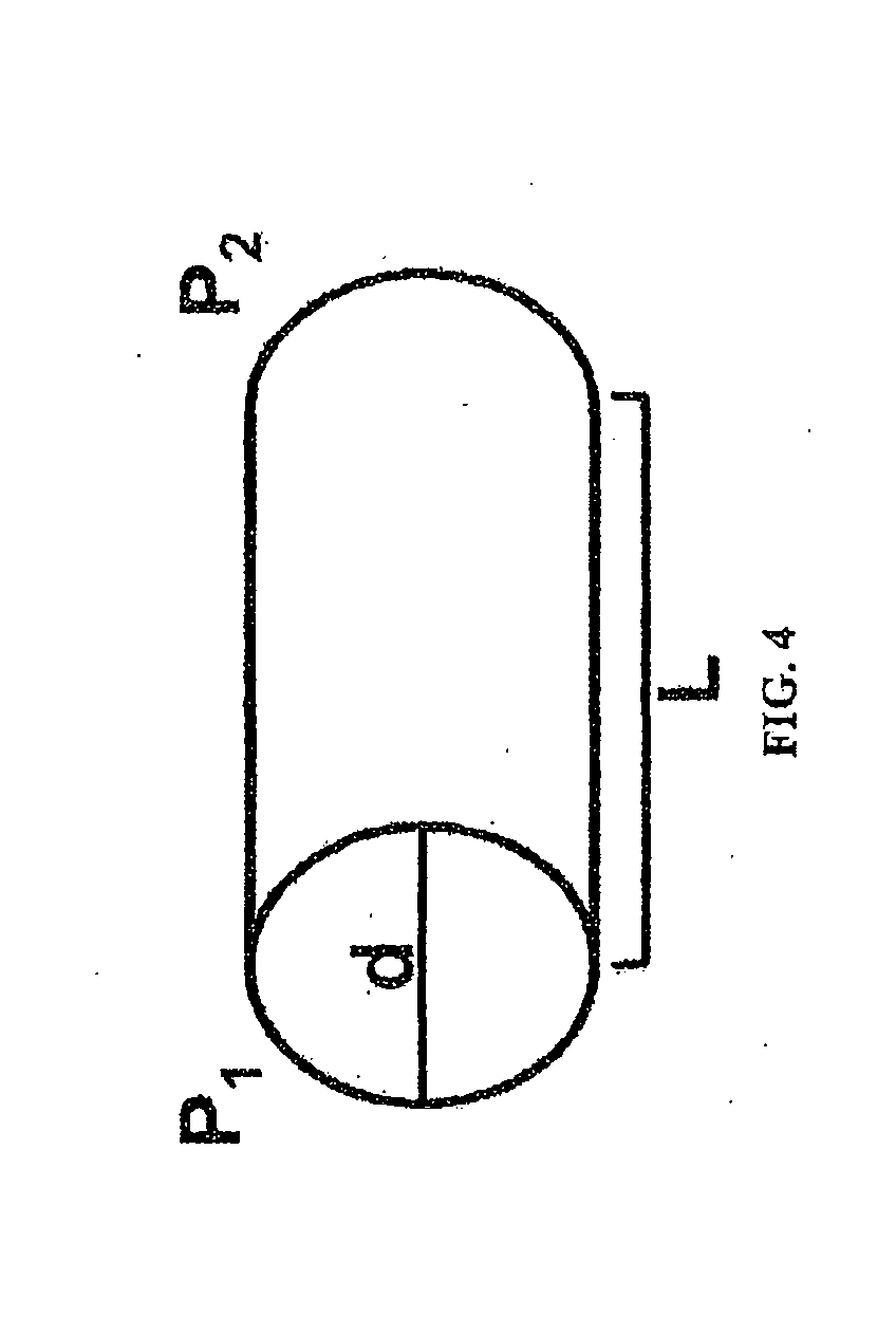
[0152]PBS was flowed at different flow rates at room temperature over the period of one day through the stents made as described in Example 1, and the actual pressure change (i.e., pressure difference between inlet end and outlet end of the stent) of the fluid was measured. It was unlikely that core degradation was observed or it would affect the result of pressure change within the relatively short test period. The measured values were compared with the predicted pressure change according to the Hagen-Poiseuille (HP) equation.

[0153]Alternatively, the pressure changes related to a wide range of variables (e.g., lengths of stents, radii of stents, and flow rates of a fluid) were mathematically modeled.

[0154]Results

[0155]As shown in Table 2, the flow of PBS through these stents obeyed the HP equation.

TABLE 2Predicted and actual pressure change with flow of PBS atdifferen...

example 3

Coating of Stents to Prevent Leakage

[0159]Materials and Methods

[0160]Polyurethane was dissolved in glacial acetic acid at 0.2 wt %, and the solution was pipetted onto stents as a thin coating and then snap-frozen. The externally coated stents were examined under SEM.

[0161]Results

[0162]This coating appeared to make the tube impermeable and prevent leakage. It could also be loaded with agents to provide for drug delivery functionality. SEM confirmed the surface smoothness of polyurethane-coated stents was different from that of uncoated stents.


PUM

PropertyMeasurementUnit
timeaaaaaaaaaa
timeaaaaaaaaaa
diameteraaaaaaaaaa

Abstract

Partially degradable stents formed of electrospun polymeric fibers have been developed. The lumen of the stent enlarges as the degradable polymeric inner luminal wall of the stents degrades over. When inserted in the eye of a subject connecting anterior chamber to the subconjunctival space, the stents allows for controlled outflow of aqueous humor, providing controlled of the rate of change in the intraocular pressure (TOP) and sustained decrease of TOP to treat eye disorders such as glaucoma. Methods of making these fibers via electrospinning and heat processing, as well as their uses in medical applications, are also provided.

Description

CROSS-REFERENCE TO RELATED APPLICATIONS[0001]This application claims benefit of and priority to U.S. Provisional Application No. 62 / 306,848, filed Mar. 11, 2016, which is hereby incorporated herein by reference in its entirety.STATEMENT REGARDING FEDERALLY SPONSORED RESEARCH OR DEVELOPMENT[0002]This invention was made with government support under grant numbers BGE-1232825, K08EY024952, and UL1 TR 001079, awarded by the National Science Foundation, National Institutes of Health, and National Center for Advancing Translational Sciences, respectively. The government has certain rights in the invention.FIELD OF THE INVENTION[0003]The present invention relates to the field of biodegradable stents, and more particularly, to tube-shunt implants.BACKGROUND OF THE INVENTION[0004]Glaucoma is a progressive optic neuropathy characterized by changes to the optic nerve and visual field loss. It is the second-leading cause of irreversible blindness and affects more than 60 million people worldwid...

Claims


Application Information

Patent Timeline
Patent Type & Authority Applications(United States)
IPC IPC(8): A61L31/14A61L31/04A61L31/16A61F9/007
CPCA61L31/148A61L31/048A61L31/16A61F9/00781A61L2430/32A61L2430/16A61L31/14
Inventor PARIKH, KUNAL S.PITHA, IANHANES, JUSTIN
Owner THE JOHN HOPKINS UNIV SCHOOL OF MEDICINE
Why Eureka
Try Eureka